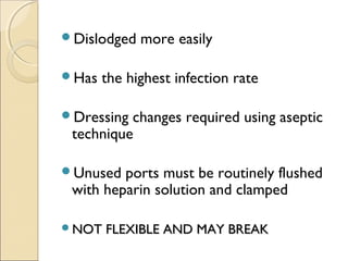
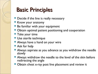
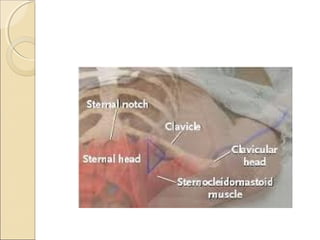
CCeennttrraall VVeennoouuss 
CCaatthheetteerriizzaattiioonn 
by, 
Dr G.RAJASEKHAR 
MBBS,DCH 
MODERATORS 
Dr SREEDEVI 
MD 
Dr PADMAJA 
MD
CCeennttrraall vveennoouuss ccaatthheetteerr 
Central venous access is the placement 
of a venous catheter in a vein that leads 
directly to the heart.
OObbjjeeccttiivveess 
Types of catheters 
Indications and Contraindications 
Technique 
Basic principles 
Specifics by Site 
complicatons 
Tips
TTYYPPEE OOFF CCEENNTTRRAALL VVEENNOOUUSS 
CCAATTHHEETTEERR 
Patient’s condition 
Anticipated length of therapy
TTyyppeess OOff CCeennttrraall VVeennoouuss CCaatthheetteerrss 
Nontunneled central catheters 
Tunneled central catheters 
Peripherally inserted central catheters 
(PICC) 
Implantable ports
NNOONNTTUUNNNNEELLEEDD CCEENNTTRRAALL 
CCAATTHHEETTEERRTTSS
NNOONNTTUUNNNNEELLEEDD CCEENNTTRRAALL 
CCAATTHHEETTEERRTTSS 
POLYURETHANE 
SINGLE OR MULTIPLE LUMENS 
USED FOR SHORT TERM THEARAPY 
EASIER PLACEMENT,REMOVAL AND 
REPLACEMENT 
USUALLY 6to8 INCHES IN LENGTH 
CAN BE QUICKLY INSERTED
Dislodged more easily 
Has the highest infection rate 
Dressing changes required using aseptic 
technique 
Unused ports must be routinely flushed 
with heparin solution and clamped 
NNOOTT FFLLEEXXIIBBLLEE AANNDD MMAAYY BBRREEAAKK
TTUUNNNNEELLEEDD CCEENNTTRRAALL VVEENNOOUUSS 
CCAATTHHEETTEERRSS
Single or multiple lumens 
Flow variable 
Log term 
Inserted surgically 
Cuff –dacron, vita 
No dressing is required after cuff heals 
unless the patient isimmunocompromised
Tunneled catheter
Peripherally IInnsseerrtteedd CCeennttrraall 
CCaatthheetteerrss ((PPIICCCC)) 
Used for intermediate to long term 
therapy 
May be single or double lumen 
Inserted percutaneously 
◦ Basalic vein 
◦ Cephalic vein 
Threaded into the superior vena cava
PPIICCCC 
SILASTIC OR POLYURETHANE 
SINGLE OR DOUBLE LUMEN 
LOW FLOW 
SHORT-LONGTERM 
EASY ACCESS
SSUUBBCCUUTTAANNEEOOUUSS PPOORRTTSS 
SINGLE OR DOUBLELUMEN 
FLOW-MOST COMMONLY SLOW 
LONG TERM 
ACCESS REQUIRES NEEDLE PUNCTURE
LESS MAINTENANCE 
ACTIVITY IS UNLIMETED AFTER SITE HEALS 
COSMETICALLY MORE APPEALING 
CONCEALED PACKET RETARDS 
INFECTION
Minimizes infection 
Huber needle must be used to access port 
Must always confirm needle placement before 
med administration 
Unused port is flushed every 28 days with 
Heparin solution
IInnddiiccaattiioonnss 
Central venous pressure monitoring 
Volume resuscitation 
Infusion of hyperalimentation 
Infusion of concentrated solutions 
Placement of transvenous pacemaker 
Cardiac catheterization & pulmonary angiography 
Temporary Hemodialysis 
Lack of peripheral access
RReellaattiivvee CCoonnttrraaiinnddiiccaattiioonnss 
Bleeding disorders 
Anticoagulation or thrombolytic therapy 
Distorted local anatomy 
Cellulitis, burns, severe dermatitis at site 
Vasculitis
TTeecchhnniiqquuee 
Seldinger technique 
◦ Use introducing needle to locate vein 
◦ Wire is threaded through the needle 
◦ Needle is removed 
◦ Skin and vessel are dilated 
◦ Catheter is placed over the wire 
◦ Wire is removed 
◦ Catheter is secured in place
BBaassiicc PPrriinncciipplleess 
Decide if the line is really necessary 
Know your anatomy 
Be familiar with your equipment 
Obtain optimal patient positioning and cooperation 
Take your time 
Use sterile technique 
Always have a hand on your wire 
Ask for help 
Always aspirate as you advance as you withdraw the needle 
slowly 
Always withdraw the needle to the level of the skin before 
redirecting the angle 
Obtain chest x-ray post line placement and review it
Location Advantage Disadvantage 
Internal 
Jugular 
• Bleeding can be recognized 
and controlled 
• Malposition is rare 
• Less risk of pneumothorax 
• Risk of carotid artery puncture 
• PTX possible 
Femoral • Easy to find vein 
• No risk of pneumothorax 
• Preferred site for 
emergencies and CPR 
• Fewer bad complications 
• Highest risk of infection 
• Risk of DVT 
• Not good for ambulatory 
patients 
Subclavian • Most comfortable for 
conscious patients 
• Highest risk of PTX, should 
not do on intubated pts 
• Should not be done if < 2 years 
• Vein is non-compressible
SSuubbccllaavviiaann AApppprrooaacchh 
Positioning 
◦ Right side preferred 
◦ Supine position, head neutral, arm abducted 
◦ Trendelenburg (10-15 degrees) 
◦ Shoulders neutral with mild retraction 
Needle placement 
◦ Junction of middle and medial thirds of clavicle 
◦ At the small tubercle in the medial deltopectoral groove 
◦ Needle should be parallel to skin 
◦ Aim towards the suprasternal notch and just under the clavicle
IInntteerrnnaall JJuugguullaarr AApppprrooaacchh 
Positioning 
◦ Right side preferred 
◦ Trendelenburg position 
◦ Head turned slightly away from side of venipuncture 
Needle placement: Central approach 
◦ Locate the triangle formed by the clavicle and the sternal and 
clavicular heads of the SCM muscle 
◦ Gently place three fingers of left hand on carotid artery 
◦ Place needle at 30 to 40 degrees to the skin, lateral to the carotid 
artery 
◦ Aim toward the ipsilateral nipple under the medial border of the 
lateral head of the SCM muscle 
◦ Vein should be 1-1.5 cm deep, avoid deep probing in the neck
Internal Jugular Central Approach
FFeemmoorraall AApppprrooaacchh 
Positioning 
◦ Supine 
Needle placement 
◦ Medial to femoral artery 
◦ Needle held at 45 degree angle 
◦ Skin insertion 2 cm below inguinal ligament 
◦ Aim toward umbilicus
Femoral nerve 
Femoral artery 
Femoral Vein 
NAVEL
PPoosstt--CCaatthheetteerr PPllaacceemmeenntt 
Aspirate blood from each port 
Flush with saline or sterile water 
(heparinised) 
Secure catheter with sutures 
Cover with sterile dressing (tega-derm) 
Obtain chest x-ray for IJ and SC lines 
Write a procedure note
CCoommpplliiccaattiioonnss 
Vascular 
◦ Air embolus 
◦ Arterial puncture 
◦ Arteriovenous fistula 
◦ Hematoma 
◦ Blood clot 
Infectious 
◦ Sepsis, cellulitis, osteomyelitis, septic arthritis 
Miscellaneous 
◦ Dysrhythmias 
◦ Catheter knotting or malposition 
◦ Nerve injury 
◦ Pneumothorax, hemothorax, hydrothorax, 
hemomediastinum 
◦ Bowel or bladder perforation
TTiippss 
After 3-4 tries, let someone else try 
Get cheast x-ray after unsuccessful attempt 
If attempt at one site fails, try new site on same side to 
avoid bilateral complications 
Halt positive pressure ventilation as the needle penetrates 
the chest wall in subclavian approach 
If you meet resistance while inserting the guide wire, 
withdraw slightly and rotate the wire and re-advance 
Align the bevel with the syringe markings 
Withdraw slowly, you will often hit the vein on the way out
Ultrasound-GGuuiiddeedd CCeennttrraall VVeennoouuss 
AAcccceessss 
 Becoming standard of care 
Vein is compressible 
Vein is not always larger 
Vein is accessed under direct 
visualization 
Helpful in patients with difficult 
anatomy
Needle entering IJ
Femoral 
Vein 
Femoral 
Artery 
Compression of vein 
with US probe
CCaatthheetteerriizzaattiioonn KKiittss
Central venous catheterization

Central venous catheterization

  • 1.
    CCeennttrraall VVeennoouuss CCaatthheetteerriizzaattiioonn by, Dr G.RAJASEKHAR MBBS,DCH MODERATORS Dr SREEDEVI MD Dr PADMAJA MD
  • 2.
    CCeennttrraall vveennoouuss ccaatthheetteerr Central venous access is the placement of a venous catheter in a vein that leads directly to the heart.
  • 3.
    OObbjjeeccttiivveess Types ofcatheters Indications and Contraindications Technique Basic principles Specifics by Site complicatons Tips
  • 4.
    TTYYPPEE OOFF CCEENNTTRRAALLVVEENNOOUUSS CCAATTHHEETTEERR Patient’s condition Anticipated length of therapy
  • 5.
    TTyyppeess OOff CCeennttrraallVVeennoouuss CCaatthheetteerrss Nontunneled central catheters Tunneled central catheters Peripherally inserted central catheters (PICC) Implantable ports
  • 6.
  • 7.
    NNOONNTTUUNNNNEELLEEDD CCEENNTTRRAALL CCAATTHHEETTEERRTTSS POLYURETHANE SINGLE OR MULTIPLE LUMENS USED FOR SHORT TERM THEARAPY EASIER PLACEMENT,REMOVAL AND REPLACEMENT USUALLY 6to8 INCHES IN LENGTH CAN BE QUICKLY INSERTED
  • 8.
    Dislodged more easily Has the highest infection rate Dressing changes required using aseptic technique Unused ports must be routinely flushed with heparin solution and clamped NNOOTT FFLLEEXXIIBBLLEE AANNDD MMAAYY BBRREEAAKK
  • 10.
  • 11.
    Single or multiplelumens Flow variable Log term Inserted surgically Cuff –dacron, vita No dressing is required after cuff heals unless the patient isimmunocompromised
  • 12.
  • 15.
    Peripherally IInnsseerrtteedd CCeennttrraall CCaatthheetteerrss ((PPIICCCC)) Used for intermediate to long term therapy May be single or double lumen Inserted percutaneously ◦ Basalic vein ◦ Cephalic vein Threaded into the superior vena cava
  • 16.
    PPIICCCC SILASTIC ORPOLYURETHANE SINGLE OR DOUBLE LUMEN LOW FLOW SHORT-LONGTERM EASY ACCESS
  • 18.
    SSUUBBCCUUTTAANNEEOOUUSS PPOORRTTSS SINGLEOR DOUBLELUMEN FLOW-MOST COMMONLY SLOW LONG TERM ACCESS REQUIRES NEEDLE PUNCTURE
  • 19.
    LESS MAINTENANCE ACTIVITYIS UNLIMETED AFTER SITE HEALS COSMETICALLY MORE APPEALING CONCEALED PACKET RETARDS INFECTION
  • 20.
    Minimizes infection Huberneedle must be used to access port Must always confirm needle placement before med administration Unused port is flushed every 28 days with Heparin solution
  • 22.
    IInnddiiccaattiioonnss Central venouspressure monitoring Volume resuscitation Infusion of hyperalimentation Infusion of concentrated solutions Placement of transvenous pacemaker Cardiac catheterization & pulmonary angiography Temporary Hemodialysis Lack of peripheral access
  • 23.
    RReellaattiivvee CCoonnttrraaiinnddiiccaattiioonnss Bleedingdisorders Anticoagulation or thrombolytic therapy Distorted local anatomy Cellulitis, burns, severe dermatitis at site Vasculitis
  • 24.
    TTeecchhnniiqquuee Seldinger technique ◦ Use introducing needle to locate vein ◦ Wire is threaded through the needle ◦ Needle is removed ◦ Skin and vessel are dilated ◦ Catheter is placed over the wire ◦ Wire is removed ◦ Catheter is secured in place
  • 26.
    BBaassiicc PPrriinncciipplleess Decideif the line is really necessary Know your anatomy Be familiar with your equipment Obtain optimal patient positioning and cooperation Take your time Use sterile technique Always have a hand on your wire Ask for help Always aspirate as you advance as you withdraw the needle slowly Always withdraw the needle to the level of the skin before redirecting the angle Obtain chest x-ray post line placement and review it
  • 27.
    Location Advantage Disadvantage Internal Jugular • Bleeding can be recognized and controlled • Malposition is rare • Less risk of pneumothorax • Risk of carotid artery puncture • PTX possible Femoral • Easy to find vein • No risk of pneumothorax • Preferred site for emergencies and CPR • Fewer bad complications • Highest risk of infection • Risk of DVT • Not good for ambulatory patients Subclavian • Most comfortable for conscious patients • Highest risk of PTX, should not do on intubated pts • Should not be done if < 2 years • Vein is non-compressible
  • 28.
    SSuubbccllaavviiaann AApppprrooaacchh Positioning ◦ Right side preferred ◦ Supine position, head neutral, arm abducted ◦ Trendelenburg (10-15 degrees) ◦ Shoulders neutral with mild retraction Needle placement ◦ Junction of middle and medial thirds of clavicle ◦ At the small tubercle in the medial deltopectoral groove ◦ Needle should be parallel to skin ◦ Aim towards the suprasternal notch and just under the clavicle
  • 30.
    IInntteerrnnaall JJuugguullaarr AApppprrooaacchh Positioning ◦ Right side preferred ◦ Trendelenburg position ◦ Head turned slightly away from side of venipuncture Needle placement: Central approach ◦ Locate the triangle formed by the clavicle and the sternal and clavicular heads of the SCM muscle ◦ Gently place three fingers of left hand on carotid artery ◦ Place needle at 30 to 40 degrees to the skin, lateral to the carotid artery ◦ Aim toward the ipsilateral nipple under the medial border of the lateral head of the SCM muscle ◦ Vein should be 1-1.5 cm deep, avoid deep probing in the neck
  • 32.
  • 33.
    FFeemmoorraall AApppprrooaacchh Positioning ◦ Supine Needle placement ◦ Medial to femoral artery ◦ Needle held at 45 degree angle ◦ Skin insertion 2 cm below inguinal ligament ◦ Aim toward umbilicus
  • 34.
    Femoral nerve Femoralartery Femoral Vein NAVEL
  • 35.
    PPoosstt--CCaatthheetteerr PPllaacceemmeenntt Aspirateblood from each port Flush with saline or sterile water (heparinised) Secure catheter with sutures Cover with sterile dressing (tega-derm) Obtain chest x-ray for IJ and SC lines Write a procedure note
  • 36.
    CCoommpplliiccaattiioonnss Vascular ◦Air embolus ◦ Arterial puncture ◦ Arteriovenous fistula ◦ Hematoma ◦ Blood clot Infectious ◦ Sepsis, cellulitis, osteomyelitis, septic arthritis Miscellaneous ◦ Dysrhythmias ◦ Catheter knotting or malposition ◦ Nerve injury ◦ Pneumothorax, hemothorax, hydrothorax, hemomediastinum ◦ Bowel or bladder perforation
  • 37.
    TTiippss After 3-4tries, let someone else try Get cheast x-ray after unsuccessful attempt If attempt at one site fails, try new site on same side to avoid bilateral complications Halt positive pressure ventilation as the needle penetrates the chest wall in subclavian approach If you meet resistance while inserting the guide wire, withdraw slightly and rotate the wire and re-advance Align the bevel with the syringe markings Withdraw slowly, you will often hit the vein on the way out
  • 38.
    Ultrasound-GGuuiiddeedd CCeennttrraall VVeennoouuss AAcccceessss  Becoming standard of care Vein is compressible Vein is not always larger Vein is accessed under direct visualization Helpful in patients with difficult anatomy
  • 39.
  • 40.
    Femoral Vein Femoral Artery Compression of vein with US probe
  • 42.

Editor's Notes

  • #4 Basic materials section involves going through an actual catheter kit with them and demonstrating technique
  • #23 Central venous pressure monitoring – for those whose volume status needs to be managed closely Volume loading – flow rate through a 14 gauge peripheral line is twice that of a 20cm 16 gauge central venous catheter Concentrated solutions – potassium chloride, hyperosmolar saline, chemo agents. Or vasoactive substances like epi, dopamine. All can cause tissue irritation or necrosis if extravasated in peripheral line
  • #24 Bleeding disorders – even with platelet counts &amp;lt;50,000, bleeding is uncommon and easily managed, in the absence of arterial puncture Distorted local anatomy – ultrasound may help
  • #25 Seldinger originally described this technique in 1953 for percutaneous arteriography.
  • #28 UNC preferred site – in the hospital manual
  • #29 Arm abduction flattens the deltoid bulge Trendelenburg reduces incidence of air embolism Shoulders – as the shoulder falls backward, the space between the clavicle and first rib narrows, making the subclavian vein less accessible Right side preferred – lower pleural dome and thoracic duct on left Junction of the middle and medial thirds of the clavicle – here the vein in just posterior to the clavicle and just above the first rib which acts as a barrier to the pleura.
  • #31 Right side preferred – left IJ is more circuitous, thoracic duct on left Trendelenburg – IJ is distensible Central approach is most common Anterior approach has highest risk of puncturing carotid artery
  • #34 The more distal you are from the inguinal ligament, the closer the vein is to the artery as the femoral vein begins to dive behind the artery and the saphenous vein comes off the femoral vein.
  • #35 NAVEL – N = nerve, A = artery, V = vein, E = empty space, L = lymphatics (must be read from right side of body, L is always medial. So it is spelled backwards from the left side approach
  • #38 Resistance during wire advancement comes from incorrect placement or from valves and tortuous vessels which can be overcome with GENTLE manipulation of the guide wire
  • #43 Go over kits and demonstrate procedure with students