Download as PDF, PPTX


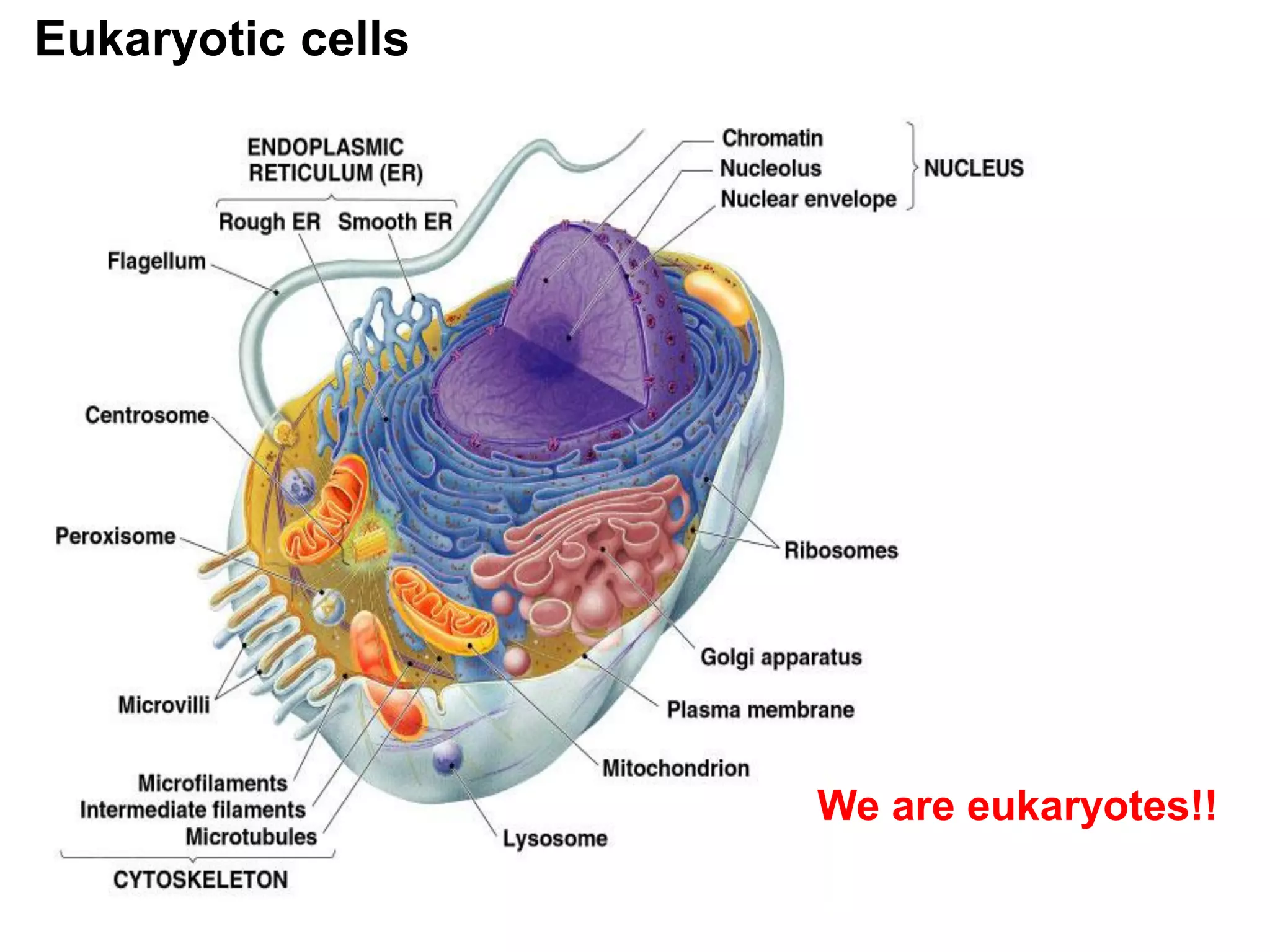
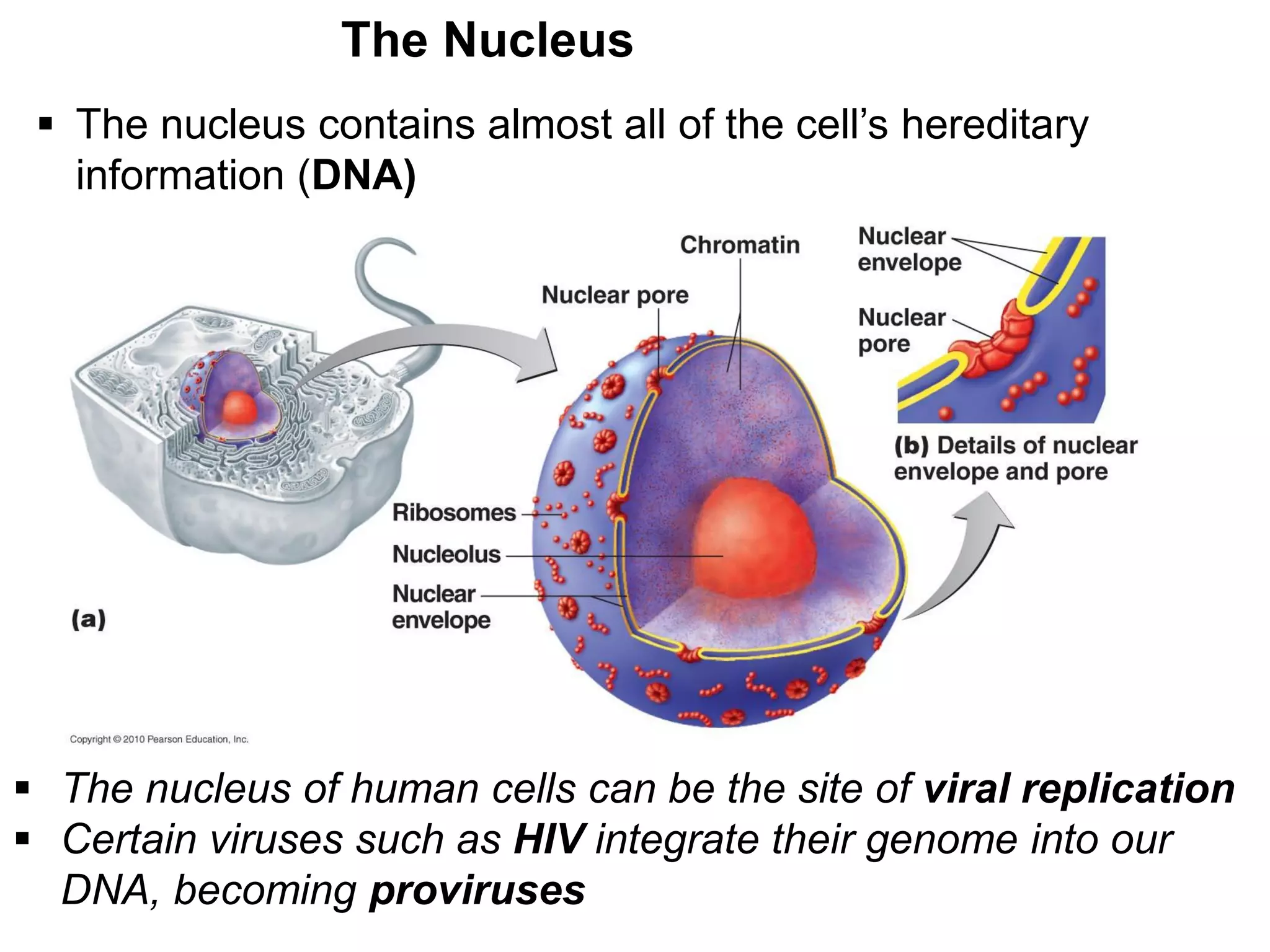
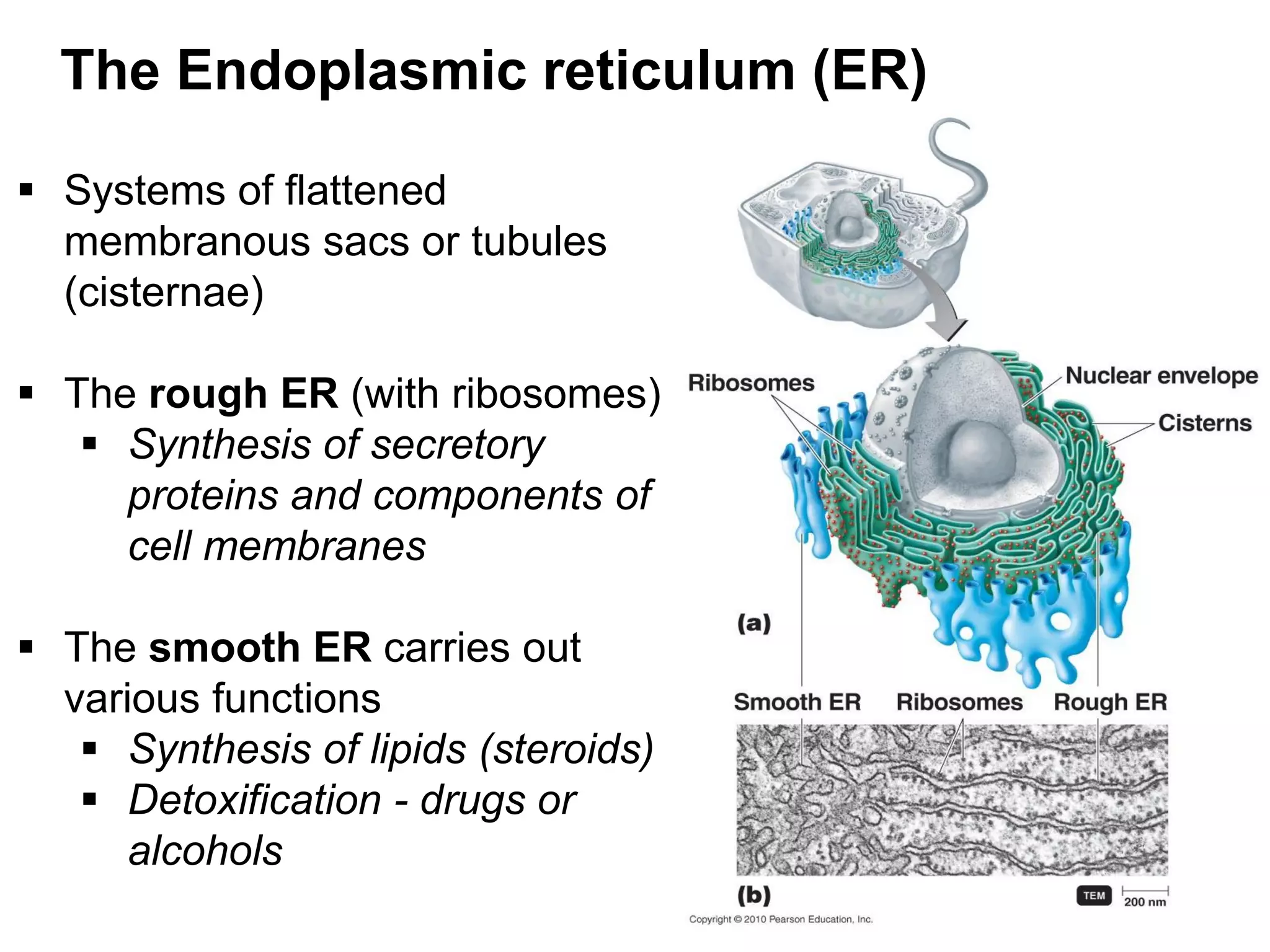


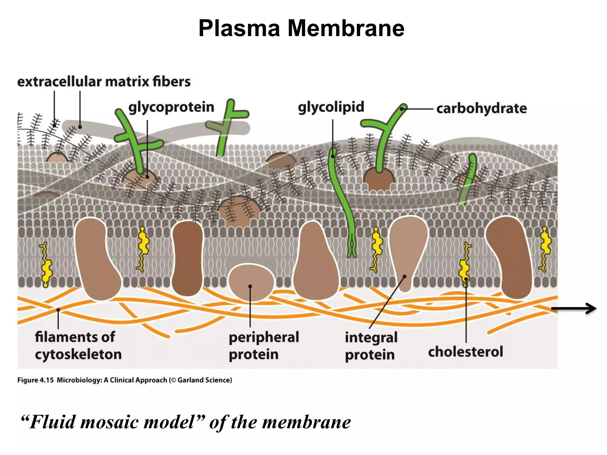
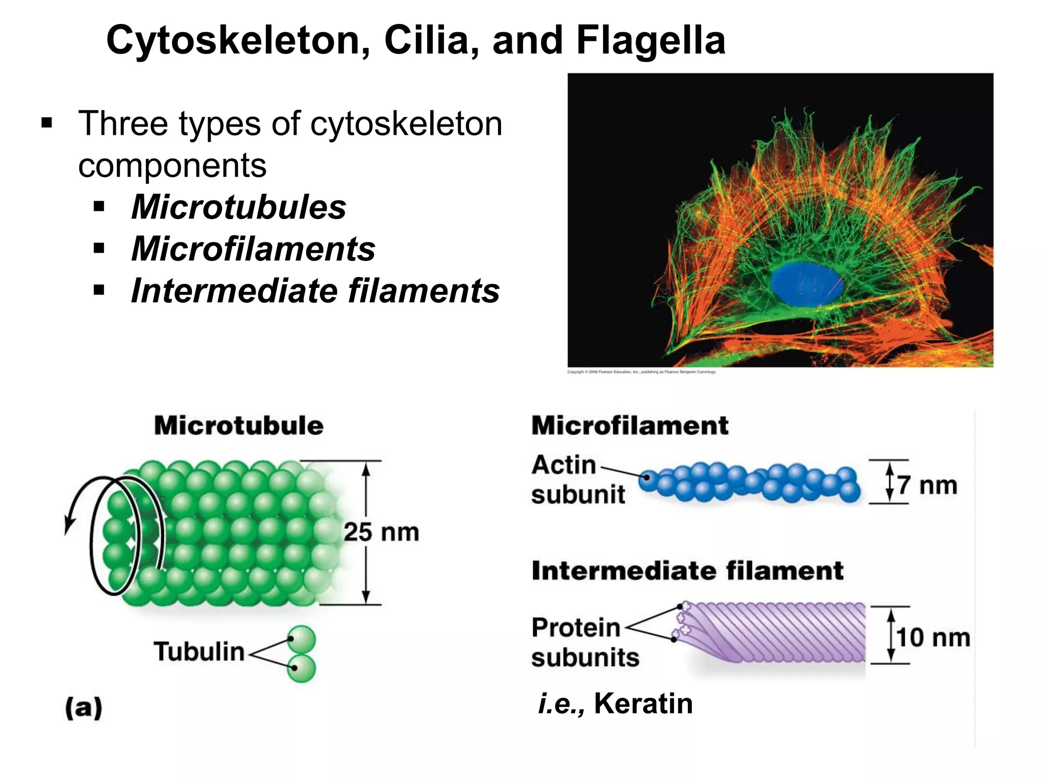
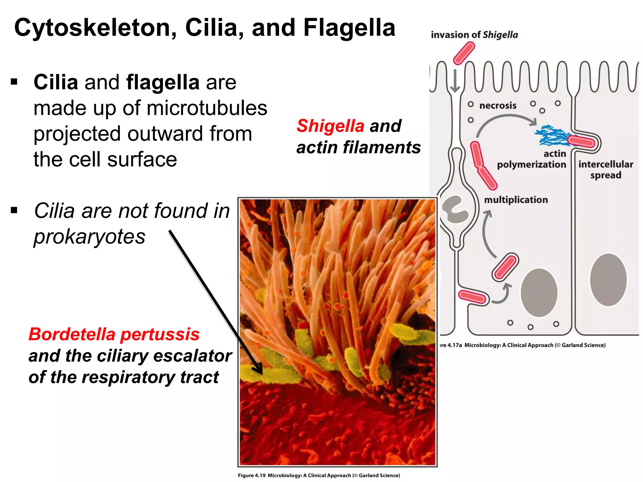
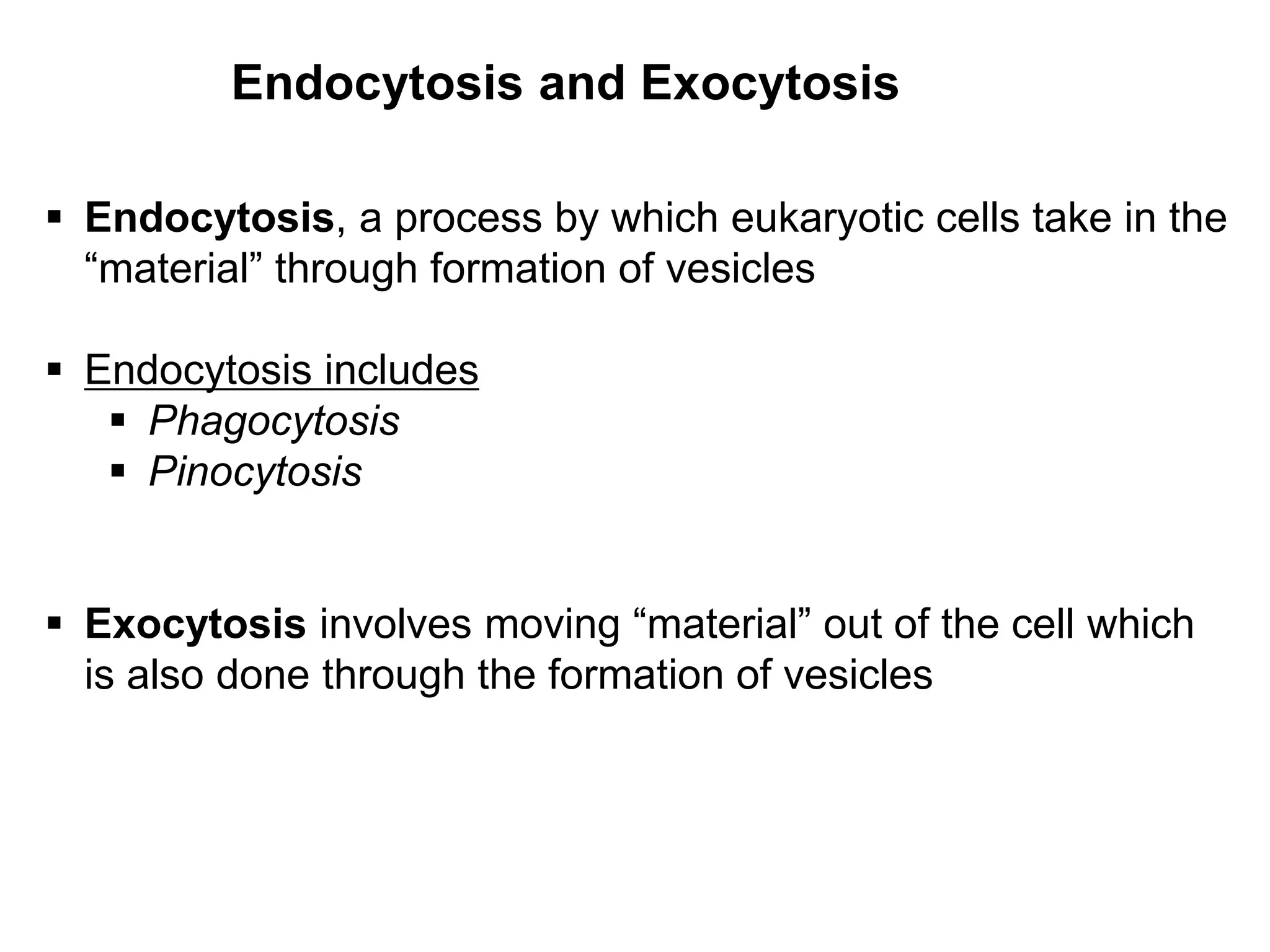
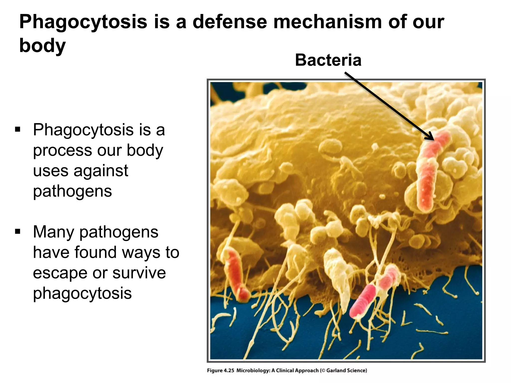


This document provides an overview comparison of prokaryotic and eukaryotic cells. It describes several key structures and organelles found in eukaryotic cells that are not present in prokaryotes, including the nucleus, endoplasmic reticulum, Golgi apparatus, mitochondria, lysosomes, peroxisomes, and cytoskeleton components like microtubules, microfilaments, and intermediate filaments. The document also discusses differences in cell membranes, ribosomes, and processes like endocytosis, exocytosis, phagocytosis, and the use of flagella and cilia for motility. Overall, the document outlines some of the major distinguishing cellular features of eukaryotes versus prokaryotes.


![BACTERIA STRUCTURE AND FUNCTION [Autosaved].pptx](https://cdn.slidesharecdn.com/ss_thumbnails/bacteriastructureandfunctionautosaved-230206013850-80c9a713-thumbnail.jpg?width=640&height=640&fit=bounds)