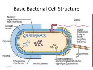
BACTERIA STRUCTURE
AND FUNCTION
Basic Bacterial Cell Structure
2/6/2023 2
INTRODUCTION
• Bacteria form a large group of single-celled, parasitic,
saprophytic and free-living microorganisms.
• They are prokaryotic and reproduce by binary fission
• Varying size from 0.1- 10um in length.
• The extreme small size makes room for high surface
area to volume ratio.
• They have a simple cell structure, contain both DNA
and RNA and lack a nuclear membrane
2/6/2023 3
• Most bacteria are capable of independent metabolic
existence and growth.
Exceptions: Chlamydia and Rickettsia are obligately
intracellular organisms
• They exist with different shapes – spherical(cocci),
rod(bacilli), comma or spiral shapes.
• Various arrangement – chains, pairs, clusters etc
• Their importance in medicine cannot be over-emphasized
as they are responsible for various diseases.
2/6/2023 4
2/6/2023 5
2/6/2023 6
2/6/2023 7
BACTERIA CELL STRUCTURE
3 basic components:
1. Surface appendages
2. Surface layers
3. Intracytoplasmic structures
2/6/2023 8
SURFACE
APPENDAGES
2/6/2023 9
SURFACE APPENDAGES
• 2 basic types of surface appendages:
1.FLAGELLA [organs of locomotion]
- thread-like appendages present on both Gram
positive and Gram negative bacteria
- their presence is useful for identification.
2.FIMBRIAE (PILI)
- found mostly on Gram negative bacteria and a few
Gram positives .e.g. Corynebacterium renale.
Some bacteria have both fimbriae and flagella. e.g.
E.coli which has numerous flagella and 2 types of
pili.
2/6/2023 10
Flagella
• Chemically, flagella are composed of many
subunits of a single protein called flagellin.
• A flagellum consists of 3 parts:
1. The long filament, which lies external to the cell
surface
2. The hook structure at the end of the filament
3. The basal body, to which the hook is anchored and
which imparts motion to the flagellum
2/6/2023 11
12
2/6/2023
• Functions of flagella:
1. Allow organisms to migrate towards favorable
growth environment(nutrients and other
chemoattractants via chemotaxis) and away from
those that might be harmful.
2. They play a role in the pathogenesis of UTIs by
propelling the bacteria up the urethra into the
bladder.
3. Some species of bacteria(e.g. Salmonella sp.)
are identified in the clinical laboratory by the use
of specific antibodies against flagella proteins
2/6/2023 13
2/6/2023 14
• Flagella can be sheared from the cell surface
without affecting the viability of the cell
The cell then becomes temporarily non motile
In time it synthesizes new flagella and regains motility.
• The protein synthesis inhibitor chloramphenicol,
however, blocks regeneration of flagella
• NB: motility is observed in young cultures (18-24
hours or less); bacteria tend to become non motile
in older cultures because of production of acid and
other toxic products
2/6/2023 15
Motility
• Types of bacterial motility
– run or swim - when a bacterium moves in
one direction for a length of time
- tumbles - periodic, abrupt random changes
in direction
– swarming - rapid wavelike growth across a
solid culture medium
2/6/2023 16
Detection of Motility
• Direct- Hanging drop
method
• Indirect
2/6/2023 17
SWARMING
2/6/2023 18
FIMBRIAE (PILI)
• Fimbriae [pili] are slender, hair-like, proteinaceous
filaments on the surface of many Gram negative
bacteria
• Shorter, finer and more rigid than flagella, 4-8nm
long and 1-5nm thick
• Composed of protein subunits referred to as pilins
• Minor proteins termed adhesins are located on
the tips of the pili and are responsible for the
attachment properties.
2/6/2023 19
Pili
2/6/2023 20
Pili cont’d
• Pili are anchored on the outer membrane of the
Gram negative bacteria
• 2types of pili are recognised:
1. Ordinary pili- short, abundant, for adhesion
2. Sex pili – a small number (one to six), very long,
for DNA transfer during conjugation
2/6/2023 21
22
F-Pili in Conjugation
2/6/2023
• Pili have two important roles;
• 1. they mediate the attachment of bacteria to
specific receptors on the human cell surface.
• 2. specialized sex pilus-------conjugation
2/6/2023 23
SURFACE LAYERS
2/6/2023 24
SURFACE LAYERS
• The principal surface layers are:
- Capsules and Loose Slime
- Cell wall of Gram positive bacteria
- Complex cell envelope of Gram negative bacteria
- Cytoplasmic membranes
- Mesosomal membrane vesicles
2/6/2023 25
CAPSULE
• High molecular weight extracellular condensed
polymers surrounding bacteria (up to 10um thick)
• Synthesized by enzymes located on the surface of
the cells
• For most bacteria, it is made up of polysaccharides
(except B. anthracis- D-glutamic acid)
2/6/2023 26
• LOOSE SLIME- formed when the extracellular polymers
are detached from the cell but the cell is entrapped in it
• GLYCOCALYX- loose meshwork of fibrils extending from
the cell. This form the basis for the formation of biofilm
- Plays a role in adherence of bacteria to
surfaces. This form the basis for the formation of
biofilm
Dental caries – Strept mutans forms biofilm on which
other bacteria adhere to form plaque, the precondition
for the destruction of the enamel and ultimate
formation of caries
2/6/2023 27
Dental plaque
2/6/2023 29
IMPORTANCE OF CAPSULES
• Capsules are known to confer resistance to phagocytosis
• Specific identification of an organism can be made by using
antiserum against the capsular polysaccharide.(quellung
reaction)
• The adherence role of glycocalyx is the basis for the formation
of biofilm
• Capsular polysaccharides are used as the antigens in certain
vaccines.
• The capsules may play a role in adherence of bacteria to
human tissues.
2/6/2023 30
CELL WALL
• Lies between the cytoplasmic membrane and capsule
• Has a high tensile strength due to peptidoglycan (aka
murein or mucopeptide)
• Important for bacterial ID- bacteria are classified as
Gram +ve or Gram –ve
2/6/2023 31
Cell wall
2/6/2023 32
Importance of cell wall:
• Protects protoplasm from external noxious stimuli
• Withstands and maintains the osmotic pressure
gradient between the cell interior and extracellular
environment
• Gives the cell its structural support and maintains its
characteristic shape.
• Facilitates communication with the environment
• Plays a role in cell division
• It is a good target for antibacterial agents
2/6/2023 33
CELL WALL OF GRAM POSITIVE
BACTERIA
Gram +ve bacterial cell wall composed of:
• Peptidoglycan- may be as much as 40-50 layers
• Teichoic acid; peculiar to Gram positive bacteria. They are
composed of polymers of glycerol phosphate or ribitol
phosphate. Two types:-
– Membrane teichoic acid- covalently linked to membrane
glycolipid therefore called lipoteichoic acid.
– Wall teichoic acid- attached covalently to peptidoglycan
• Cell wall associated protein
– In Streptococcus pyogenes LTA is associated with the Mprotein
that protrudes from the cell membrane through the
peptidoglycan layer.
34
Wall of Gram +ve Bacteria
2/6/2023 35
• PEPTIDOGLYCAN LAYER made up of 3 parts
1. A glycan back bone composed of alternating N-
acetylmuramic acid (NAM) and N-
acetylglucosamine (NAG) linked together by B-
1,4 glycosidic linkages.
2. A set of identical tetrapeptide side chains
attached to N-acetylmuramic acid ( L-Alanine, D-
Isoglutamine, L-Lysine and D-Alanine)
3. A set of identical cross-bridges between
neighbouring tetrapeptide side chains
2/6/2023 36
The Structure of Peptidoglycan
2/6/2023 37
2/6/2023 38
CELL ENVELOPE OF GRAM NEGATIVE BACTERIA
• Highly complex and multilayered of abt 5-10nm thickness
• Composed of:
1. A peptidoglycan layer- relatively thinner
abt 2nm thick (5-10% of dry mass)
2. Outer membrane- bilayered structure
3. Lipopolysaccharides – attached to outer membrane
4. Lipoprotein – cross-link outer membrane & peptidoglycan
5. Periplasmic space – space bw inner and outer membrane
2/6/2023 39
Cell Envelope of Gram –ve Bacteria
2/6/2023 40
OUTER MEMBRANE
• Lipopolysaccharides is attached to it by a weak
cohesive forces
• Lipoprotein and ompA anchor outer membrane to
the peptidoglycan layer
2/6/2023 41
LIPOPOLYSACCHARIDE
• Only one Gram +ve bacteria contains LPS- Listeria
monocytogenes
• LPS (endotoxins) is released when the cells are lysed
• The LPS molecule have 3 covalently linked regions:
(1) Lipid A, (2) Core polysaccharide, (3) O- specific
polysaccharide chain (O-Antigen)
2/6/2023 42
Lipopolysaccharide complex
2/6/2023 43
2/6/2023 44
BACTERIA LACKING CELL WALL
• Bacteria lacking cell walls- 4 groups have been identified:
1. Mycoplasma spp- Mycoplasma pneumonia, Ureaplasma
urealyticum
2. L-forms- discovered in Lister Institute, London. Produced in
lab but could be found in the body of patients being treated
with penicillin
3. Protoplasts- Gram positive bacteria lost cell wall to lysozyme.
4. Spheroplasts- Gram negative bacteria loss cell wall to
lysozymes
2/6/2023 45
Bacterial Identification
India ink technique -
– Bacteria is suspended in diluted India ink
– Bacterial cells appear to lie in a lacunae
Quellung reaction - Homologous antibody is added to a
preparation of capsule.
– microprecipitation at the periphery of the capsule altering its
refractive index rendering the capsule to be visible
2/6/2023 46
Quellung reaction
2/6/2023 47
Gram stain technique
2/6/2023 48
2/6/2023 49
PERIPLASMIC SPACE
• Region between outer and inner membrane
(cytoplasmic membrane)
• Approximately 20-40% of cell volume
• Periplasmic space contains peptidoglycan layer and
gel-like solution of protein including antibiotics
degrading enzymes (e.g B-lactamase and
aminoglycoside phosphorylase)
2/6/2023 50
CYTOPLASMIC MEMBRANE
• Plasma [cytoplasmic ]membrane: composed
primarily of lipids bilayer with interspersing proteins
• Protein to lipid ratio is usually 3:1
• Thickness about 4-5nm
• Lack sterols (except for Mycoplasma )
2/6/2023 51
Functions of the cytoplasmic membrane
• Transport of nutrients into and waste products out of
the cell by providing high SA/volume ratio
• Site of electron transport and oxidative
phosphorylation
• Anchoring site for DNA
• Provides the cell with mechanism for the separation
of sister chromosomes.
• Excretion of hydrolytic exoenzymes for the digestion
of macromolecular organic polymer nutrients
2/6/2023 52
INTRACYTOPLASMIC
STRUCTURES
2/6/2023 53
Cytoplasm
• 80% Water {20% Salts-Proteins)
• DNA is circular, Haploid
• Plasmids; extra circular DNA
–Antibiotic Resistance
• No organelles ( no mitochondria, golgi, etc.)
• Inclusion bodies/granules; e.g. glycogen,
polyphosphates
2/6/2023 54
NUCLEOID
• Contain a single double stranded DNA located in the
cytoplasm
• The DNA can be visualized with the aid of Feulgen
staining, and is seen as a single, continuous, giant
circular molecule with molecular weight of approx.
3 x 10⁹. The unfolded nuclear DNA would be about
1mm long.
• Nucleoid lacks nuclear membrane and mitotic
apparatus
2/6/2023 55
2/6/2023 56
2/6/2023 57
PLASMIDS
• Extrachromosomal genetic apparatus capable of
replication within prokaryotic cell line
• Circular twisted DNA molecule that transfer genetic
information from one cell to another
• Drug resistance plasmids renders bacteria
resistance to antibiotic treatment
2/6/2023 58
Cell division in Prokaryotes
• Prokaryotes use a relatively
simple form of cell division -
binary fission.
• The cell wall and membrane
are in red,
• the bacterial chromosome in
blue,
• the cytoplasm in light green,
• the yellow dot represents a
point of attachment of the
chromosome to the cell
membrane.
2/6/2023 59
RIBOSOMES
• Densely packed ribosomes of 70S in size
• Each 70S ribosome has 30S and 50S subunits
• Ribosomes are involved in the synthesis of various
bacteria proteins
2/6/2023 60
2/6/2023 61
BACTERIA STRUCTURE AND FUNCTION [Autosaved].pptx

BACTERIA STRUCTURE AND FUNCTION [Autosaved].pptx

  • 1.
  • 2.
    Basic Bacterial CellStructure 2/6/2023 2
  • 3.
    INTRODUCTION • Bacteria forma large group of single-celled, parasitic, saprophytic and free-living microorganisms. • They are prokaryotic and reproduce by binary fission • Varying size from 0.1- 10um in length. • The extreme small size makes room for high surface area to volume ratio. • They have a simple cell structure, contain both DNA and RNA and lack a nuclear membrane 2/6/2023 3
  • 4.
    • Most bacteriaare capable of independent metabolic existence and growth. Exceptions: Chlamydia and Rickettsia are obligately intracellular organisms • They exist with different shapes – spherical(cocci), rod(bacilli), comma or spiral shapes. • Various arrangement – chains, pairs, clusters etc • Their importance in medicine cannot be over-emphasized as they are responsible for various diseases. 2/6/2023 4
  • 5.
  • 6.
  • 7.
  • 8.
    BACTERIA CELL STRUCTURE 3basic components: 1. Surface appendages 2. Surface layers 3. Intracytoplasmic structures 2/6/2023 8
  • 9.
  • 10.
    SURFACE APPENDAGES • 2basic types of surface appendages: 1.FLAGELLA [organs of locomotion] - thread-like appendages present on both Gram positive and Gram negative bacteria - their presence is useful for identification. 2.FIMBRIAE (PILI) - found mostly on Gram negative bacteria and a few Gram positives .e.g. Corynebacterium renale. Some bacteria have both fimbriae and flagella. e.g. E.coli which has numerous flagella and 2 types of pili. 2/6/2023 10
  • 11.
    Flagella • Chemically, flagellaare composed of many subunits of a single protein called flagellin. • A flagellum consists of 3 parts: 1. The long filament, which lies external to the cell surface 2. The hook structure at the end of the filament 3. The basal body, to which the hook is anchored and which imparts motion to the flagellum 2/6/2023 11
  • 12.
  • 13.
    • Functions offlagella: 1. Allow organisms to migrate towards favorable growth environment(nutrients and other chemoattractants via chemotaxis) and away from those that might be harmful. 2. They play a role in the pathogenesis of UTIs by propelling the bacteria up the urethra into the bladder. 3. Some species of bacteria(e.g. Salmonella sp.) are identified in the clinical laboratory by the use of specific antibodies against flagella proteins 2/6/2023 13
  • 14.
  • 15.
    • Flagella canbe sheared from the cell surface without affecting the viability of the cell The cell then becomes temporarily non motile In time it synthesizes new flagella and regains motility. • The protein synthesis inhibitor chloramphenicol, however, blocks regeneration of flagella • NB: motility is observed in young cultures (18-24 hours or less); bacteria tend to become non motile in older cultures because of production of acid and other toxic products 2/6/2023 15
  • 16.
    Motility • Types ofbacterial motility – run or swim - when a bacterium moves in one direction for a length of time - tumbles - periodic, abrupt random changes in direction – swarming - rapid wavelike growth across a solid culture medium 2/6/2023 16
  • 17.
    Detection of Motility •Direct- Hanging drop method • Indirect 2/6/2023 17
  • 18.
  • 19.
    FIMBRIAE (PILI) • Fimbriae[pili] are slender, hair-like, proteinaceous filaments on the surface of many Gram negative bacteria • Shorter, finer and more rigid than flagella, 4-8nm long and 1-5nm thick • Composed of protein subunits referred to as pilins • Minor proteins termed adhesins are located on the tips of the pili and are responsible for the attachment properties. 2/6/2023 19
  • 20.
  • 21.
    Pili cont’d • Piliare anchored on the outer membrane of the Gram negative bacteria • 2types of pili are recognised: 1. Ordinary pili- short, abundant, for adhesion 2. Sex pili – a small number (one to six), very long, for DNA transfer during conjugation 2/6/2023 21
  • 22.
  • 23.
    • Pili havetwo important roles; • 1. they mediate the attachment of bacteria to specific receptors on the human cell surface. • 2. specialized sex pilus-------conjugation 2/6/2023 23
  • 24.
  • 25.
    SURFACE LAYERS • Theprincipal surface layers are: - Capsules and Loose Slime - Cell wall of Gram positive bacteria - Complex cell envelope of Gram negative bacteria - Cytoplasmic membranes - Mesosomal membrane vesicles 2/6/2023 25
  • 26.
    CAPSULE • High molecularweight extracellular condensed polymers surrounding bacteria (up to 10um thick) • Synthesized by enzymes located on the surface of the cells • For most bacteria, it is made up of polysaccharides (except B. anthracis- D-glutamic acid) 2/6/2023 26
  • 27.
    • LOOSE SLIME-formed when the extracellular polymers are detached from the cell but the cell is entrapped in it • GLYCOCALYX- loose meshwork of fibrils extending from the cell. This form the basis for the formation of biofilm - Plays a role in adherence of bacteria to surfaces. This form the basis for the formation of biofilm Dental caries – Strept mutans forms biofilm on which other bacteria adhere to form plaque, the precondition for the destruction of the enamel and ultimate formation of caries 2/6/2023 27
  • 28.
  • 29.
  • 30.
    IMPORTANCE OF CAPSULES •Capsules are known to confer resistance to phagocytosis • Specific identification of an organism can be made by using antiserum against the capsular polysaccharide.(quellung reaction) • The adherence role of glycocalyx is the basis for the formation of biofilm • Capsular polysaccharides are used as the antigens in certain vaccines. • The capsules may play a role in adherence of bacteria to human tissues. 2/6/2023 30
  • 31.
    CELL WALL • Liesbetween the cytoplasmic membrane and capsule • Has a high tensile strength due to peptidoglycan (aka murein or mucopeptide) • Important for bacterial ID- bacteria are classified as Gram +ve or Gram –ve 2/6/2023 31
  • 32.
  • 33.
    Importance of cellwall: • Protects protoplasm from external noxious stimuli • Withstands and maintains the osmotic pressure gradient between the cell interior and extracellular environment • Gives the cell its structural support and maintains its characteristic shape. • Facilitates communication with the environment • Plays a role in cell division • It is a good target for antibacterial agents 2/6/2023 33
  • 34.
    CELL WALL OFGRAM POSITIVE BACTERIA Gram +ve bacterial cell wall composed of: • Peptidoglycan- may be as much as 40-50 layers • Teichoic acid; peculiar to Gram positive bacteria. They are composed of polymers of glycerol phosphate or ribitol phosphate. Two types:- – Membrane teichoic acid- covalently linked to membrane glycolipid therefore called lipoteichoic acid. – Wall teichoic acid- attached covalently to peptidoglycan • Cell wall associated protein – In Streptococcus pyogenes LTA is associated with the Mprotein that protrudes from the cell membrane through the peptidoglycan layer. 34
  • 35.
    Wall of Gram+ve Bacteria 2/6/2023 35
  • 36.
    • PEPTIDOGLYCAN LAYERmade up of 3 parts 1. A glycan back bone composed of alternating N- acetylmuramic acid (NAM) and N- acetylglucosamine (NAG) linked together by B- 1,4 glycosidic linkages. 2. A set of identical tetrapeptide side chains attached to N-acetylmuramic acid ( L-Alanine, D- Isoglutamine, L-Lysine and D-Alanine) 3. A set of identical cross-bridges between neighbouring tetrapeptide side chains 2/6/2023 36
  • 37.
    The Structure ofPeptidoglycan 2/6/2023 37
  • 38.
  • 39.
    CELL ENVELOPE OFGRAM NEGATIVE BACTERIA • Highly complex and multilayered of abt 5-10nm thickness • Composed of: 1. A peptidoglycan layer- relatively thinner abt 2nm thick (5-10% of dry mass) 2. Outer membrane- bilayered structure 3. Lipopolysaccharides – attached to outer membrane 4. Lipoprotein – cross-link outer membrane & peptidoglycan 5. Periplasmic space – space bw inner and outer membrane 2/6/2023 39
  • 40.
    Cell Envelope ofGram –ve Bacteria 2/6/2023 40
  • 41.
    OUTER MEMBRANE • Lipopolysaccharidesis attached to it by a weak cohesive forces • Lipoprotein and ompA anchor outer membrane to the peptidoglycan layer 2/6/2023 41
  • 42.
    LIPOPOLYSACCHARIDE • Only oneGram +ve bacteria contains LPS- Listeria monocytogenes • LPS (endotoxins) is released when the cells are lysed • The LPS molecule have 3 covalently linked regions: (1) Lipid A, (2) Core polysaccharide, (3) O- specific polysaccharide chain (O-Antigen) 2/6/2023 42
  • 43.
  • 44.
  • 45.
    BACTERIA LACKING CELLWALL • Bacteria lacking cell walls- 4 groups have been identified: 1. Mycoplasma spp- Mycoplasma pneumonia, Ureaplasma urealyticum 2. L-forms- discovered in Lister Institute, London. Produced in lab but could be found in the body of patients being treated with penicillin 3. Protoplasts- Gram positive bacteria lost cell wall to lysozyme. 4. Spheroplasts- Gram negative bacteria loss cell wall to lysozymes 2/6/2023 45
  • 46.
    Bacterial Identification India inktechnique - – Bacteria is suspended in diluted India ink – Bacterial cells appear to lie in a lacunae Quellung reaction - Homologous antibody is added to a preparation of capsule. – microprecipitation at the periphery of the capsule altering its refractive index rendering the capsule to be visible 2/6/2023 46
  • 47.
  • 48.
  • 49.
  • 50.
    PERIPLASMIC SPACE • Regionbetween outer and inner membrane (cytoplasmic membrane) • Approximately 20-40% of cell volume • Periplasmic space contains peptidoglycan layer and gel-like solution of protein including antibiotics degrading enzymes (e.g B-lactamase and aminoglycoside phosphorylase) 2/6/2023 50
  • 51.
    CYTOPLASMIC MEMBRANE • Plasma[cytoplasmic ]membrane: composed primarily of lipids bilayer with interspersing proteins • Protein to lipid ratio is usually 3:1 • Thickness about 4-5nm • Lack sterols (except for Mycoplasma ) 2/6/2023 51
  • 52.
    Functions of thecytoplasmic membrane • Transport of nutrients into and waste products out of the cell by providing high SA/volume ratio • Site of electron transport and oxidative phosphorylation • Anchoring site for DNA • Provides the cell with mechanism for the separation of sister chromosomes. • Excretion of hydrolytic exoenzymes for the digestion of macromolecular organic polymer nutrients 2/6/2023 52
  • 53.
  • 54.
    Cytoplasm • 80% Water{20% Salts-Proteins) • DNA is circular, Haploid • Plasmids; extra circular DNA –Antibiotic Resistance • No organelles ( no mitochondria, golgi, etc.) • Inclusion bodies/granules; e.g. glycogen, polyphosphates 2/6/2023 54
  • 55.
    NUCLEOID • Contain asingle double stranded DNA located in the cytoplasm • The DNA can be visualized with the aid of Feulgen staining, and is seen as a single, continuous, giant circular molecule with molecular weight of approx. 3 x 10⁹. The unfolded nuclear DNA would be about 1mm long. • Nucleoid lacks nuclear membrane and mitotic apparatus 2/6/2023 55
  • 56.
  • 57.
  • 58.
    PLASMIDS • Extrachromosomal geneticapparatus capable of replication within prokaryotic cell line • Circular twisted DNA molecule that transfer genetic information from one cell to another • Drug resistance plasmids renders bacteria resistance to antibiotic treatment 2/6/2023 58
  • 59.
    Cell division inProkaryotes • Prokaryotes use a relatively simple form of cell division - binary fission. • The cell wall and membrane are in red, • the bacterial chromosome in blue, • the cytoplasm in light green, • the yellow dot represents a point of attachment of the chromosome to the cell membrane. 2/6/2023 59
  • 60.
    RIBOSOMES • Densely packedribosomes of 70S in size • Each 70S ribosome has 30S and 50S subunits • Ribosomes are involved in the synthesis of various bacteria proteins 2/6/2023 60
  • 61.

Editor's Notes

  • #4 Surface Area to Volume is 3:1 Typical Eukaryote Cell SA/Vol is 0.3:1
  • #13 The hook is a short curved structure that appears to serve as a universal joint between the motor in the basal structure and the flagellum. The basal body bears a set of rings, one pair in Gram positive bacteria and two pairs in Gram negative bacteria.
  • #20 Shigella species and E. coli- pili are distributed profusely over the cell surface, with as many as 200 per cell.
  • #23 Conjugation is the process by which bacterium transfers genetic material to one another via direct cell-to-cell contact.
  • #30 S. pyogenes- hyaluronic acid
  • #31  Capsules are known to confer resistance to phagocytosis cos complement cannot penetrate it, hence protecting bacteria against host defence to invasion
  • #37 D form of Amino acids used not L form cos it is hard to break down
  • #43 Lipid A responsible for endotoxic effect Core polysaccharide of 5 sugars linked through ketodeoxyoctanoate(KDO ) TO LIPIDA An outer polysaccharide consisting of up to 25 repeating units of three to five sugars.
  • #46 Protoplast and spheroplasts- Metabolically active but cannot reproduce