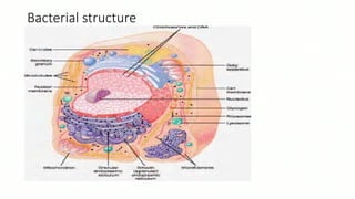
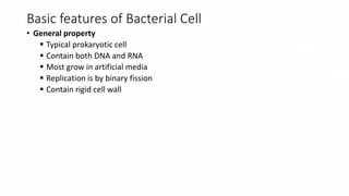
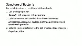
BACTRERIAL CELL STRUCTURE
DAVID MANYIEL AGOK (DMLT@RHSI)
Bacterial structure
Basic features of Bacterial Cell
• General property
 Typical prokaryotic cell
 Contain both DNA and RNA
 Most grow in artificial media
 Replication is by binary fission
 Contain rigid cell wall
Structure of Bacteria
Bacterial structure is considered at three levels.
1. Cell envelope proper:
Capsule, cell wall and cell membrane
2. Cellular element enclosed with in the cell envelope:
Mesosomes, ribosome, nuclear material, polyamines and
cytoplasmic granules.
3. Cellular element external to the cell envelope (appendages) :
Flagellum, Pilus
Cell wall
• This is the outer most layer that encases the protoplasm
Functions of cell wall
• It is porous and therefore permeable to salt molecules
• It maintains the bacterial shape
• It protects the bacteria against mechanical damage
• It supports the plasma membrane.
Types of cell wall
1. Gram-Positive Cell Wall
2. Gram negative cell wall of bacteria
Flagella.
• These are organs for locomotion. They occur on both Gram positive
and Gram negative bacteria.
• They can be used in the identification of bacteria
• Structurally, the bacterial flagella are long measuring 3-12 µm in
diameter.
• Chemically, the flagella are composed of a class of proteins called
Flagellins that is very important in the locomotion.
• There are four major arrangements of flagellum
A. Monotrichous - Single flagellum e.g. Vibryo cholerae
B. Lophotrichous - A tuft of flagella found at one of the cell pole e.g.
Spirillum serpens
C. Amphitrichous - Single flagellum found at each of two opposite poles.
D. Peritrichous - Multiple flagella found at several locations about the cell
e.g. Proteus species
Pilli.
• They are thin hair- like surface appendages on many gram- negative
bacteria. They are made up of proteins called Pilli that enable them to
perform their function.
Function
• Pilli have adhesive properties on the bacterial cells enabling them to
adhere to many epithelial surfaces, red blood cells, yeasts and
phagocytic cells which is important in bacterial colonization of the
epithelial surfaces and they are therefore referred to as colonization
factor i.e. they enable the organisms to adhere to the host tissues and
to one another.
Capsule
• This is a gelatinous outer- most layer of the bacterial cell. It is
composed of viscous polysaccharides that are retained as a thick gel
outside the cell wall envelope.
• It is up to 10µm thick
• A true capsule is a discrete detectable layer deposited outside the cell
wall. While a less discrete structure is called slime layer.(soft/sliperly)
Functions
• It offers resistance to phagocytosis and hence provides the bacterial
cell with protection against host defenses to invasion i.e. protects
bacterial cell from engulfment.
• Also often mediates adherence of cells to surfaces.
Cytoplasmic Membrane
• They are composed of proteins and lipids (Phospholipids). It is extremely a
thin membrane.
Functions.
• They are the sites of active transport, i.e. it enables selective transport of
specific solutes into the cell and waste products out of the cell.
• It permits passive diffusion in and outwards of water and other small
molecular substances.
• It is also an anchoring site for DNA and provides the cell with a mechanism
for separation of sister chromosomes.
• It passes larger molecules or any charged substances by means of special
membrane transport systems.
• Constitutes an osmotic barrier impermeable to many molecular solutes
hence maintains the differences in solute content between the cytoplasm
and external environment
Nuclear material
• This is an elongated dense body found in the middle of the cell. It
contains the genetic material i.e. DNA in form of chromosomes that
controls the cell activity.
Cytoplasm.
• It contains all the internal structure of the cell. The cytoplasmic
constituents include the prokaryotic chromosome and ribosomes
SPORE FORMATION.
• When conditions of vegetative growth are not favorable especially
when carbon and nitrogen become unavailable, some bacteria are
able to survive by forming resistant endospores by a process of
sporulation which are highly heat-resistant.
• A spore is unable to multiply but when conditions for vegetative
growth return, it’s able to produce a bacterial cell which is capable of
reproducing.
• Spore formation involves a change in enzyme activity and
morphology.
CONT...............
• They resist heating by autoclaving at 1210c for 15 minutes or in a hot
air oven at 160-1800c for 1 hour and can withstand dehydration, cold
and the action of disinfectants.
• Some spores may be positioned at the end of the bacterium called
Terminal Spores or can be sub terminal positioned near to one end or
centrally positioned.
• Shape: It can be circular, oval and central
• Size: It can be large, longer than the width of the cell and cause
bulging of the cell wall.
• Can be small and fit within the width of the cell.
CONT...........
• Examples of organisms that form spores include;
Plasmolysis
• This is the contraction of a cell due to loss of water through osmosis
in a bacterium when a cell is placed in a more concentrated salt
(hypertonic) solution.
Plasmoptysis.
• This describes the expansion in volume of the protoplasm of the cells
in hypotonic solutions with rapture of the cell walls and extrusion of
protoplasmic contents.
Fig. Different arrangements of spores in the bacteria
GENERAL PROPERTIES OF PATHOGENIC MICROORGANISMS
• Basing on cellular structure microorganisms can be categorized into
two groups:
• Prokaryotes
• Eukaryotes
• The table below show the differences between Prokaryotes and
Eukaryotes.
Prokaryotic Group. Eukaryotic group.
They are micro-organisms with very simple cell structure that have no nuclear
membrane thus no definite nucleus e.g.,
They are micro-organisms with a complex cell structure similar to that of
higher organisms. They have a nuclear membrane which forms a defined
nucleus
Prokaryotes multiply by simple division known as binary fission They multiply by a process known as mitosis
The cell contains simple enzyme systems that lack mitochondria The cell is able to make its own energy because it processes complex
enzyme systems, mitochondria and other organelles.
The genetic material is not organized into chromosomes inside a nuclear membrane
but lies as a single piece of double stranded DNA inside the cytoplasm.
The genetic material is differentiated into chromosomes which are
contained in a nuclear membrane to form a definite nucleus.
They include bacteria Rickettsiae, Chlamydia, Mycoplasma They include fungi and algae
BACTERIA PATHOGENICTY AND VIRULENCE
• Pathogens: are microorganisms that cause disease.
• Two major factors that help microbes to cause a disease are.
Virulence
Pathogenicity
Definition of terminologies
• Aetiology: The cause of a disease or condition (the known occurrence
and existence of micro organisms responsible for a particular
condition/disease)
• Infection: This is the invasion of the body by a pathogen.
CONT...................
• Disease:It’s the impairment of the body or unwell being of the body.
• Pathogenicity: It is the degree by which pathogens enter the body,
multiply and cause a disease. i.e.the ability to produce toxins causing
undesirable effects.
• Pathogenesis: It is the process of disease development with its
pathogenic mechanism
• Factors for Virulence and Pathogenicity
Factors for Virulence and Pathogenicity
• Some factors that assist pathogens to get established in the tissues
multiply and cause a disease. Are;
Capsule
Production of toxins
Possession of pilli
Possession of flagella
Production of extracellular enzymes
The extracellular enzymes produced include
A. Kinase:
• Kinase enzymes include Streptokinase which is produced by streptococcus
species and staphylokinase produced by staphylococcus species.
• This kinase assists the organisms to spread by breaking down the fibrin clot
which acts as a protective barrier to the body.
B. Coagulase:
• Coagulase enzyme is produced by staphylococcus aureus.
• It assists the pathogen to spread by coagulating plasma.
• This forms fibrin, which is thought to protect staphylococcus aureus from
phagocytosis by the host cells (macrophages).
• When organisms are held inside the fibrin clot, they are able to multiply
more easily and are less likely to be removed from the circulation by
reticuloendothelial cells.
CONT............
C. Hyaluronidase:
• This enzyme is Produced by clostridium perfringens, some
streptococci and staphylococci species.
• This enzyme helps the organisms to spread through the body by
breaking down the hyaluronic acid of connective tissue.
D. Beta-lactamase (B-Lactamase):
• This enzyme is Produced by many bacteria e.g. Staph.aureus,Neisseria
gonorrhoae.
• This enzyme confers to the organism resistance penicillin antibiotic.
They destroy the β-lactam ring of penicillin.
Capsule secretion
• Bacteria such as streptococci pneumonia, Klebssiella pneumonia and
Haemophilus Influenza are able to secret around their cell wall a thick
protective capsule.
• It helps prevents such organisms from being engulfed and being
destroyed by the phagocytic cells of the host.
Possession pilli /Fimbriae
• Virulent strains of the organisms such as Neisseria gonorrhea and
Escherichia coli are covered with pilli which helps them to adhere to
another and to the tissue of the host.
Possession of Flagella
• Some bacteria such as Salmonella species have flagella that assist in
the movement of the organisms.
• Production of toxins
• They are two types of toxins produced by micro organism i.e. the
endotoxin and exotoxins.
• Endotoxins are secreted within the microbial cells when the organism
is dead exotoxins are secreted outside the microbial cells when
organisms is still alive.
ENDOTOXINS-EXOTOXINS PRODUCTION
Properties
EXOTOXINS ENDOTOXINS
 Secreted by live cell and found in high concentrations in liquid media.
 They are polypeptides with molecular weight of 10,000-900,000.
 They are relatively thermo-labile
(destroyed at 60oc)
 They are highly antigenic i.e. stimulates the production of antitoxins.
 They are highly toxic i.e. stimulates the production of antitoxins.
 They are highly toxic and fatal to laboratory animals.
 They do not produce fever in the host.
 Examples include: Clostridium tetani neurotoxins producer, which affects the cells
of the central nervous system.
Others Include; Clostridium species, corynebacterium diptheriae, E.coli, produced by
enteric pathogens are known as Enterotoxic E. coli
 Integral parts of the microbial cells and are liberated upon their
disintegration.
 They are polysaccharide with molecular weight 10,000.
 Relatively heat-stable and withstands temperature.
 They induce production of antibodies.
 Weakly toxic and fatal to laboratory animals in 100 of
microorganisms.
 Usually produce fever in the host by release of interleukin-1 and
other mediators. Interleukin-1 acts on thermoregulatory centre.
 Examples include: Streptococcus species.
Other factors for pathogenicity include;
• Transmission route: for a pathogen to cause disease, it must enter the
body by route which will enable it to reach a site where it can
establish itself and multiply e.g. an organism which cause gas
gangrene must reach deep tissues to find the anaerobic conditions
necessary for its growth.
• Number of bacteria that invade. The bigger the number of organisms,
the faster the pathogenicity.
• Status of health of the person infected. The lower the immunity of
the patients, the easier the organism to spread into the host’s tissues.
THE END.
• THE NEXT TOPIC IS MICROSCOPICAL TECHNIQUES USED IN MICROBIOLOGY

BACTERIAL CELL STRUCTURE DavidManyiel.pptx

  • 1.
    BACTRERIAL CELL STRUCTURE DAVIDMANYIEL AGOK (DMLT@RHSI)
  • 2.
  • 3.
    Basic features ofBacterial Cell • General property  Typical prokaryotic cell  Contain both DNA and RNA  Most grow in artificial media  Replication is by binary fission  Contain rigid cell wall
  • 4.
    Structure of Bacteria Bacterialstructure is considered at three levels. 1. Cell envelope proper: Capsule, cell wall and cell membrane 2. Cellular element enclosed with in the cell envelope: Mesosomes, ribosome, nuclear material, polyamines and cytoplasmic granules. 3. Cellular element external to the cell envelope (appendages) : Flagellum, Pilus
  • 5.
    Cell wall • Thisis the outer most layer that encases the protoplasm Functions of cell wall • It is porous and therefore permeable to salt molecules • It maintains the bacterial shape • It protects the bacteria against mechanical damage • It supports the plasma membrane.
  • 6.
    Types of cellwall 1. Gram-Positive Cell Wall
  • 7.
    2. Gram negativecell wall of bacteria
  • 8.
    Flagella. • These areorgans for locomotion. They occur on both Gram positive and Gram negative bacteria. • They can be used in the identification of bacteria • Structurally, the bacterial flagella are long measuring 3-12 µm in diameter. • Chemically, the flagella are composed of a class of proteins called Flagellins that is very important in the locomotion.
  • 9.
    • There arefour major arrangements of flagellum A. Monotrichous - Single flagellum e.g. Vibryo cholerae B. Lophotrichous - A tuft of flagella found at one of the cell pole e.g. Spirillum serpens C. Amphitrichous - Single flagellum found at each of two opposite poles. D. Peritrichous - Multiple flagella found at several locations about the cell e.g. Proteus species
  • 10.
    Pilli. • They arethin hair- like surface appendages on many gram- negative bacteria. They are made up of proteins called Pilli that enable them to perform their function. Function • Pilli have adhesive properties on the bacterial cells enabling them to adhere to many epithelial surfaces, red blood cells, yeasts and phagocytic cells which is important in bacterial colonization of the epithelial surfaces and they are therefore referred to as colonization factor i.e. they enable the organisms to adhere to the host tissues and to one another.
  • 11.
    Capsule • This isa gelatinous outer- most layer of the bacterial cell. It is composed of viscous polysaccharides that are retained as a thick gel outside the cell wall envelope. • It is up to 10µm thick • A true capsule is a discrete detectable layer deposited outside the cell wall. While a less discrete structure is called slime layer.(soft/sliperly) Functions • It offers resistance to phagocytosis and hence provides the bacterial cell with protection against host defenses to invasion i.e. protects bacterial cell from engulfment. • Also often mediates adherence of cells to surfaces.
  • 12.
    Cytoplasmic Membrane • Theyare composed of proteins and lipids (Phospholipids). It is extremely a thin membrane. Functions. • They are the sites of active transport, i.e. it enables selective transport of specific solutes into the cell and waste products out of the cell. • It permits passive diffusion in and outwards of water and other small molecular substances. • It is also an anchoring site for DNA and provides the cell with a mechanism for separation of sister chromosomes. • It passes larger molecules or any charged substances by means of special membrane transport systems. • Constitutes an osmotic barrier impermeable to many molecular solutes hence maintains the differences in solute content between the cytoplasm and external environment
  • 13.
    Nuclear material • Thisis an elongated dense body found in the middle of the cell. It contains the genetic material i.e. DNA in form of chromosomes that controls the cell activity. Cytoplasm. • It contains all the internal structure of the cell. The cytoplasmic constituents include the prokaryotic chromosome and ribosomes
  • 14.
    SPORE FORMATION. • Whenconditions of vegetative growth are not favorable especially when carbon and nitrogen become unavailable, some bacteria are able to survive by forming resistant endospores by a process of sporulation which are highly heat-resistant. • A spore is unable to multiply but when conditions for vegetative growth return, it’s able to produce a bacterial cell which is capable of reproducing. • Spore formation involves a change in enzyme activity and morphology.
  • 15.
    CONT............... • They resistheating by autoclaving at 1210c for 15 minutes or in a hot air oven at 160-1800c for 1 hour and can withstand dehydration, cold and the action of disinfectants. • Some spores may be positioned at the end of the bacterium called Terminal Spores or can be sub terminal positioned near to one end or centrally positioned. • Shape: It can be circular, oval and central • Size: It can be large, longer than the width of the cell and cause bulging of the cell wall. • Can be small and fit within the width of the cell.
  • 16.
    CONT........... • Examples oforganisms that form spores include; Plasmolysis • This is the contraction of a cell due to loss of water through osmosis in a bacterium when a cell is placed in a more concentrated salt (hypertonic) solution. Plasmoptysis. • This describes the expansion in volume of the protoplasm of the cells in hypotonic solutions with rapture of the cell walls and extrusion of protoplasmic contents.
  • 17.
    Fig. Different arrangementsof spores in the bacteria
  • 18.
    GENERAL PROPERTIES OFPATHOGENIC MICROORGANISMS • Basing on cellular structure microorganisms can be categorized into two groups: • Prokaryotes • Eukaryotes • The table below show the differences between Prokaryotes and Eukaryotes.
  • 19.
    Prokaryotic Group. Eukaryoticgroup. They are micro-organisms with very simple cell structure that have no nuclear membrane thus no definite nucleus e.g., They are micro-organisms with a complex cell structure similar to that of higher organisms. They have a nuclear membrane which forms a defined nucleus Prokaryotes multiply by simple division known as binary fission They multiply by a process known as mitosis The cell contains simple enzyme systems that lack mitochondria The cell is able to make its own energy because it processes complex enzyme systems, mitochondria and other organelles. The genetic material is not organized into chromosomes inside a nuclear membrane but lies as a single piece of double stranded DNA inside the cytoplasm. The genetic material is differentiated into chromosomes which are contained in a nuclear membrane to form a definite nucleus. They include bacteria Rickettsiae, Chlamydia, Mycoplasma They include fungi and algae
  • 20.
    BACTERIA PATHOGENICTY ANDVIRULENCE • Pathogens: are microorganisms that cause disease. • Two major factors that help microbes to cause a disease are. Virulence Pathogenicity Definition of terminologies • Aetiology: The cause of a disease or condition (the known occurrence and existence of micro organisms responsible for a particular condition/disease) • Infection: This is the invasion of the body by a pathogen.
  • 21.
    CONT................... • Disease:It’s theimpairment of the body or unwell being of the body. • Pathogenicity: It is the degree by which pathogens enter the body, multiply and cause a disease. i.e.the ability to produce toxins causing undesirable effects. • Pathogenesis: It is the process of disease development with its pathogenic mechanism • Factors for Virulence and Pathogenicity
  • 22.
    Factors for Virulenceand Pathogenicity • Some factors that assist pathogens to get established in the tissues multiply and cause a disease. Are; Capsule Production of toxins Possession of pilli Possession of flagella Production of extracellular enzymes
  • 23.
    The extracellular enzymesproduced include A. Kinase: • Kinase enzymes include Streptokinase which is produced by streptococcus species and staphylokinase produced by staphylococcus species. • This kinase assists the organisms to spread by breaking down the fibrin clot which acts as a protective barrier to the body. B. Coagulase: • Coagulase enzyme is produced by staphylococcus aureus. • It assists the pathogen to spread by coagulating plasma. • This forms fibrin, which is thought to protect staphylococcus aureus from phagocytosis by the host cells (macrophages). • When organisms are held inside the fibrin clot, they are able to multiply more easily and are less likely to be removed from the circulation by reticuloendothelial cells.
  • 24.
    CONT............ C. Hyaluronidase: • Thisenzyme is Produced by clostridium perfringens, some streptococci and staphylococci species. • This enzyme helps the organisms to spread through the body by breaking down the hyaluronic acid of connective tissue. D. Beta-lactamase (B-Lactamase): • This enzyme is Produced by many bacteria e.g. Staph.aureus,Neisseria gonorrhoae. • This enzyme confers to the organism resistance penicillin antibiotic. They destroy the β-lactam ring of penicillin.
  • 25.
    Capsule secretion • Bacteriasuch as streptococci pneumonia, Klebssiella pneumonia and Haemophilus Influenza are able to secret around their cell wall a thick protective capsule. • It helps prevents such organisms from being engulfed and being destroyed by the phagocytic cells of the host. Possession pilli /Fimbriae • Virulent strains of the organisms such as Neisseria gonorrhea and Escherichia coli are covered with pilli which helps them to adhere to another and to the tissue of the host. Possession of Flagella • Some bacteria such as Salmonella species have flagella that assist in the movement of the organisms.
  • 26.
    • Production oftoxins • They are two types of toxins produced by micro organism i.e. the endotoxin and exotoxins. • Endotoxins are secreted within the microbial cells when the organism is dead exotoxins are secreted outside the microbial cells when organisms is still alive.
  • 27.
    ENDOTOXINS-EXOTOXINS PRODUCTION Properties EXOTOXINS ENDOTOXINS Secreted by live cell and found in high concentrations in liquid media.  They are polypeptides with molecular weight of 10,000-900,000.  They are relatively thermo-labile (destroyed at 60oc)  They are highly antigenic i.e. stimulates the production of antitoxins.  They are highly toxic i.e. stimulates the production of antitoxins.  They are highly toxic and fatal to laboratory animals.  They do not produce fever in the host.  Examples include: Clostridium tetani neurotoxins producer, which affects the cells of the central nervous system. Others Include; Clostridium species, corynebacterium diptheriae, E.coli, produced by enteric pathogens are known as Enterotoxic E. coli  Integral parts of the microbial cells and are liberated upon their disintegration.  They are polysaccharide with molecular weight 10,000.  Relatively heat-stable and withstands temperature.  They induce production of antibodies.  Weakly toxic and fatal to laboratory animals in 100 of microorganisms.  Usually produce fever in the host by release of interleukin-1 and other mediators. Interleukin-1 acts on thermoregulatory centre.  Examples include: Streptococcus species.
  • 28.
    Other factors forpathogenicity include; • Transmission route: for a pathogen to cause disease, it must enter the body by route which will enable it to reach a site where it can establish itself and multiply e.g. an organism which cause gas gangrene must reach deep tissues to find the anaerobic conditions necessary for its growth. • Number of bacteria that invade. The bigger the number of organisms, the faster the pathogenicity. • Status of health of the person infected. The lower the immunity of the patients, the easier the organism to spread into the host’s tissues.
  • 29.
    THE END. • THENEXT TOPIC IS MICROSCOPICAL TECHNIQUES USED IN MICROBIOLOGY