Download to read offline
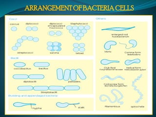
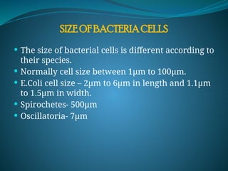
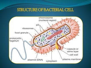
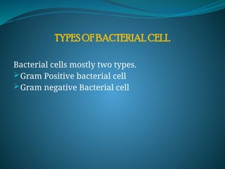
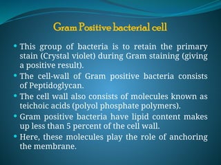
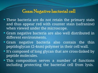
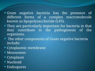
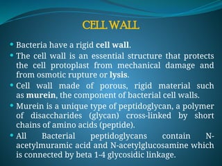
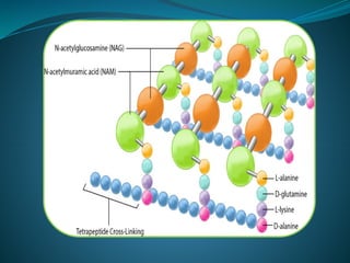
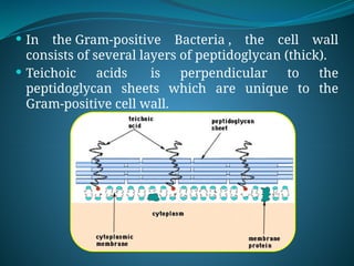
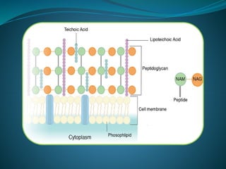
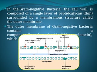
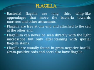
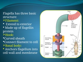
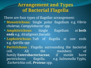
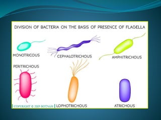
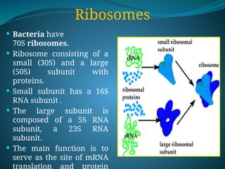
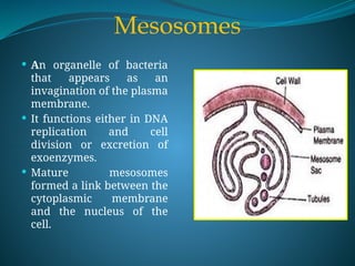
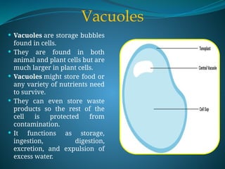


The document provides an overview of bacteria, highlighting their structure, function, and various types, including gram-positive and gram-negative bacteria. It describes the morphology, cell wall composition, and key organelles like the nucleoid, ribosomes, and vacuoles, as well as appendages such as flagella and pili essential for movement and adhesion. Additionally, it addresses the concepts of spores and cysts, detailing their roles in bacterial survival and reproduction under adverse conditions.