SlideShare a Scribd company logo
Pulmonary Infections -Pulmonary Infections - PneumoniaPneumonia
PneumoniaPneumonia
 Pneumonia is defined as an inflammation of the pulmonaryPneumonia is defined as an inflammation of the pulmonary
parenchymaparenchyma
  
ClassificationClassification
A) According to immune statusA) According to immune status
 Primary Pneumonia: Community acquiredPrimary Pneumonia: Community acquired
 Secondary Pneumonia (occurs in compromised host or Lungs)Secondary Pneumonia (occurs in compromised host or Lungs)
 BronchopneumoniaBronchopneumonia
 Nosocomial Pneumonia (hospital acquired)Nosocomial Pneumonia (hospital acquired)
 Aspiration pneumoniaAspiration pneumonia
 Pneumonia in the immmunocompromised patientPneumonia in the immmunocompromised patient
   
ClassificationClassification
B) Anatomical classificationB) Anatomical classification
 Lobar PneumoniaLobar Pneumonia
 Lobular Pneumonia (bronchopneumonia if bilateral)Lobular Pneumonia (bronchopneumonia if bilateral)
 Interstitial PneumoniaInterstitial Pneumonia
C)Aetiological classificationC)Aetiological classification
 Infective-Bacteria, Virus, Fungi, Protozoa etc.Infective-Bacteria, Virus, Fungi, Protozoa etc.
 AllergicAllergic
 Chemical agents: Irritant gases and liquid e.g. ammonia, vomitusChemical agents: Irritant gases and liquid e.g. ammonia, vomitus
 Physical agents: RadiationPhysical agents: Radiation
Predisposing factorsPredisposing factors
• A number of potent defense mechanisms clear or destroy anyA number of potent defense mechanisms clear or destroy any
bacteria inhaled with air or accidently deposited in the airwaybacteria inhaled with air or accidently deposited in the airway
passage.passage.
 The following 3 defense mechanisms play vital role in preventing fromThe following 3 defense mechanisms play vital role in preventing from
any sort of micro organisms infection that reaches our airwayany sort of micro organisms infection that reaches our airway
passages.passages.
a. Nasal clearancea. Nasal clearance
b. Tracheobronchial clearanceb. Tracheobronchial clearance
c. Alveolar clearancec. Alveolar clearance
a.a.Nasal clearanceNasal clearance
Particles including inhaled microorganisms are cleared byParticles including inhaled microorganisms are cleared by
sneezing or blowing from the nose. Those microorganisms whichsneezing or blowing from the nose. Those microorganisms which
manage to escape down are swallowed.manage to escape down are swallowed.
b.b.Tracheobronchial clearanceTracheobronchial clearance
Due to mucociliary action any particles or mocroorganismsDue to mucociliary action any particles or mocroorganisms
deposited in the tracheobronchial tree are eventually either swalloweddeposited in the tracheobronchial tree are eventually either swallowed
or expectorated.or expectorated.
c. Alveolar clearancec. Alveolar clearance
 Any bacteria or solid particles deposited in the alveoli areAny bacteria or solid particles deposited in the alveoli are
phagocytosed by alveolar macrophages.phagocytosed by alveolar macrophages.
 Some of the particles are carried to regional lymph nodes and viaSome of the particles are carried to regional lymph nodes and via
the blood stream, they reach other parts of the body.the blood stream, they reach other parts of the body.
Pathogenesis of PneumoniaPathogenesis of Pneumonia
Pneumonia may occurPneumonia may occur
 When these defense mechanisms are impaired Or whenever theWhen these defense mechanisms are impaired Or whenever the
resistant of the host in general is lowered.resistant of the host in general is lowered.
Infection of lung tissueInfection of lung tissue
Following factors may interfere with clearing mechanisms.Following factors may interfere with clearing mechanisms.
Loss or suppression of cough reflexLoss or suppression of cough reflex
- due to coma, drugs, anaesthesia, neuromuscular disorders.- due to coma, drugs, anaesthesia, neuromuscular disorders.
These condition can lead to aspiration of gastric contents too.These condition can lead to aspiration of gastric contents too.
Injury to mucociliary apparatusInjury to mucociliary apparatus
This can be due to cigarette smoking, due to inhalation of hot orThis can be due to cigarette smoking, due to inhalation of hot or
corrosive gasses, viral diseases, or genetic diseases.corrosive gasses, viral diseases, or genetic diseases.
Interference with phagocytic or bactericidal action ofInterference with phagocytic or bactericidal action of
alveolar macrophagesalveolar macrophages
This can be due to alcohol, tobacco, smoke, anoxia, or oxygenThis can be due to alcohol, tobacco, smoke, anoxia, or oxygen
intoxication.intoxication.
Pulmonary congestion & oedemaPulmonary congestion & oedema
Accumulations of secretionsAccumulations of secretions – cystic fibrosis &– cystic fibrosis &
bronchial obstructionbronchial obstruction
LOBAR PNEUMONIALOBAR PNEUMONIA : -: -
When a part of lobe, entire lobe or lobes of one or both lungs areWhen a part of lobe, entire lobe or lobes of one or both lungs are
involvedinvolved
Etiology: -Etiology: -
 Streptococcus PneumoniaeStreptococcus Pneumoniae
 StaphylococcalStaphylococcal
 H. InfluenzaeH. Influenzae
 Pseudomonas.Pseudomonas.
4 sequences or stages4 sequences or stages
1. Stage of Congestion1. Stage of Congestion : - Lasts for 1 – 2 days.: - Lasts for 1 – 2 days.
MicroMicro
Dilated & congested blood vessels with oedema fluid filled in air sacsDilated & congested blood vessels with oedema fluid filled in air sacs
(alveoli) with plenty of bacteria & a few neutrophil(alveoli) with plenty of bacteria & a few neutrophil
Gross : -Gross : -Lobe is enlarged, heavy, congested & exudes pink frothyLobe is enlarged, heavy, congested & exudes pink frothy
fluid on cut surface.fluid on cut surface.
2. Stage of Red Hepatization2. Stage of Red Hepatization : - Lasts – 2 – 4 days: - Lasts – 2 – 4 days
 MICRO : -MICRO : - Oedema fluid replaced by fibrin threads with markedOedema fluid replaced by fibrin threads with marked
neutrophils & R.B.C., Neutrophils with engulfed bacteria & theneutrophils & R.B.C., Neutrophils with engulfed bacteria & the
alveolar septa is less prominent than stage I.alveolar septa is less prominent than stage I.
 GROSS : -GROSS : - Liver like red, firm, airless. On cut section, red – pink,Liver like red, firm, airless. On cut section, red – pink,
dry, granular to look at.dry, granular to look at.
3. Stage of Grey Hepatization3. Stage of Grey Hepatization : - 4 – 8 days: - 4 – 8 days
 MICRO : -MICRO : - Due to R.B.C. disintegration and numerous fibrinDue to R.B.C. disintegration and numerous fibrin
strands and their contraction causes a separation of exudates fromstrands and their contraction causes a separation of exudates from
the alveolar walls.the alveolar walls.
Exudates are now less rich in bacteria, Rbc and inflammatory cells.Exudates are now less rich in bacteria, Rbc and inflammatory cells.
 GROSS : -GROSS : - Firm, heavy with dry, granular and grey liver like.Firm, heavy with dry, granular and grey liver like. LessLess
hyperaemiahyperaemia
4. Stage of Resolution:4. Stage of Resolution: - 8 – 16 days.- 8 – 16 days.
MICRO : -MICRO : -
 Consolidated exudate within the alveolar spaces undergoesConsolidated exudate within the alveolar spaces undergoes
progressive enzymatic digestion to produce a granular semisolidprogressive enzymatic digestion to produce a granular semisolid
debris that is either resorbed, ingested by macrophages or cougheddebris that is either resorbed, ingested by macrophages or coughed
upup
 Lungs return to normalLungs return to normal
 This is a lobarThis is a lobar
pneumonia in whichpneumonia in which
consolidation of theconsolidation of the
entire left upper lobeentire left upper lobe
has occurred.has occurred.
Clinical signs and symptomsClinical signs and symptoms
i. Fever and chillsi. Fever and chills
ii. Productive cough with yellow-green (pus) or rusty (bloody) sputumii. Productive cough with yellow-green (pus) or rusty (bloody) sputum
iii. Tachypnea(abnormal rapid breathing)iii. Tachypnea(abnormal rapid breathing)
iv. Pleuritic chest painiv. Pleuritic chest pain
v. Decreased breath sounds, bronchial breath sounds ,v. Decreased breath sounds, bronchial breath sounds ,
crepitations(crackling sound), and dullness to percussioncrepitations(crackling sound), and dullness to percussion
Nonspecific symptoms Most patients also have:Nonspecific symptoms Most patients also have:
 Fatigue, Myalgia(muscles pain), Abdominal pain, Anorexia,Fatigue, Myalgia(muscles pain), Abdominal pain, Anorexia,
HeadacheHeadache
Lab: Elevated WBCLab: Elevated WBC countcount
Chest x-rayChest x-ray
i. Lobar: lobar or segmental consolidation (opacification)i. Lobar: lobar or segmental consolidation (opacification)
ii. Bronchopneumonia: patchy opacificationii. Bronchopneumonia: patchy opacification
iii. Pleural effusioniii. Pleural effusion
Clinical keys: identification of the organism and early treatment withClinical keys: identification of the organism and early treatment with
antibioticsantibiotics
X-Ray showing Right middle lobe pneumoniaX-Ray showing Right middle lobe pneumonia
Chest x-ray demonstrating complete right upper lobe consolidation, consistent with aChest x-ray demonstrating complete right upper lobe consolidation, consistent with a
lobar pneumonia.lobar pneumonia.
CT Air-bronchogram
LOBAR PNEUMONIA
CT
air-bronchogram
BronchopneumoniaBronchopneumonia
This is the inflammatory consolidation of the surrounding alveoli of theThis is the inflammatory consolidation of the surrounding alveoli of the
terminal bronchioles leading to patchy solidification of lung.terminal bronchioles leading to patchy solidification of lung.
Etiology : -Etiology : -
 StaphylococcusStaphylococcus
 Haemophilus influenza,Haemophilus influenza,
 pseudomonaspseudomonas
 & E.coli.& E.coli.
This is more common in elderly or children i.e extremes of age due toThis is more common in elderly or children i.e extremes of age due to
viral or other bacterial infections.viral or other bacterial infections.
Usually secondary to other conditions associated with local andUsually secondary to other conditions associated with local and
general defense mechanisms:general defense mechanisms:
- viral infections (influenza, measles)- viral infections (influenza, measles)
- aspiration of food or vomitus- aspiration of food or vomitus
- obstruction of a bronchus (foreign body or neoplasm)- obstruction of a bronchus (foreign body or neoplasm)
- inhalation of irritant gases- inhalation of irritant gases
- major surgery- major surgery
- malnutrition- malnutrition
  PATHOLOGYPATHOLOGY : -: -
GROSS : -GROSS : -
Patchy areas of consolidation in one or more lobes frequently bilateral andPatchy areas of consolidation in one or more lobes frequently bilateral and
more often involving the lower zone due to gravitational reasons.more often involving the lower zone due to gravitational reasons.
Here is example of aHere is example of a
bronchopneumonia. Thebronchopneumonia. The
lighter areas that appearlighter areas that appear
to be raised on cutto be raised on cut
surface from thesurface from the
surrounding lung are thesurrounding lung are the
areas of consolidation ofareas of consolidation of
the lung.the lung.
BRONCHOPNEUMONIA
Bilateral
multifocal
 This radiographThis radiograph
demonstrates patchydemonstrates patchy
infiltrates consistent with ainfiltrates consistent with a
bronchopneumonia from abronchopneumonia from a
bacterial infection. Typicalbacterial infection. Typical
organisms includeorganisms include
Streptococcus pneumoniae,Streptococcus pneumoniae,
Staphylococcus aureus,Staphylococcus aureus,
Pseudomonas aeruginosa,Pseudomonas aeruginosa,
Hemophilus influenzae,Hemophilus influenzae,
Klebsiella pneumoniae,Klebsiella pneumoniae,
among others.among others.
BRONCHOPNEUMONIA
Patchy
Bilateral
BRONCHOPNEUMONIA
Patchy
Bilateral
Bronchopneumonia
1. Usually occurs in persons
with pre existing lung
disease
2. Common in extreme of age
3. Usually by staphylococci
4. Patchy consolidation
involving one lobe or but
often multilobular
5. Usually bi lateral & basal
6. Complete resolution
uncommon, may lead to
complications
Lobar PneumoniaLobar Pneumonia
1.1. Usually occurs in healthyUsually occurs in healthy
young adultyoung adult
2.2. Common in young ageCommon in young age
3.3. Usually by PneumococciUsually by Pneumococci
4.4. Lobar consolidationLobar consolidation
involving large portion ofinvolving large portion of
one lobe or entire lobeone lobe or entire lobe
5.5. Can effect any lobe – uni orCan effect any lobe – uni or
bi lateralbi lateral
6.6. Complete resolution usuallyComplete resolution usually
resolution takes placeresolution takes place
 INTERSTITIAL PNEUMONIAINTERSTITIAL PNEUMONIA : -Atypical Pneumonia: -Atypical Pneumonia
This is characterized by patchy inflammatory changes largelyThis is characterized by patchy inflammatory changes largely
confined to interstitial tissues of lung without any alveolar exudate.confined to interstitial tissues of lung without any alveolar exudate.
ETIOLOGYETIOLOGY : -: -
Mycoplasma pneumonae, Respiratory syncytial virus (RV)Mycoplasma pneumonae, Respiratory syncytial virus (RV)
Influenza virus, cytomegalo, pneumocystis jiroveci (pneumocystisInfluenza virus, cytomegalo, pneumocystis jiroveci (pneumocystis
carinii )carinii )
More common in immunosuppressed state.More common in immunosuppressed state.
PATHOLOGYPATHOLOGY : -: - red blue colourred blue colour
GROSS : -GROSS : - It may be patchy to massive involvement with heavyIt may be patchy to massive involvement with heavy
congested &congested & subcrepitant lungs.subcrepitant lungs.
Histo :Histo :
 Mainly interstitial infiltration of mononuclear cells with thickening ofMainly interstitial infiltration of mononuclear cells with thickening of
alveolar septa.alveolar septa.
 Necrosis of bronchial epithelial lining & filled bronchiolar lumen withNecrosis of bronchial epithelial lining & filled bronchiolar lumen with
secretions.secretions.
 A reactive change by multinuceated giant cells formation is shownA reactive change by multinuceated giant cells formation is shown
by lining bronchiolar epithelium.by lining bronchiolar epithelium.
Complications : -Complications : -
Main complication is secondary bacterial infection or a reactiveMain complication is secondary bacterial infection or a reactive
fibrosis of interstitial tissue.fibrosis of interstitial tissue.
Secondary bacterial infectionSecondary bacterial infection
staphylococcistaphylococci
Nosocomial infection or Hospital acquired pneumoniaNosocomial infection or Hospital acquired pneumonia
(HAP)(HAP)::
- After 48 hours of admission to the hospital, often patients in ICU- After 48 hours of admission to the hospital, often patients in ICU
- ↓ Local resistance to infection in lungs- ↓ Local resistance to infection in lungs
- Intubation of respiratory tract- Intubation of respiratory tract
- Altered normal flora due to antibiotics- Altered normal flora due to antibiotics
- Mostly gram – negative enterobacteria or Staph aureus, E.coli,- Mostly gram – negative enterobacteria or Staph aureus, E.coli,
Klebsiella, Proteus, Pseudomonas, Bacteroides,Klebsiella, Proteus, Pseudomonas, Bacteroides,
Community acquired pneumoniaCommunity acquired pneumonia
Commonest cause is due toCommonest cause is due to
 Streptococcus pneumoniae,Streptococcus pneumoniae,
other areother are
 H. Influenzae,H. Influenzae,
 Mycoplasma pneumoniae,Mycoplasma pneumoniae,
 Moraxella catarrhalis ,Moraxella catarrhalis ,
 Staph. Aureus,Staph. Aureus,
 clamydia, etc.clamydia, etc.
The most common causes forThe most common causes for viral pneumoniaviral pneumonia are:are:
 InfluenzaInfluenza
 ParainfluenzaParainfluenza
 AdenovirusAdenovirus
 Respiratory syncytial virus (RSV)Respiratory syncytial virus (RSV)
- appears mostly in children- appears mostly in children
 CytomegalovirusCytomegalovirus
- in immunocompromised hosts- in immunocompromised hosts
Modes of transmission of PneumoniaModes of transmission of Pneumonia
 Aspiration of organisms that colonize the oropharynx orAspiration of organisms that colonize the oropharynx or
nasopharynxnasopharynx
 Inhalation of infectious agentsInhalation of infectious agents
 Haematogenous{formation of blood} spread from extra pulmonaryHaematogenous{formation of blood} spread from extra pulmonary
sitesite
 Direct inoculation (tracheal intubations or stab wound to the chest) &Direct inoculation (tracheal intubations or stab wound to the chest) &
contiguous spread from an adjacent site of infectioncontiguous spread from an adjacent site of infection
Complications of pneumoniaComplications of pneumonia
 Lung abscess formationLung abscess formation
 Pleural effusionPleural effusion
 Empyema{collection of pus in a cavity in the body}Empyema{collection of pus in a cavity in the body}
 Failure of resolution ⇒ intra-alveolar scarringFailure of resolution ⇒ intra-alveolar scarring
 Respiratory failureRespiratory failure
 Septicaemia and bacteraemiaSepticaemia and bacteraemia
 - Infective endocarditis- Infective endocarditis
 - Cerebral abscess / meningitis- Cerebral abscess / meningitis
 - Septic arthritis{pus formation in joints}- Septic arthritis{pus formation in joints}
 Fibrous scarring and pleural adhesionsFibrous scarring and pleural adhesions
 Define Pneumonia & classify or causes of PneumoniaDefine Pneumonia & classify or causes of Pneumonia
 What are the complications of PneumoniaWhat are the complications of Pneumonia
 Give the pathogenesis of Lobar PneumoniaGive the pathogenesis of Lobar Pneumonia
 Give the morphology / stages of pneumoniaGive the morphology / stages of pneumonia
 Difference between lobar & bronchopneumoniaDifference between lobar & bronchopneumonia

More Related Content

What's hot

COPD -- GOLD guidelines REPORT 2021
COPD -- GOLD guidelines REPORT 2021COPD -- GOLD guidelines REPORT 2021
COPD -- GOLD guidelines REPORT 2021
Dr. Mishal Saleem
 
Pulmonary artery Hypertension
Pulmonary artery HypertensionPulmonary artery Hypertension
Pulmonary artery Hypertension
Rikin Hasnani
 
Autosomal Dominant Polycystic Kidney Disease
Autosomal Dominant Polycystic Kidney DiseaseAutosomal Dominant Polycystic Kidney Disease
Autosomal Dominant Polycystic Kidney Disease
Wisit Cheungpasitporn
 
Aortic regurgitation
Aortic regurgitationAortic regurgitation
Aortic regurgitation
Vitrag Shah
 
FlashPath - Lung - Cryptogenic Organizing Pneumonia (Bronchiolitis Obliterans...
FlashPath - Lung - Cryptogenic Organizing Pneumonia (Bronchiolitis Obliterans...FlashPath - Lung - Cryptogenic Organizing Pneumonia (Bronchiolitis Obliterans...
FlashPath - Lung - Cryptogenic Organizing Pneumonia (Bronchiolitis Obliterans...
Hazem Ali
 
Renal Artery Stenosis
Renal Artery StenosisRenal Artery Stenosis
Renal Artery Stenosis
KhalafAlGhamdi
 
Chronic Stable Angina- Diagnosis & management
Chronic Stable Angina- Diagnosis & managementChronic Stable Angina- Diagnosis & management
Chronic Stable Angina- Diagnosis & management
LPS Institute of Cardiology Kanpur UP India
 
Tubulointerstitial nephritis
Tubulointerstitial nephritisTubulointerstitial nephritis
Tubulointerstitial nephritis
Swati Wadhai
 
Aortic regurgitation
Aortic regurgitationAortic regurgitation
Aortic regurgitation
Pratap Tiwari
 
Aspiration Pneumonia
Aspiration PneumoniaAspiration Pneumonia
Aspiration Pneumoniatjsiddiqui
 
necrotizing pneumonia.ppt
necrotizing pneumonia.pptnecrotizing pneumonia.ppt
necrotizing pneumonia.ppt
farhanashafie3
 
Bronchiectasis.
Bronchiectasis.Bronchiectasis.
Bronchiectasis.
Davis Kurian
 
Renal artery stenosis
Renal artery stenosisRenal artery stenosis
Renal artery stenosis
NAJEEB ULLAH SOFI
 
Acute Respiratory Distress Syndrome
Acute Respiratory Distress SyndromeAcute Respiratory Distress Syndrome
Acute Respiratory Distress Syndrome
PERCY ARPITHA JENNIFER
 
Update on Scleroderma and the Lung
Update on Scleroderma and the LungUpdate on Scleroderma and the Lung
Update on Scleroderma and the Lung
Scleroderma Foundation of Greater Chicago
 
RHD- Rheumatic Heart Disease , VHD - Valvular Heart Disease
RHD- Rheumatic Heart Disease , VHD - Valvular Heart Disease RHD- Rheumatic Heart Disease , VHD - Valvular Heart Disease
RHD- Rheumatic Heart Disease , VHD - Valvular Heart Disease
Mathew Varghese V
 
Sepsis
SepsisSepsis
Pulmonary Renal Syndorme
Pulmonary Renal Syndorme Pulmonary Renal Syndorme
Pulmonary Renal Syndorme
Farhan Tahir MD, FACR, Diplomate ABIHM
 
Pulmonary Aspergillosis-1.pptx
Pulmonary Aspergillosis-1.pptxPulmonary Aspergillosis-1.pptx
Pulmonary Aspergillosis-1.pptx
Kemi Adaramola
 
Cardiorenal Syndrome
Cardiorenal SyndromeCardiorenal Syndrome
Cardiorenal Syndrome
Sujay Iyer
 

What's hot (20)

COPD -- GOLD guidelines REPORT 2021
COPD -- GOLD guidelines REPORT 2021COPD -- GOLD guidelines REPORT 2021
COPD -- GOLD guidelines REPORT 2021
 
Pulmonary artery Hypertension
Pulmonary artery HypertensionPulmonary artery Hypertension
Pulmonary artery Hypertension
 
Autosomal Dominant Polycystic Kidney Disease
Autosomal Dominant Polycystic Kidney DiseaseAutosomal Dominant Polycystic Kidney Disease
Autosomal Dominant Polycystic Kidney Disease
 
Aortic regurgitation
Aortic regurgitationAortic regurgitation
Aortic regurgitation
 
FlashPath - Lung - Cryptogenic Organizing Pneumonia (Bronchiolitis Obliterans...
FlashPath - Lung - Cryptogenic Organizing Pneumonia (Bronchiolitis Obliterans...FlashPath - Lung - Cryptogenic Organizing Pneumonia (Bronchiolitis Obliterans...
FlashPath - Lung - Cryptogenic Organizing Pneumonia (Bronchiolitis Obliterans...
 
Renal Artery Stenosis
Renal Artery StenosisRenal Artery Stenosis
Renal Artery Stenosis
 
Chronic Stable Angina- Diagnosis & management
Chronic Stable Angina- Diagnosis & managementChronic Stable Angina- Diagnosis & management
Chronic Stable Angina- Diagnosis & management
 
Tubulointerstitial nephritis
Tubulointerstitial nephritisTubulointerstitial nephritis
Tubulointerstitial nephritis
 
Aortic regurgitation
Aortic regurgitationAortic regurgitation
Aortic regurgitation
 
Aspiration Pneumonia
Aspiration PneumoniaAspiration Pneumonia
Aspiration Pneumonia
 
necrotizing pneumonia.ppt
necrotizing pneumonia.pptnecrotizing pneumonia.ppt
necrotizing pneumonia.ppt
 
Bronchiectasis.
Bronchiectasis.Bronchiectasis.
Bronchiectasis.
 
Renal artery stenosis
Renal artery stenosisRenal artery stenosis
Renal artery stenosis
 
Acute Respiratory Distress Syndrome
Acute Respiratory Distress SyndromeAcute Respiratory Distress Syndrome
Acute Respiratory Distress Syndrome
 
Update on Scleroderma and the Lung
Update on Scleroderma and the LungUpdate on Scleroderma and the Lung
Update on Scleroderma and the Lung
 
RHD- Rheumatic Heart Disease , VHD - Valvular Heart Disease
RHD- Rheumatic Heart Disease , VHD - Valvular Heart Disease RHD- Rheumatic Heart Disease , VHD - Valvular Heart Disease
RHD- Rheumatic Heart Disease , VHD - Valvular Heart Disease
 
Sepsis
SepsisSepsis
Sepsis
 
Pulmonary Renal Syndorme
Pulmonary Renal Syndorme Pulmonary Renal Syndorme
Pulmonary Renal Syndorme
 
Pulmonary Aspergillosis-1.pptx
Pulmonary Aspergillosis-1.pptxPulmonary Aspergillosis-1.pptx
Pulmonary Aspergillosis-1.pptx
 
Cardiorenal Syndrome
Cardiorenal SyndromeCardiorenal Syndrome
Cardiorenal Syndrome
 

Similar to 1pneumonia

LUNG ABSCESS.pdf
LUNG ABSCESS.pdfLUNG ABSCESS.pdf
LUNG ABSCESS.pdf
Ashraf Shaik
 
6 bronchiectasis
6 bronchiectasis6 bronchiectasis
6 bronchiectasis
PNK SINGH
 
5 pneumonia
5 pneumonia5 pneumonia
5 pneumonia
Claudiu Cucu
 
Respiratory Tract Infections- A Pharmacotherapeutic Approach
Respiratory Tract Infections- A Pharmacotherapeutic ApproachRespiratory Tract Infections- A Pharmacotherapeutic Approach
Respiratory Tract Infections- A Pharmacotherapeutic Approach
Dr. Ankit Gaur
 
4.copd
4.copd4.copd
4.copd
PNK SINGH
 
Pneumonia in children by dr. sundar karki
Pneumonia in children  by dr. sundar karkiPneumonia in children  by dr. sundar karki
Pneumonia in children by dr. sundar karki
Dr. Sundar Karki
 
Pneumonia last
Pneumonia lastPneumonia last
Pneumonia last
es al
 
7 examination of respiratory system
7 examination of respiratory system 7 examination of respiratory system
7 examination of respiratory system
karima Akool AlSalihi
 
Tb path & pathogenesis
Tb path & pathogenesisTb path & pathogenesis
Tb path & pathogenesis
Dr.Manish Kumar
 
Tb path & pathogenesis
Tb path & pathogenesis Tb path & pathogenesis
Tb path & pathogenesis
Dr.Manish Kumar
 
8 lung abscess
8 lung abscess8 lung abscess
8 lung abscess
Claudiu Cucu
 
Veterinary Pathology of Respiratory System
Veterinary Pathology of Respiratory SystemVeterinary Pathology of Respiratory System
Veterinary Pathology of Respiratory System
Muhammad Amir Sohail
 
Respiratory system pathology lab
Respiratory system pathology labRespiratory system pathology lab
Respiratory system pathology lab
Mohammad Ihmeidan
 
Pulmonary Infections
Pulmonary InfectionsPulmonary Infections
Pulmonary Infections
Prasad CSBR
 
Bronchectasis DISEASE MFM faculty 2017 .pdf
Bronchectasis DISEASE MFM faculty 2017 .pdfBronchectasis DISEASE MFM faculty 2017 .pdf
Bronchectasis DISEASE MFM faculty 2017 .pdf
ahmednasser648712
 
Bronchiectasis
BronchiectasisBronchiectasis
Bronchiectasis
Yatheendra Vasanth
 
Pneumonia
Pneumonia Pneumonia
Pneumonia
Mukhtar Alam
 
community acquired pneumonia (CAP)
community acquired pneumonia (CAP) community acquired pneumonia (CAP)
community acquired pneumonia (CAP)
Hamdi Turkey
 
Intrestitial lung disease 16 5-2016
Intrestitial  lung disease 16 5-2016Intrestitial  lung disease 16 5-2016
Intrestitial lung disease 16 5-2016
pathologydept
 
Intrestitial lung disease 9 5-2016
Intrestitial  lung disease 9 5-2016Intrestitial  lung disease 9 5-2016
Intrestitial lung disease 9 5-2016
pathologydept
 

Similar to 1pneumonia (20)

LUNG ABSCESS.pdf
LUNG ABSCESS.pdfLUNG ABSCESS.pdf
LUNG ABSCESS.pdf
 
6 bronchiectasis
6 bronchiectasis6 bronchiectasis
6 bronchiectasis
 
5 pneumonia
5 pneumonia5 pneumonia
5 pneumonia
 
Respiratory Tract Infections- A Pharmacotherapeutic Approach
Respiratory Tract Infections- A Pharmacotherapeutic ApproachRespiratory Tract Infections- A Pharmacotherapeutic Approach
Respiratory Tract Infections- A Pharmacotherapeutic Approach
 
4.copd
4.copd4.copd
4.copd
 
Pneumonia in children by dr. sundar karki
Pneumonia in children  by dr. sundar karkiPneumonia in children  by dr. sundar karki
Pneumonia in children by dr. sundar karki
 
Pneumonia last
Pneumonia lastPneumonia last
Pneumonia last
 
7 examination of respiratory system
7 examination of respiratory system 7 examination of respiratory system
7 examination of respiratory system
 
Tb path & pathogenesis
Tb path & pathogenesisTb path & pathogenesis
Tb path & pathogenesis
 
Tb path & pathogenesis
Tb path & pathogenesis Tb path & pathogenesis
Tb path & pathogenesis
 
8 lung abscess
8 lung abscess8 lung abscess
8 lung abscess
 
Veterinary Pathology of Respiratory System
Veterinary Pathology of Respiratory SystemVeterinary Pathology of Respiratory System
Veterinary Pathology of Respiratory System
 
Respiratory system pathology lab
Respiratory system pathology labRespiratory system pathology lab
Respiratory system pathology lab
 
Pulmonary Infections
Pulmonary InfectionsPulmonary Infections
Pulmonary Infections
 
Bronchectasis DISEASE MFM faculty 2017 .pdf
Bronchectasis DISEASE MFM faculty 2017 .pdfBronchectasis DISEASE MFM faculty 2017 .pdf
Bronchectasis DISEASE MFM faculty 2017 .pdf
 
Bronchiectasis
BronchiectasisBronchiectasis
Bronchiectasis
 
Pneumonia
Pneumonia Pneumonia
Pneumonia
 
community acquired pneumonia (CAP)
community acquired pneumonia (CAP) community acquired pneumonia (CAP)
community acquired pneumonia (CAP)
 
Intrestitial lung disease 16 5-2016
Intrestitial  lung disease 16 5-2016Intrestitial  lung disease 16 5-2016
Intrestitial lung disease 16 5-2016
 
Intrestitial lung disease 9 5-2016
Intrestitial  lung disease 9 5-2016Intrestitial  lung disease 9 5-2016
Intrestitial lung disease 9 5-2016
 

More from PNK SINGH

Burnsss
BurnsssBurnsss
Burnsss
PNK SINGH
 
Section ii a biochemistry carbohydrate
Section ii a biochemistry carbohydrateSection ii a biochemistry carbohydrate
Section ii a biochemistry carbohydrate
PNK SINGH
 
Anemia 1
Anemia 1Anemia 1
Anemia 1
PNK SINGH
 
Anemia
Anemia Anemia
Anemia
PNK SINGH
 
Cancer cell growth
Cancer cell growthCancer cell growth
Cancer cell growth
PNK SINGH
 
Other renal pathology
Other renal pathologyOther renal pathology
Other renal pathology
PNK SINGH
 
1.glomerulonephritis
1.glomerulonephritis1.glomerulonephritis
1.glomerulonephritis
PNK SINGH
 
Tumour of cns
Tumour of cnsTumour of cns
Tumour of cns
PNK SINGH
 
Central nervous system
Central nervous systemCentral nervous system
Central nervous system
PNK SINGH
 
11.Vascular pathology
11.Vascular pathology11.Vascular pathology
11.Vascular pathology
PNK SINGH
 
10.Diseases of pericardium
10.Diseases of pericardium10.Diseases of pericardium
10.Diseases of pericardium
PNK SINGH
 
9.Congenital heart disease
9.Congenital heart disease9.Congenital heart disease
9.Congenital heart disease
PNK SINGH
 
8. Disease of myocardium
8. Disease of  myocardium8. Disease of  myocardium
8. Disease of myocardium
PNK SINGH
 
7.Valvular heart disease pathology
7.Valvular heart disease pathology7.Valvular heart disease pathology
7.Valvular heart disease pathology
PNK SINGH
 
6.Infective endocarditis pathology
6.Infective endocarditis pathology6.Infective endocarditis pathology
6.Infective endocarditis pathology
PNK SINGH
 
5.Rheumatic heart disease pathology
5.Rheumatic heart disease pathology5.Rheumatic heart disease pathology
5.Rheumatic heart disease pathology
PNK SINGH
 
4heart failure pathology
4heart failure pathology4heart failure pathology
4heart failure pathology
PNK SINGH
 
3.Ischemic heart disease( coronary artery disease)
3.Ischemic heart disease( coronary artery disease)3.Ischemic heart disease( coronary artery disease)
3.Ischemic heart disease( coronary artery disease)
PNK SINGH
 
2 atherosclerosis pathology
2 atherosclerosis pathology2 atherosclerosis pathology
2 atherosclerosis pathology
PNK SINGH
 
1 hypertenson
1 hypertenson1 hypertenson
1 hypertenson
PNK SINGH
 

More from PNK SINGH (20)

Burnsss
BurnsssBurnsss
Burnsss
 
Section ii a biochemistry carbohydrate
Section ii a biochemistry carbohydrateSection ii a biochemistry carbohydrate
Section ii a biochemistry carbohydrate
 
Anemia 1
Anemia 1Anemia 1
Anemia 1
 
Anemia
Anemia Anemia
Anemia
 
Cancer cell growth
Cancer cell growthCancer cell growth
Cancer cell growth
 
Other renal pathology
Other renal pathologyOther renal pathology
Other renal pathology
 
1.glomerulonephritis
1.glomerulonephritis1.glomerulonephritis
1.glomerulonephritis
 
Tumour of cns
Tumour of cnsTumour of cns
Tumour of cns
 
Central nervous system
Central nervous systemCentral nervous system
Central nervous system
 
11.Vascular pathology
11.Vascular pathology11.Vascular pathology
11.Vascular pathology
 
10.Diseases of pericardium
10.Diseases of pericardium10.Diseases of pericardium
10.Diseases of pericardium
 
9.Congenital heart disease
9.Congenital heart disease9.Congenital heart disease
9.Congenital heart disease
 
8. Disease of myocardium
8. Disease of  myocardium8. Disease of  myocardium
8. Disease of myocardium
 
7.Valvular heart disease pathology
7.Valvular heart disease pathology7.Valvular heart disease pathology
7.Valvular heart disease pathology
 
6.Infective endocarditis pathology
6.Infective endocarditis pathology6.Infective endocarditis pathology
6.Infective endocarditis pathology
 
5.Rheumatic heart disease pathology
5.Rheumatic heart disease pathology5.Rheumatic heart disease pathology
5.Rheumatic heart disease pathology
 
4heart failure pathology
4heart failure pathology4heart failure pathology
4heart failure pathology
 
3.Ischemic heart disease( coronary artery disease)
3.Ischemic heart disease( coronary artery disease)3.Ischemic heart disease( coronary artery disease)
3.Ischemic heart disease( coronary artery disease)
 
2 atherosclerosis pathology
2 atherosclerosis pathology2 atherosclerosis pathology
2 atherosclerosis pathology
 
1 hypertenson
1 hypertenson1 hypertenson
1 hypertenson
 

Recently uploaded

Knee anatomy and clinical tests 2024.pdf
Knee anatomy and clinical tests 2024.pdfKnee anatomy and clinical tests 2024.pdf
Knee anatomy and clinical tests 2024.pdf
vimalpl1234
 
micro teaching on communication m.sc nursing.pdf
micro teaching on communication m.sc nursing.pdfmicro teaching on communication m.sc nursing.pdf
micro teaching on communication m.sc nursing.pdf
Anurag Sharma
 
BRACHYTHERAPY OVERVIEW AND APPLICATORS
BRACHYTHERAPY OVERVIEW  AND  APPLICATORSBRACHYTHERAPY OVERVIEW  AND  APPLICATORS
BRACHYTHERAPY OVERVIEW AND APPLICATORS
Krishan Murari
 
basicmodesofventilation2022-220313203758.pdf
basicmodesofventilation2022-220313203758.pdfbasicmodesofventilation2022-220313203758.pdf
basicmodesofventilation2022-220313203758.pdf
aljamhori teaching hospital
 
Physiology of Special Chemical Sensation of Taste
Physiology of Special Chemical Sensation of TastePhysiology of Special Chemical Sensation of Taste
Physiology of Special Chemical Sensation of Taste
MedicoseAcademics
 
Charaka Samhita Sutra sthana Chapter 15 Upakalpaniyaadhyaya
Charaka Samhita Sutra sthana Chapter 15 UpakalpaniyaadhyayaCharaka Samhita Sutra sthana Chapter 15 Upakalpaniyaadhyaya
Charaka Samhita Sutra sthana Chapter 15 Upakalpaniyaadhyaya
Dr KHALID B.M
 
Tom Selleck Health: A Comprehensive Look at the Iconic Actor’s Wellness Journey
Tom Selleck Health: A Comprehensive Look at the Iconic Actor’s Wellness JourneyTom Selleck Health: A Comprehensive Look at the Iconic Actor’s Wellness Journey
Tom Selleck Health: A Comprehensive Look at the Iconic Actor’s Wellness Journey
greendigital
 
heat stroke and heat exhaustion in children
heat stroke and heat exhaustion in childrenheat stroke and heat exhaustion in children
heat stroke and heat exhaustion in children
SumeraAhmad5
 
New Directions in Targeted Therapeutic Approaches for Older Adults With Mantl...
New Directions in Targeted Therapeutic Approaches for Older Adults With Mantl...New Directions in Targeted Therapeutic Approaches for Older Adults With Mantl...
New Directions in Targeted Therapeutic Approaches for Older Adults With Mantl...
i3 Health
 
TEST BANK for Operations Management, 14th Edition by William J. Stevenson, Ve...
TEST BANK for Operations Management, 14th Edition by William J. Stevenson, Ve...TEST BANK for Operations Management, 14th Edition by William J. Stevenson, Ve...
TEST BANK for Operations Management, 14th Edition by William J. Stevenson, Ve...
kevinkariuki227
 
Ophthalmology Clinical Tests for OSCE exam
Ophthalmology Clinical Tests for OSCE examOphthalmology Clinical Tests for OSCE exam
Ophthalmology Clinical Tests for OSCE exam
KafrELShiekh University
 
POST OPERATIVE OLIGURIA and its management
POST OPERATIVE OLIGURIA and its managementPOST OPERATIVE OLIGURIA and its management
POST OPERATIVE OLIGURIA and its management
touseefaziz1
 
Surat @ℂall @Girls ꧁❤8527049040❤꧂@ℂall @Girls Service Vip Top Model Safe
Surat @ℂall @Girls ꧁❤8527049040❤꧂@ℂall @Girls Service Vip Top Model SafeSurat @ℂall @Girls ꧁❤8527049040❤꧂@ℂall @Girls Service Vip Top Model Safe
Surat @ℂall @Girls ꧁❤8527049040❤꧂@ℂall @Girls Service Vip Top Model Safe
Savita Shen $i11
 
Couples presenting to the infertility clinic- Do they really have infertility...
Couples presenting to the infertility clinic- Do they really have infertility...Couples presenting to the infertility clinic- Do they really have infertility...
Couples presenting to the infertility clinic- Do they really have infertility...
Sujoy Dasgupta
 
24 Upakrama.pptx class ppt useful in all
24 Upakrama.pptx class ppt useful in all24 Upakrama.pptx class ppt useful in all
24 Upakrama.pptx class ppt useful in all
DrSathishMS1
 
Maxilla, Mandible & Hyoid Bone & Clinical Correlations by Dr. RIG.pptx
Maxilla, Mandible & Hyoid Bone & Clinical Correlations by Dr. RIG.pptxMaxilla, Mandible & Hyoid Bone & Clinical Correlations by Dr. RIG.pptx
Maxilla, Mandible & Hyoid Bone & Clinical Correlations by Dr. RIG.pptx
Dr. Rabia Inam Gandapore
 
Ocular injury ppt Upendra pal optometrist upums saifai etawah
Ocular injury  ppt  Upendra pal  optometrist upums saifai etawahOcular injury  ppt  Upendra pal  optometrist upums saifai etawah
Ocular injury ppt Upendra pal optometrist upums saifai etawah
pal078100
 
Lung Cancer: Artificial Intelligence, Synergetics, Complex System Analysis, S...
Lung Cancer: Artificial Intelligence, Synergetics, Complex System Analysis, S...Lung Cancer: Artificial Intelligence, Synergetics, Complex System Analysis, S...
Lung Cancer: Artificial Intelligence, Synergetics, Complex System Analysis, S...
Oleg Kshivets
 
Novas diretrizes da OMS para os cuidados perinatais de mais qualidade
Novas diretrizes da OMS para os cuidados perinatais de mais qualidadeNovas diretrizes da OMS para os cuidados perinatais de mais qualidade
Novas diretrizes da OMS para os cuidados perinatais de mais qualidade
Prof. Marcus Renato de Carvalho
 
263778731218 Abortion Clinic /Pills In Harare ,
263778731218 Abortion Clinic /Pills In Harare ,263778731218 Abortion Clinic /Pills In Harare ,
263778731218 Abortion Clinic /Pills In Harare ,
sisternakatoto
 

Recently uploaded (20)

Knee anatomy and clinical tests 2024.pdf
Knee anatomy and clinical tests 2024.pdfKnee anatomy and clinical tests 2024.pdf
Knee anatomy and clinical tests 2024.pdf
 
micro teaching on communication m.sc nursing.pdf
micro teaching on communication m.sc nursing.pdfmicro teaching on communication m.sc nursing.pdf
micro teaching on communication m.sc nursing.pdf
 
BRACHYTHERAPY OVERVIEW AND APPLICATORS
BRACHYTHERAPY OVERVIEW  AND  APPLICATORSBRACHYTHERAPY OVERVIEW  AND  APPLICATORS
BRACHYTHERAPY OVERVIEW AND APPLICATORS
 
basicmodesofventilation2022-220313203758.pdf
basicmodesofventilation2022-220313203758.pdfbasicmodesofventilation2022-220313203758.pdf
basicmodesofventilation2022-220313203758.pdf
 
Physiology of Special Chemical Sensation of Taste
Physiology of Special Chemical Sensation of TastePhysiology of Special Chemical Sensation of Taste
Physiology of Special Chemical Sensation of Taste
 
Charaka Samhita Sutra sthana Chapter 15 Upakalpaniyaadhyaya
Charaka Samhita Sutra sthana Chapter 15 UpakalpaniyaadhyayaCharaka Samhita Sutra sthana Chapter 15 Upakalpaniyaadhyaya
Charaka Samhita Sutra sthana Chapter 15 Upakalpaniyaadhyaya
 
Tom Selleck Health: A Comprehensive Look at the Iconic Actor’s Wellness Journey
Tom Selleck Health: A Comprehensive Look at the Iconic Actor’s Wellness JourneyTom Selleck Health: A Comprehensive Look at the Iconic Actor’s Wellness Journey
Tom Selleck Health: A Comprehensive Look at the Iconic Actor’s Wellness Journey
 
heat stroke and heat exhaustion in children
heat stroke and heat exhaustion in childrenheat stroke and heat exhaustion in children
heat stroke and heat exhaustion in children
 
New Directions in Targeted Therapeutic Approaches for Older Adults With Mantl...
New Directions in Targeted Therapeutic Approaches for Older Adults With Mantl...New Directions in Targeted Therapeutic Approaches for Older Adults With Mantl...
New Directions in Targeted Therapeutic Approaches for Older Adults With Mantl...
 
TEST BANK for Operations Management, 14th Edition by William J. Stevenson, Ve...
TEST BANK for Operations Management, 14th Edition by William J. Stevenson, Ve...TEST BANK for Operations Management, 14th Edition by William J. Stevenson, Ve...
TEST BANK for Operations Management, 14th Edition by William J. Stevenson, Ve...
 
Ophthalmology Clinical Tests for OSCE exam
Ophthalmology Clinical Tests for OSCE examOphthalmology Clinical Tests for OSCE exam
Ophthalmology Clinical Tests for OSCE exam
 
POST OPERATIVE OLIGURIA and its management
POST OPERATIVE OLIGURIA and its managementPOST OPERATIVE OLIGURIA and its management
POST OPERATIVE OLIGURIA and its management
 
Surat @ℂall @Girls ꧁❤8527049040❤꧂@ℂall @Girls Service Vip Top Model Safe
Surat @ℂall @Girls ꧁❤8527049040❤꧂@ℂall @Girls Service Vip Top Model SafeSurat @ℂall @Girls ꧁❤8527049040❤꧂@ℂall @Girls Service Vip Top Model Safe
Surat @ℂall @Girls ꧁❤8527049040❤꧂@ℂall @Girls Service Vip Top Model Safe
 
Couples presenting to the infertility clinic- Do they really have infertility...
Couples presenting to the infertility clinic- Do they really have infertility...Couples presenting to the infertility clinic- Do they really have infertility...
Couples presenting to the infertility clinic- Do they really have infertility...
 
24 Upakrama.pptx class ppt useful in all
24 Upakrama.pptx class ppt useful in all24 Upakrama.pptx class ppt useful in all
24 Upakrama.pptx class ppt useful in all
 
Maxilla, Mandible & Hyoid Bone & Clinical Correlations by Dr. RIG.pptx
Maxilla, Mandible & Hyoid Bone & Clinical Correlations by Dr. RIG.pptxMaxilla, Mandible & Hyoid Bone & Clinical Correlations by Dr. RIG.pptx
Maxilla, Mandible & Hyoid Bone & Clinical Correlations by Dr. RIG.pptx
 
Ocular injury ppt Upendra pal optometrist upums saifai etawah
Ocular injury  ppt  Upendra pal  optometrist upums saifai etawahOcular injury  ppt  Upendra pal  optometrist upums saifai etawah
Ocular injury ppt Upendra pal optometrist upums saifai etawah
 
Lung Cancer: Artificial Intelligence, Synergetics, Complex System Analysis, S...
Lung Cancer: Artificial Intelligence, Synergetics, Complex System Analysis, S...Lung Cancer: Artificial Intelligence, Synergetics, Complex System Analysis, S...
Lung Cancer: Artificial Intelligence, Synergetics, Complex System Analysis, S...
 
Novas diretrizes da OMS para os cuidados perinatais de mais qualidade
Novas diretrizes da OMS para os cuidados perinatais de mais qualidadeNovas diretrizes da OMS para os cuidados perinatais de mais qualidade
Novas diretrizes da OMS para os cuidados perinatais de mais qualidade
 
263778731218 Abortion Clinic /Pills In Harare ,
263778731218 Abortion Clinic /Pills In Harare ,263778731218 Abortion Clinic /Pills In Harare ,
263778731218 Abortion Clinic /Pills In Harare ,
 

1pneumonia

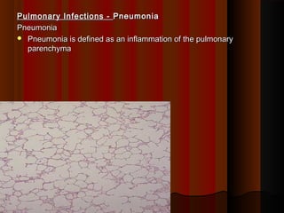
  • 1. Pulmonary Infections -Pulmonary Infections - PneumoniaPneumonia PneumoniaPneumonia  Pneumonia is defined as an inflammation of the pulmonaryPneumonia is defined as an inflammation of the pulmonary parenchymaparenchyma   
  • 2. ClassificationClassification A) According to immune statusA) According to immune status  Primary Pneumonia: Community acquiredPrimary Pneumonia: Community acquired  Secondary Pneumonia (occurs in compromised host or Lungs)Secondary Pneumonia (occurs in compromised host or Lungs)  BronchopneumoniaBronchopneumonia  Nosocomial Pneumonia (hospital acquired)Nosocomial Pneumonia (hospital acquired)  Aspiration pneumoniaAspiration pneumonia  Pneumonia in the immmunocompromised patientPneumonia in the immmunocompromised patient    
  • 3. ClassificationClassification B) Anatomical classificationB) Anatomical classification  Lobar PneumoniaLobar Pneumonia  Lobular Pneumonia (bronchopneumonia if bilateral)Lobular Pneumonia (bronchopneumonia if bilateral)  Interstitial PneumoniaInterstitial Pneumonia C)Aetiological classificationC)Aetiological classification  Infective-Bacteria, Virus, Fungi, Protozoa etc.Infective-Bacteria, Virus, Fungi, Protozoa etc.  AllergicAllergic  Chemical agents: Irritant gases and liquid e.g. ammonia, vomitusChemical agents: Irritant gases and liquid e.g. ammonia, vomitus  Physical agents: RadiationPhysical agents: Radiation
  • 4. Predisposing factorsPredisposing factors • A number of potent defense mechanisms clear or destroy anyA number of potent defense mechanisms clear or destroy any bacteria inhaled with air or accidently deposited in the airwaybacteria inhaled with air or accidently deposited in the airway passage.passage.  The following 3 defense mechanisms play vital role in preventing fromThe following 3 defense mechanisms play vital role in preventing from any sort of micro organisms infection that reaches our airwayany sort of micro organisms infection that reaches our airway passages.passages. a. Nasal clearancea. Nasal clearance b. Tracheobronchial clearanceb. Tracheobronchial clearance c. Alveolar clearancec. Alveolar clearance
  • 5. a.a.Nasal clearanceNasal clearance Particles including inhaled microorganisms are cleared byParticles including inhaled microorganisms are cleared by sneezing or blowing from the nose. Those microorganisms whichsneezing or blowing from the nose. Those microorganisms which manage to escape down are swallowed.manage to escape down are swallowed. b.b.Tracheobronchial clearanceTracheobronchial clearance Due to mucociliary action any particles or mocroorganismsDue to mucociliary action any particles or mocroorganisms deposited in the tracheobronchial tree are eventually either swalloweddeposited in the tracheobronchial tree are eventually either swallowed or expectorated.or expectorated.
  • 6. c. Alveolar clearancec. Alveolar clearance  Any bacteria or solid particles deposited in the alveoli areAny bacteria or solid particles deposited in the alveoli are phagocytosed by alveolar macrophages.phagocytosed by alveolar macrophages.  Some of the particles are carried to regional lymph nodes and viaSome of the particles are carried to regional lymph nodes and via the blood stream, they reach other parts of the body.the blood stream, they reach other parts of the body.
  • 7. Pathogenesis of PneumoniaPathogenesis of Pneumonia Pneumonia may occurPneumonia may occur  When these defense mechanisms are impaired Or whenever theWhen these defense mechanisms are impaired Or whenever the resistant of the host in general is lowered.resistant of the host in general is lowered. Infection of lung tissueInfection of lung tissue Following factors may interfere with clearing mechanisms.Following factors may interfere with clearing mechanisms. Loss or suppression of cough reflexLoss or suppression of cough reflex - due to coma, drugs, anaesthesia, neuromuscular disorders.- due to coma, drugs, anaesthesia, neuromuscular disorders. These condition can lead to aspiration of gastric contents too.These condition can lead to aspiration of gastric contents too.
  • 8. Injury to mucociliary apparatusInjury to mucociliary apparatus This can be due to cigarette smoking, due to inhalation of hot orThis can be due to cigarette smoking, due to inhalation of hot or corrosive gasses, viral diseases, or genetic diseases.corrosive gasses, viral diseases, or genetic diseases. Interference with phagocytic or bactericidal action ofInterference with phagocytic or bactericidal action of alveolar macrophagesalveolar macrophages This can be due to alcohol, tobacco, smoke, anoxia, or oxygenThis can be due to alcohol, tobacco, smoke, anoxia, or oxygen intoxication.intoxication.
  • 9. Pulmonary congestion & oedemaPulmonary congestion & oedema Accumulations of secretionsAccumulations of secretions – cystic fibrosis &– cystic fibrosis & bronchial obstructionbronchial obstruction
  • 10. LOBAR PNEUMONIALOBAR PNEUMONIA : -: - When a part of lobe, entire lobe or lobes of one or both lungs areWhen a part of lobe, entire lobe or lobes of one or both lungs are involvedinvolved Etiology: -Etiology: -  Streptococcus PneumoniaeStreptococcus Pneumoniae  StaphylococcalStaphylococcal  H. InfluenzaeH. Influenzae  Pseudomonas.Pseudomonas.
  • 11. 4 sequences or stages4 sequences or stages 1. Stage of Congestion1. Stage of Congestion : - Lasts for 1 – 2 days.: - Lasts for 1 – 2 days. MicroMicro Dilated & congested blood vessels with oedema fluid filled in air sacsDilated & congested blood vessels with oedema fluid filled in air sacs (alveoli) with plenty of bacteria & a few neutrophil(alveoli) with plenty of bacteria & a few neutrophil Gross : -Gross : -Lobe is enlarged, heavy, congested & exudes pink frothyLobe is enlarged, heavy, congested & exudes pink frothy fluid on cut surface.fluid on cut surface.
  • 12. 2. Stage of Red Hepatization2. Stage of Red Hepatization : - Lasts – 2 – 4 days: - Lasts – 2 – 4 days  MICRO : -MICRO : - Oedema fluid replaced by fibrin threads with markedOedema fluid replaced by fibrin threads with marked neutrophils & R.B.C., Neutrophils with engulfed bacteria & theneutrophils & R.B.C., Neutrophils with engulfed bacteria & the alveolar septa is less prominent than stage I.alveolar septa is less prominent than stage I.  GROSS : -GROSS : - Liver like red, firm, airless. On cut section, red – pink,Liver like red, firm, airless. On cut section, red – pink, dry, granular to look at.dry, granular to look at.
  • 13. 3. Stage of Grey Hepatization3. Stage of Grey Hepatization : - 4 – 8 days: - 4 – 8 days  MICRO : -MICRO : - Due to R.B.C. disintegration and numerous fibrinDue to R.B.C. disintegration and numerous fibrin strands and their contraction causes a separation of exudates fromstrands and their contraction causes a separation of exudates from the alveolar walls.the alveolar walls. Exudates are now less rich in bacteria, Rbc and inflammatory cells.Exudates are now less rich in bacteria, Rbc and inflammatory cells.  GROSS : -GROSS : - Firm, heavy with dry, granular and grey liver like.Firm, heavy with dry, granular and grey liver like. LessLess hyperaemiahyperaemia
  • 14. 4. Stage of Resolution:4. Stage of Resolution: - 8 – 16 days.- 8 – 16 days. MICRO : -MICRO : -  Consolidated exudate within the alveolar spaces undergoesConsolidated exudate within the alveolar spaces undergoes progressive enzymatic digestion to produce a granular semisolidprogressive enzymatic digestion to produce a granular semisolid debris that is either resorbed, ingested by macrophages or cougheddebris that is either resorbed, ingested by macrophages or coughed upup  Lungs return to normalLungs return to normal
  • 15.
  • 16.  This is a lobarThis is a lobar pneumonia in whichpneumonia in which consolidation of theconsolidation of the entire left upper lobeentire left upper lobe has occurred.has occurred.
  • 17.
  • 18. Clinical signs and symptomsClinical signs and symptoms i. Fever and chillsi. Fever and chills ii. Productive cough with yellow-green (pus) or rusty (bloody) sputumii. Productive cough with yellow-green (pus) or rusty (bloody) sputum iii. Tachypnea(abnormal rapid breathing)iii. Tachypnea(abnormal rapid breathing) iv. Pleuritic chest painiv. Pleuritic chest pain v. Decreased breath sounds, bronchial breath sounds ,v. Decreased breath sounds, bronchial breath sounds , crepitations(crackling sound), and dullness to percussioncrepitations(crackling sound), and dullness to percussion Nonspecific symptoms Most patients also have:Nonspecific symptoms Most patients also have:  Fatigue, Myalgia(muscles pain), Abdominal pain, Anorexia,Fatigue, Myalgia(muscles pain), Abdominal pain, Anorexia, HeadacheHeadache
  • 19. Lab: Elevated WBCLab: Elevated WBC countcount Chest x-rayChest x-ray i. Lobar: lobar or segmental consolidation (opacification)i. Lobar: lobar or segmental consolidation (opacification) ii. Bronchopneumonia: patchy opacificationii. Bronchopneumonia: patchy opacification iii. Pleural effusioniii. Pleural effusion Clinical keys: identification of the organism and early treatment withClinical keys: identification of the organism and early treatment with antibioticsantibiotics
  • 20. X-Ray showing Right middle lobe pneumoniaX-Ray showing Right middle lobe pneumonia
  • 21. Chest x-ray demonstrating complete right upper lobe consolidation, consistent with aChest x-ray demonstrating complete right upper lobe consolidation, consistent with a lobar pneumonia.lobar pneumonia.
  • 24. BronchopneumoniaBronchopneumonia This is the inflammatory consolidation of the surrounding alveoli of theThis is the inflammatory consolidation of the surrounding alveoli of the terminal bronchioles leading to patchy solidification of lung.terminal bronchioles leading to patchy solidification of lung. Etiology : -Etiology : -  StaphylococcusStaphylococcus  Haemophilus influenza,Haemophilus influenza,  pseudomonaspseudomonas  & E.coli.& E.coli.
  • 25. This is more common in elderly or children i.e extremes of age due toThis is more common in elderly or children i.e extremes of age due to viral or other bacterial infections.viral or other bacterial infections. Usually secondary to other conditions associated with local andUsually secondary to other conditions associated with local and general defense mechanisms:general defense mechanisms: - viral infections (influenza, measles)- viral infections (influenza, measles) - aspiration of food or vomitus- aspiration of food or vomitus - obstruction of a bronchus (foreign body or neoplasm)- obstruction of a bronchus (foreign body or neoplasm) - inhalation of irritant gases- inhalation of irritant gases - major surgery- major surgery - malnutrition- malnutrition
  • 26.   PATHOLOGYPATHOLOGY : -: - GROSS : -GROSS : - Patchy areas of consolidation in one or more lobes frequently bilateral andPatchy areas of consolidation in one or more lobes frequently bilateral and more often involving the lower zone due to gravitational reasons.more often involving the lower zone due to gravitational reasons.
  • 27. Here is example of aHere is example of a bronchopneumonia. Thebronchopneumonia. The lighter areas that appearlighter areas that appear to be raised on cutto be raised on cut surface from thesurface from the surrounding lung are thesurrounding lung are the areas of consolidation ofareas of consolidation of the lung.the lung.
  • 29.  This radiographThis radiograph demonstrates patchydemonstrates patchy infiltrates consistent with ainfiltrates consistent with a bronchopneumonia from abronchopneumonia from a bacterial infection. Typicalbacterial infection. Typical organisms includeorganisms include Streptococcus pneumoniae,Streptococcus pneumoniae, Staphylococcus aureus,Staphylococcus aureus, Pseudomonas aeruginosa,Pseudomonas aeruginosa, Hemophilus influenzae,Hemophilus influenzae, Klebsiella pneumoniae,Klebsiella pneumoniae, among others.among others.
  • 32. Bronchopneumonia 1. Usually occurs in persons with pre existing lung disease 2. Common in extreme of age 3. Usually by staphylococci 4. Patchy consolidation involving one lobe or but often multilobular 5. Usually bi lateral & basal 6. Complete resolution uncommon, may lead to complications Lobar PneumoniaLobar Pneumonia 1.1. Usually occurs in healthyUsually occurs in healthy young adultyoung adult 2.2. Common in young ageCommon in young age 3.3. Usually by PneumococciUsually by Pneumococci 4.4. Lobar consolidationLobar consolidation involving large portion ofinvolving large portion of one lobe or entire lobeone lobe or entire lobe 5.5. Can effect any lobe – uni orCan effect any lobe – uni or bi lateralbi lateral 6.6. Complete resolution usuallyComplete resolution usually resolution takes placeresolution takes place
  • 33.  INTERSTITIAL PNEUMONIAINTERSTITIAL PNEUMONIA : -Atypical Pneumonia: -Atypical Pneumonia This is characterized by patchy inflammatory changes largelyThis is characterized by patchy inflammatory changes largely confined to interstitial tissues of lung without any alveolar exudate.confined to interstitial tissues of lung without any alveolar exudate. ETIOLOGYETIOLOGY : -: - Mycoplasma pneumonae, Respiratory syncytial virus (RV)Mycoplasma pneumonae, Respiratory syncytial virus (RV) Influenza virus, cytomegalo, pneumocystis jiroveci (pneumocystisInfluenza virus, cytomegalo, pneumocystis jiroveci (pneumocystis carinii )carinii ) More common in immunosuppressed state.More common in immunosuppressed state.
  • 34. PATHOLOGYPATHOLOGY : -: - red blue colourred blue colour GROSS : -GROSS : - It may be patchy to massive involvement with heavyIt may be patchy to massive involvement with heavy congested &congested & subcrepitant lungs.subcrepitant lungs. Histo :Histo :  Mainly interstitial infiltration of mononuclear cells with thickening ofMainly interstitial infiltration of mononuclear cells with thickening of alveolar septa.alveolar septa.  Necrosis of bronchial epithelial lining & filled bronchiolar lumen withNecrosis of bronchial epithelial lining & filled bronchiolar lumen with secretions.secretions.  A reactive change by multinuceated giant cells formation is shownA reactive change by multinuceated giant cells formation is shown by lining bronchiolar epithelium.by lining bronchiolar epithelium. Complications : -Complications : - Main complication is secondary bacterial infection or a reactiveMain complication is secondary bacterial infection or a reactive fibrosis of interstitial tissue.fibrosis of interstitial tissue.
  • 35. Secondary bacterial infectionSecondary bacterial infection staphylococcistaphylococci
  • 36. Nosocomial infection or Hospital acquired pneumoniaNosocomial infection or Hospital acquired pneumonia (HAP)(HAP):: - After 48 hours of admission to the hospital, often patients in ICU- After 48 hours of admission to the hospital, often patients in ICU - ↓ Local resistance to infection in lungs- ↓ Local resistance to infection in lungs - Intubation of respiratory tract- Intubation of respiratory tract - Altered normal flora due to antibiotics- Altered normal flora due to antibiotics - Mostly gram – negative enterobacteria or Staph aureus, E.coli,- Mostly gram – negative enterobacteria or Staph aureus, E.coli, Klebsiella, Proteus, Pseudomonas, Bacteroides,Klebsiella, Proteus, Pseudomonas, Bacteroides,
  • 37. Community acquired pneumoniaCommunity acquired pneumonia Commonest cause is due toCommonest cause is due to  Streptococcus pneumoniae,Streptococcus pneumoniae, other areother are  H. Influenzae,H. Influenzae,  Mycoplasma pneumoniae,Mycoplasma pneumoniae,  Moraxella catarrhalis ,Moraxella catarrhalis ,  Staph. Aureus,Staph. Aureus,  clamydia, etc.clamydia, etc.
  • 38. The most common causes forThe most common causes for viral pneumoniaviral pneumonia are:are:  InfluenzaInfluenza  ParainfluenzaParainfluenza  AdenovirusAdenovirus  Respiratory syncytial virus (RSV)Respiratory syncytial virus (RSV) - appears mostly in children- appears mostly in children  CytomegalovirusCytomegalovirus - in immunocompromised hosts- in immunocompromised hosts
  • 39. Modes of transmission of PneumoniaModes of transmission of Pneumonia  Aspiration of organisms that colonize the oropharynx orAspiration of organisms that colonize the oropharynx or nasopharynxnasopharynx  Inhalation of infectious agentsInhalation of infectious agents  Haematogenous{formation of blood} spread from extra pulmonaryHaematogenous{formation of blood} spread from extra pulmonary sitesite  Direct inoculation (tracheal intubations or stab wound to the chest) &Direct inoculation (tracheal intubations or stab wound to the chest) & contiguous spread from an adjacent site of infectioncontiguous spread from an adjacent site of infection
  • 40. Complications of pneumoniaComplications of pneumonia  Lung abscess formationLung abscess formation  Pleural effusionPleural effusion  Empyema{collection of pus in a cavity in the body}Empyema{collection of pus in a cavity in the body}  Failure of resolution ⇒ intra-alveolar scarringFailure of resolution ⇒ intra-alveolar scarring  Respiratory failureRespiratory failure  Septicaemia and bacteraemiaSepticaemia and bacteraemia  - Infective endocarditis- Infective endocarditis  - Cerebral abscess / meningitis- Cerebral abscess / meningitis  - Septic arthritis{pus formation in joints}- Septic arthritis{pus formation in joints}  Fibrous scarring and pleural adhesionsFibrous scarring and pleural adhesions
  • 41.  Define Pneumonia & classify or causes of PneumoniaDefine Pneumonia & classify or causes of Pneumonia  What are the complications of PneumoniaWhat are the complications of Pneumonia  Give the pathogenesis of Lobar PneumoniaGive the pathogenesis of Lobar Pneumonia  Give the morphology / stages of pneumoniaGive the morphology / stages of pneumonia  Difference between lobar & bronchopneumoniaDifference between lobar & bronchopneumonia