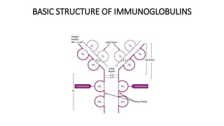
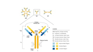
Antibody ̶ structure, types,
properties and functions of
Immunoglobulins
DEFINITION
• Immunoglobulin (Ig)
• Immunoglobulins are glycoprotein molecules that are produced by
plasma cells in response to an immunogen and which function as
antibodies.
• The immunoglobulins derive their name from the finding that they
migrate with globular proteins when antibody-containing serum is
placed in an electrical field
BASIC STRUCTURE OF IMMUNOGLOBULINS
• The basic structure
• A. Heavy and Light Chains
• All immunoglobulins have a four chain structure as their basic unit. They are
composed of two identical light chains (23kD) and two identical heavy
chains (50-70kD)
• B. Disulfide bonds
• 1. Inter-chain disulfide bonds - The heavy and light chains and the two
heavy chains are held together by inter-chain disulfide bonds and by non-
covalent interactions The number of inter-chain disulfide bonds varies
among different immunoglobulin molecules.
• 2. Intra-chain disulfide binds - Within each of the polypeptide chains there
are also intra-chain disulfide
• C. Variable (V) and Constant (C) Regions
• When the amino acid sequences of many different heavy chains and light
chains were compared, it became clear that both the heavy and light chain
could be divided into two regions based on variability in the amino acid
sequences. These are the:
• 1. Light Chain - VL (110 amino acids) and CL (110 amino acids)
• 2. Heavy Chain - VH (110 amino acids) and CH (330-440 amino acids)
• D. Hinge Region
• This is the region at which the arms of the antibody molecule forms a Y. It is
called the hinge region because there is some flexibility in the molecule at this
point.
• E. Domains
• Three dimensional images of the immunoglobulin molecule show that it is
not straight as depicted in figure 2A. Rather, it is folded into globular
regions each of which contains an intra-chain disulfide bond (figure 2B-D).
These regions are called domains.
• 1. Light Chain Domains - VL and CL
• 2. Heavy Chain Domains - VH, CH1 - CH3 (or CH4)
• F. Oligosaccharides
• Carbohydrates are attached to the CH2 domain in most immunoglobulins.
However, in some cases carbohydrates may also be attached at other
locations.
Hypervariable (HVR) or complementarity determining regions
(CDR)
• Comparisons of the amino acid sequences of the variable regions of
immunoglobulins show that most of the variability resides in three regions
called the hypervariable regions or the complementarity determining
regions.
•
• Antibodies with different specificities (i.e. different combining sites) have
different complementarity determining regions while antibodies of the
exact same specificity have identical complementarity determining regions
(i.e. CDR is the antibody combining site).
• Complementarity determining regions are found in both the H and the L
chains.
Fab
• Digestion with papain breaks the immunoglobulin molecule in the hinge
region before the H-H inter-chain disulfide bond.
• This results in the formation of two identical fragments that contain the
light chain and the VH and CH1 domains of the heavy chain.
• Antigen binding - These fragments were called the Fab fragments because
they contained the antigen binding sites of the antibody.
• Each Fab fragment is monovalent whereas the original molecule was
divalent.
• The combining site of the antibody is created by both VH and VL.
• An antibody is able to bind a particular antigenic determinant because it
has a particular combination of VH and VL.
• Different combinations of a VH and VL result in antibodies that can bind a
different antigenic determinants.
Fc
• Digestion with papain also produces a fragment that contains the
remainder of the two heavy chains each containing a CH2 and CH3
domain. This fragment was called Fc because it was easily crystallized.
• Effector functions - The effector functions of immunoglobulins are
mediated by this part of the molecule. Different functions are mediated
by the different domains in this fragment.
• Normally the ability of an antibody to carry out an effector function
requires the prior binding of an antigen; however, there are exceptions
to this rule.
Fab’
• Treatment of immunoglobulins with pepsin results in cleavage of the
heavy chain after the H-H inter-chain disulfide bonds resulting in a
fragment that contains both antigen binding sites.
• This fragment was called F(ab')2 because it is divalent.
• The Fc region of the molecule is digested into small peptides by pepsin.
• The F(ab')2 binds antigen but it does not mediate the effector
functions of antibodies.
• The immunoglobulins can be divided into five different classes, based on
differences in the amino acid sequences in the constant region of the
heavy chains.
• All immunoglobulins within a given class will have very similar heavy
chain constant regions.
• These differences can be detected by sequence studies or more
commonly by serological means (i.e. by the use of antibodies directed to
these differences).
• 1. IgG - Gamma heavy chains
• 2. IgM - Mu heavy chains
• 3. IgA - Alpha heavy chains
• 4. IgD - Delta heavy chains
• 5. IgE - Epsilon heavy chains
Ig G
• All IgG's are monomers (7S immunoglobulin). The subclasses differ in
the number of disulfide bonds and length of the hinge region.
• IgG is the most versatile immunoglobulin because it is capable of
carrying out all of the functions of immunoglobulin molecules.
• a) IgG is the major Ig in serum - 75% of serum Ig is IgG
• b) IgG is the major Ig in extra vascular spaces
• c) Placental transfer - IgG is the only class of Ig that crosses the
placenta. Transfer is mediated by a receptor on placental cells for the
Fc region of IgG. Not all subclasses cross equally well; IgG2 does not
cross well.
• d) Fixes complement - Not all subclasses fix equally well; IgG4 does not
fix complement
• e) Binding to cells - Macrophages, monocytes, PMNs and some
lymphocytes have Fc receptors for the Fc region of IgG.
• A consequence of binding to the Fc receptors on PMNs, monocytes and
macrophages is that the cell can now internalize the antigen better.
• The antibody has prepared the antigen for eating by the phagocytic cells.
• The term opsonin is used to describe substances that enhance
phagocytosis.
• IgG is a good opsonin. Binding of IgG to Fc receptors on other types of
cells results in the activation of other functions.
Ig M
• IgM normally exists as a pentamer (19S immunoglobulin) but it can also
exist as a monomer.
• In the pentameric form all heavy chains are identical and all light chains
are identical. Thus, the valence is theoretically 10.
• IgM has an extra domain on the mu chain (CH4) and it has another
protein covalently bound via a S-S bond called the J chain.
• This chain functions in polymerization of the molecule into a pentamer.
• a) IgM is the third most common serum Ig.
• b) IgM is the first Ig to be made by the fetus and the first Ig to be
made by a virgin B cells when it is stimulated by antigen.
• c) As a consequence of its pentameric structure, IgM is a good
complement fixing Ig. Thus, IgM antibodies are very efficient in
leading to the lysis of microorganisms.
• d) As a consequence of its structure, IgM is also a good agglutinating
Ig . Thus, IgM antibodies are very good in clumping microorganisms
for eventual elimination from the body.
• e) IgM binds to some cells via Fc receptors.
• f) B cell surface Ig
Ig A
• Serum IgA is a monomer but IgA found in secretions is a dimer. When
IgA exits as a dimer, a J chain is associated with it.
• When IgA is found in secretions is also has another protein associated
with it called the secretory piece or T piece; sIgA is sometimes
referred to as 11S immunoglobulin.
• Unlike the remainder of the IgA which is made in the plasma cell, the
secretory piece is made in epithelial cells and is added to the IgA as it
passes into the secretions.
• The secretory piece helps IgA to be transported across mucosa and
also protects it from degradation in the secretions.
Properties
• a) IgA is the 2nd most common serum Ig.
• b) IgA is the major class of Ig in secretions - tears, saliva, colostrum,
mucus. Since it is found in secretions secretory IgA is important in
local (mucosal) immunity.
• c) Normally IgA does not fix complement, unless aggregated.
• d) IgA can binding to some cells - PMN's and some lymphocytes.
Ig D
• a) Ig D exists as monomer only
• B) IgD is found in low levels in serum; its role in serum uncertain.
• c) IgD is primarily found on B cell surfaces where it functions as a
receptor for antigen. IgD on the surface of B cells has extra amino
acids at C-terminal end for anchoring to the membrane. It also
associates with the Ig-alpha and Ig-beta chains.
• d) IgD does not bind complement.
•
Ig E
• a) IgE is the least common serum Ig since it binds very tightly to Fc
receptors on basophils and mast cells even before interacting with
antigen.
• b) Involved in allergic reactions - As a consequence of its binding to
basophils an mast cells, IgE is involved in allergic reactions. Binding of
the allergen to the IgE on the cells results in the release of various
pharmacological mediators that result in allergic symptoms.
• c) IgE also plays a role in parasitic helminth diseases. Since serum IgE
levels rise in parasitic diseases, measuring IgE levels is helpful in
diagnosing parasitic infections. Eosinophils have Fc receptors for IgE and
binding of eosinophils to IgE-coated helminths results in killing of the
parasite.
• d) IgE does not fix complement.
• https://www.thermofisher.com/in/en/home/life-
science/antibodies/antibodies-learning-center/antibodies-resource-
library/antibody-methods/immunoglobulin-structure-
classes.html#:~:text=The%20five%20primary%20classes%20of,chain%
20found%20in%20the%20molecule.&text=Differences%20in%20hea
• https://www.microbiologybook.org/mobile/m.immuno-4.htm

Antibodies

  • 1.
    Antibody ̶ structure,types, properties and functions of Immunoglobulins
  • 2.
    DEFINITION • Immunoglobulin (Ig) •Immunoglobulins are glycoprotein molecules that are produced by plasma cells in response to an immunogen and which function as antibodies. • The immunoglobulins derive their name from the finding that they migrate with globular proteins when antibody-containing serum is placed in an electrical field
  • 3.
    BASIC STRUCTURE OFIMMUNOGLOBULINS
  • 4.
    • The basicstructure • A. Heavy and Light Chains • All immunoglobulins have a four chain structure as their basic unit. They are composed of two identical light chains (23kD) and two identical heavy chains (50-70kD) • B. Disulfide bonds • 1. Inter-chain disulfide bonds - The heavy and light chains and the two heavy chains are held together by inter-chain disulfide bonds and by non- covalent interactions The number of inter-chain disulfide bonds varies among different immunoglobulin molecules. • 2. Intra-chain disulfide binds - Within each of the polypeptide chains there are also intra-chain disulfide
  • 5.
    • C. Variable(V) and Constant (C) Regions • When the amino acid sequences of many different heavy chains and light chains were compared, it became clear that both the heavy and light chain could be divided into two regions based on variability in the amino acid sequences. These are the: • 1. Light Chain - VL (110 amino acids) and CL (110 amino acids) • 2. Heavy Chain - VH (110 amino acids) and CH (330-440 amino acids) • D. Hinge Region • This is the region at which the arms of the antibody molecule forms a Y. It is called the hinge region because there is some flexibility in the molecule at this point.
  • 6.
    • E. Domains •Three dimensional images of the immunoglobulin molecule show that it is not straight as depicted in figure 2A. Rather, it is folded into globular regions each of which contains an intra-chain disulfide bond (figure 2B-D). These regions are called domains. • 1. Light Chain Domains - VL and CL • 2. Heavy Chain Domains - VH, CH1 - CH3 (or CH4) • F. Oligosaccharides • Carbohydrates are attached to the CH2 domain in most immunoglobulins. However, in some cases carbohydrates may also be attached at other locations.
  • 8.
    Hypervariable (HVR) orcomplementarity determining regions (CDR) • Comparisons of the amino acid sequences of the variable regions of immunoglobulins show that most of the variability resides in three regions called the hypervariable regions or the complementarity determining regions. • • Antibodies with different specificities (i.e. different combining sites) have different complementarity determining regions while antibodies of the exact same specificity have identical complementarity determining regions (i.e. CDR is the antibody combining site). • Complementarity determining regions are found in both the H and the L chains.
  • 9.
    Fab • Digestion withpapain breaks the immunoglobulin molecule in the hinge region before the H-H inter-chain disulfide bond. • This results in the formation of two identical fragments that contain the light chain and the VH and CH1 domains of the heavy chain. • Antigen binding - These fragments were called the Fab fragments because they contained the antigen binding sites of the antibody. • Each Fab fragment is monovalent whereas the original molecule was divalent. • The combining site of the antibody is created by both VH and VL. • An antibody is able to bind a particular antigenic determinant because it has a particular combination of VH and VL. • Different combinations of a VH and VL result in antibodies that can bind a different antigenic determinants.
  • 10.
    Fc • Digestion withpapain also produces a fragment that contains the remainder of the two heavy chains each containing a CH2 and CH3 domain. This fragment was called Fc because it was easily crystallized. • Effector functions - The effector functions of immunoglobulins are mediated by this part of the molecule. Different functions are mediated by the different domains in this fragment. • Normally the ability of an antibody to carry out an effector function requires the prior binding of an antigen; however, there are exceptions to this rule.
  • 11.
    Fab’ • Treatment ofimmunoglobulins with pepsin results in cleavage of the heavy chain after the H-H inter-chain disulfide bonds resulting in a fragment that contains both antigen binding sites. • This fragment was called F(ab')2 because it is divalent. • The Fc region of the molecule is digested into small peptides by pepsin. • The F(ab')2 binds antigen but it does not mediate the effector functions of antibodies.
  • 12.
    • The immunoglobulinscan be divided into five different classes, based on differences in the amino acid sequences in the constant region of the heavy chains. • All immunoglobulins within a given class will have very similar heavy chain constant regions. • These differences can be detected by sequence studies or more commonly by serological means (i.e. by the use of antibodies directed to these differences). • 1. IgG - Gamma heavy chains • 2. IgM - Mu heavy chains • 3. IgA - Alpha heavy chains • 4. IgD - Delta heavy chains • 5. IgE - Epsilon heavy chains
  • 13.
    Ig G • AllIgG's are monomers (7S immunoglobulin). The subclasses differ in the number of disulfide bonds and length of the hinge region. • IgG is the most versatile immunoglobulin because it is capable of carrying out all of the functions of immunoglobulin molecules. • a) IgG is the major Ig in serum - 75% of serum Ig is IgG • b) IgG is the major Ig in extra vascular spaces • c) Placental transfer - IgG is the only class of Ig that crosses the placenta. Transfer is mediated by a receptor on placental cells for the Fc region of IgG. Not all subclasses cross equally well; IgG2 does not cross well.
  • 14.
    • d) Fixescomplement - Not all subclasses fix equally well; IgG4 does not fix complement • e) Binding to cells - Macrophages, monocytes, PMNs and some lymphocytes have Fc receptors for the Fc region of IgG. • A consequence of binding to the Fc receptors on PMNs, monocytes and macrophages is that the cell can now internalize the antigen better. • The antibody has prepared the antigen for eating by the phagocytic cells. • The term opsonin is used to describe substances that enhance phagocytosis. • IgG is a good opsonin. Binding of IgG to Fc receptors on other types of cells results in the activation of other functions.
  • 15.
    Ig M • IgMnormally exists as a pentamer (19S immunoglobulin) but it can also exist as a monomer. • In the pentameric form all heavy chains are identical and all light chains are identical. Thus, the valence is theoretically 10. • IgM has an extra domain on the mu chain (CH4) and it has another protein covalently bound via a S-S bond called the J chain. • This chain functions in polymerization of the molecule into a pentamer.
  • 16.
    • a) IgMis the third most common serum Ig. • b) IgM is the first Ig to be made by the fetus and the first Ig to be made by a virgin B cells when it is stimulated by antigen. • c) As a consequence of its pentameric structure, IgM is a good complement fixing Ig. Thus, IgM antibodies are very efficient in leading to the lysis of microorganisms. • d) As a consequence of its structure, IgM is also a good agglutinating Ig . Thus, IgM antibodies are very good in clumping microorganisms for eventual elimination from the body. • e) IgM binds to some cells via Fc receptors. • f) B cell surface Ig
  • 17.
    Ig A • SerumIgA is a monomer but IgA found in secretions is a dimer. When IgA exits as a dimer, a J chain is associated with it. • When IgA is found in secretions is also has another protein associated with it called the secretory piece or T piece; sIgA is sometimes referred to as 11S immunoglobulin. • Unlike the remainder of the IgA which is made in the plasma cell, the secretory piece is made in epithelial cells and is added to the IgA as it passes into the secretions. • The secretory piece helps IgA to be transported across mucosa and also protects it from degradation in the secretions.
  • 18.
    Properties • a) IgAis the 2nd most common serum Ig. • b) IgA is the major class of Ig in secretions - tears, saliva, colostrum, mucus. Since it is found in secretions secretory IgA is important in local (mucosal) immunity. • c) Normally IgA does not fix complement, unless aggregated. • d) IgA can binding to some cells - PMN's and some lymphocytes.
  • 19.
    Ig D • a)Ig D exists as monomer only • B) IgD is found in low levels in serum; its role in serum uncertain. • c) IgD is primarily found on B cell surfaces where it functions as a receptor for antigen. IgD on the surface of B cells has extra amino acids at C-terminal end for anchoring to the membrane. It also associates with the Ig-alpha and Ig-beta chains. • d) IgD does not bind complement. •
  • 20.
    Ig E • a)IgE is the least common serum Ig since it binds very tightly to Fc receptors on basophils and mast cells even before interacting with antigen. • b) Involved in allergic reactions - As a consequence of its binding to basophils an mast cells, IgE is involved in allergic reactions. Binding of the allergen to the IgE on the cells results in the release of various pharmacological mediators that result in allergic symptoms. • c) IgE also plays a role in parasitic helminth diseases. Since serum IgE levels rise in parasitic diseases, measuring IgE levels is helpful in diagnosing parasitic infections. Eosinophils have Fc receptors for IgE and binding of eosinophils to IgE-coated helminths results in killing of the parasite. • d) IgE does not fix complement.
  • 21.