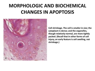
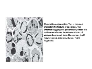
SlideShare a Scribd company logo
1 of 18
Apoptosis
Apoptosis
• is a pathway of cell death that is induced by a
tightly regulated suicide program in which
cells destined to die activate enzymes that
degrade the cells' own nuclear DNA and
nuclear and cytoplasmic proteins.
• The plasma membrane of the apoptotic cell
and bodies remains intact, but its structure is
altered in such a way that these become
“tasty” targets for phagocytes
• The dead cell and its fragments are rapidly
devoured, before the contents have leaked
out, and therefore cell death by this pathway
does not elicit an inflammatory reaction in the
host
• distinct from necrosis, which is characterized
by loss of membrane integrity, enzymatic
digestion of cells, leakage of cellular contents,
and frequently a host reaction However,
apoptosis and necrosis sometimes coexist,
and apoptosis induced by some pathologic
stimuli may progress to necrosis.
CAUSES OF APOPTOSIS
Apoptosis occurs normally both during
development and throughout adulthood, and
serves to eliminate unwanted, aged or
potentially harmful cells. It is also a pathologic
event when diseased cells become damaged
beyond repair and are eliminated.
Importance of Apoptosis in Physiologic
Conditions
• The programmed destruction of cells during
embryogenesis, including implantation,
organogenesis, developmental involution, and
metamorphosis. The term “programmed cell
death” was originally coined to denote death
of specific cell types at defined times during
the development of an organism
• Involution of hormone-dependent tissues upon
hormone withdrawal, such as endometrial cell
breakdown during the menstrual cycle,
ovarian follicular atresia in menopause, the
regression of the lactating breast after
weaning, and prostatic atrophy after
castration
• Cell loss in proliferating cell populations, such
as immature lymphocytes in the bone marrow
and thymus that fail to express useful antigen
receptors B lymphocytes in germinal centers,
and epithelial cells in intestinal crypts, so as to
maintain a constant number (homeostasis).
• Elimination of potentially harmful self-reactive
lymphocytes, either before or after they have
completed their maturation, so as to prevent
reactions against one's own tissues
• Death of host cells that have served their
useful purpose, such as neutrophils in an
acute inflammatory response, and
lymphocytes at the end of an immune
response. In these situations cells undergo
apoptosis because they are deprived of
necessary survival signals, such as growth
factors.
Apoptosis in Pathologic Conditions
• Apoptosis eliminates cells that are injured
beyond repair without eliciting a host
reaction, thus limiting collateral tissue
damage. Death by apoptosis is responsible for
loss of cells in a variety of pathologic states
• DNA damage. Radiation, cytotoxic anticancer
drugs, and hypoxia can damage DNA, either
directly or via production of free radicals. If repair
mechanisms cannot cope with the injury, the cell
triggers intrinsic mechanisms that induce
apoptosis. In these situations elimination of the
cell may be a better alternative than risking
mutations in the damaged DNA, which may result
in malignant transformation. These injurious
stimuli can cause apoptosis if the insult is mild,
but larger doses of the same stimuli may result in
necrotic cell death.
• Accumulation of misfolded proteins. Improperly
folded proteins may arise because of mutations in
the genes encoding these proteins or because of
extrinsic factors, such as damage caused by free
radicals. Excessive accumulation of these proteins
in the ER leads to a condition called ER stress,
which culminates in apoptotic cell death.
Apoptosis caused by the accumulation of
misfolded proteins has been invoked as the basis
of several degenerative diseases of the central
nervous system and other organs
• Cell death in certain infections, particularly
viral infections, in which loss of infected cells
is largely due to apoptosis that may be
induced by the virus (as in adenovirus and
HIV infections) or by the host immune
response (as in viral hepatitis).
• Pathologic atrophy in parenchymal organs
after duct obstruction, such as occurs in the
pancreas, parotid gland, and kidney
MORPHOLOGIC AND BIOCHEMICAL
CHANGES IN APOPTOSIS
Cell shrinkage. The cell is smaller in size; the
cytoplasm is dense; and the organelles,
though relatively normal, are more tightly
packed. (Recall that in other forms of cell
injury, an early feature is cell swelling, not
shrinkage.)
Chromatin condensation. This is the most
characteristic feature of apoptosis. The
chromatin aggregates peripherally, under the
nuclear membrane, into dense masses of
various shapes and sizes. The nucleus itself
may break up, producing two or more
fragments
Formation of cytoplasmic blebs and
apoptotic bodies. The apoptotic cell first
shows extensive surface blebbing, then
undergoes fragmentation into
membrane-bound apoptotic bodies
composed of cytoplasm and tightly
packed organelles, with or without
nuclear fragments

More Related Content

What's hot (20)

Apoptosis
Apoptosis Apoptosis
Apoptosis
 
Apoptosis
ApoptosisApoptosis
Apoptosis
 
Autoimmunity
AutoimmunityAutoimmunity
Autoimmunity
 
B- lymphocytes
B- lymphocytesB- lymphocytes
B- lymphocytes
 
Immunohistochemistry
Immunohistochemistry Immunohistochemistry
Immunohistochemistry
 
Apoptosis seminar
Apoptosis seminarApoptosis seminar
Apoptosis seminar
 
Apoptosis
ApoptosisApoptosis
Apoptosis
 
Apoptosis
Apoptosis Apoptosis
Apoptosis
 
The Complement System
The Complement SystemThe Complement System
The Complement System
 
Apoptosis.ppt
Apoptosis.pptApoptosis.ppt
Apoptosis.ppt
 
Autoimmune diseases
Autoimmune diseasesAutoimmune diseases
Autoimmune diseases
 
Innate immunity
Innate immunityInnate immunity
Innate immunity
 
Apoptosis basics + microphotographs
Apoptosis basics + microphotographsApoptosis basics + microphotographs
Apoptosis basics + microphotographs
 
Immunological Basis of Graft Rejection
Immunological Basis of Graft RejectionImmunological Basis of Graft Rejection
Immunological Basis of Graft Rejection
 
Apoptosis
ApoptosisApoptosis
Apoptosis
 
Apoptosis
ApoptosisApoptosis
Apoptosis
 
Apoptosis
ApoptosisApoptosis
Apoptosis
 
4.pathology apoptosis2016
4.pathology  apoptosis20164.pathology  apoptosis2016
4.pathology apoptosis2016
 
Immunohistochemistry in pathology laboratory
Immunohistochemistry in pathology laboratoryImmunohistochemistry in pathology laboratory
Immunohistochemistry in pathology laboratory
 
Cellular ageing
Cellular ageingCellular ageing
Cellular ageing
 

Viewers also liked

Apoptosis (presentation)
Apoptosis (presentation)Apoptosis (presentation)
Apoptosis (presentation)Leo Vang
 
General pathology lecture 1 introduction & cell injury
General pathology lecture 1 introduction & cell injuryGeneral pathology lecture 1 introduction & cell injury
General pathology lecture 1 introduction & cell injuryHuang Yu-Wen
 
Pathology cell injury i
Pathology   cell injury iPathology   cell injury i
Pathology cell injury iMBBS IMS MSU
 
Initiation and maintainence of endometriosis
Initiation and maintainence of endometriosisInitiation and maintainence of endometriosis
Initiation and maintainence of endometriosisKumgang Media
 
Irreversible cell injury
Irreversible cell injuryIrreversible cell injury
Irreversible cell injurySuraj Choudhary
 
Introduction To Pathology
Introduction To PathologyIntroduction To Pathology
Introduction To PathologyHeather Johnson
 
Cell death
Cell deathCell death
Cell deathSmawi GH
 
Cell injury: causes, pathogenesis, Morphology of reversible cell injury
Cell injury: causes, pathogenesis, Morphology of reversible cell injuryCell injury: causes, pathogenesis, Morphology of reversible cell injury
Cell injury: causes, pathogenesis, Morphology of reversible cell injuryVijay Shankar
 
Cell Injury Patho
Cell Injury PathoCell Injury Patho
Cell Injury Pathoaxix
 
Basic Pathology : Introduction To Cells & Tissue Damage
Basic Pathology : Introduction To Cells & Tissue DamageBasic Pathology : Introduction To Cells & Tissue Damage
Basic Pathology : Introduction To Cells & Tissue DamageSado Anatomist
 
Cell injury, adaptation, and death fix
Cell injury, adaptation, and death fix Cell injury, adaptation, and death fix
Cell injury, adaptation, and death fix Raniagaye Mansibang
 

Viewers also liked (15)

Apoptosis (presentation)
Apoptosis (presentation)Apoptosis (presentation)
Apoptosis (presentation)
 
General pathology lecture 1 introduction & cell injury
General pathology lecture 1 introduction & cell injuryGeneral pathology lecture 1 introduction & cell injury
General pathology lecture 1 introduction & cell injury
 
Pathology cell injury i
Pathology   cell injury iPathology   cell injury i
Pathology cell injury i
 
Initiation and maintainence of endometriosis
Initiation and maintainence of endometriosisInitiation and maintainence of endometriosis
Initiation and maintainence of endometriosis
 
Cell injury
Cell injuryCell injury
Cell injury
 
Apoptosis vs necrosis
Apoptosis vs necrosisApoptosis vs necrosis
Apoptosis vs necrosis
 
Irreversible cell injury
Irreversible cell injuryIrreversible cell injury
Irreversible cell injury
 
Endometriosis
EndometriosisEndometriosis
Endometriosis
 
Introduction To Pathology
Introduction To PathologyIntroduction To Pathology
Introduction To Pathology
 
Cell death
Cell deathCell death
Cell death
 
Cell injury: causes, pathogenesis, Morphology of reversible cell injury
Cell injury: causes, pathogenesis, Morphology of reversible cell injuryCell injury: causes, pathogenesis, Morphology of reversible cell injury
Cell injury: causes, pathogenesis, Morphology of reversible cell injury
 
Cell Injury Patho
Cell Injury PathoCell Injury Patho
Cell Injury Patho
 
Apoptosis
ApoptosisApoptosis
Apoptosis
 
Basic Pathology : Introduction To Cells & Tissue Damage
Basic Pathology : Introduction To Cells & Tissue DamageBasic Pathology : Introduction To Cells & Tissue Damage
Basic Pathology : Introduction To Cells & Tissue Damage
 
Cell injury, adaptation, and death fix
Cell injury, adaptation, and death fix Cell injury, adaptation, and death fix
Cell injury, adaptation, and death fix
 

Similar to Apoptosis

Cell injury apoptosis lect 08jan 2020
Cell injury apoptosis lect 08jan 2020Cell injury apoptosis lect 08jan 2020
Cell injury apoptosis lect 08jan 2020DrAbdulAzizShaikh
 
apoptosis lecture-powerpoint slides (1) (1).pdf
apoptosis lecture-powerpoint slides (1) (1).pdfapoptosis lecture-powerpoint slides (1) (1).pdf
apoptosis lecture-powerpoint slides (1) (1).pdfSrimathideviJ
 
Apoptosis-The-Programmed-Cell-Death.pptx
Apoptosis-The-Programmed-Cell-Death.pptxApoptosis-The-Programmed-Cell-Death.pptx
Apoptosis-The-Programmed-Cell-Death.pptxsaraqmc
 
Trauma and cellular injury in pathophysiology
Trauma and cellular injury in pathophysiologyTrauma and cellular injury in pathophysiology
Trauma and cellular injury in pathophysiologyMuhammad Aurangzeb
 
NECROSIS & APOTOSIS by Dr. Roopam Jain
NECROSIS & APOTOSIS by Dr. Roopam JainNECROSIS & APOTOSIS by Dr. Roopam Jain
NECROSIS & APOTOSIS by Dr. Roopam JainDr. Roopam Jain
 
Cell injury apoptosis
Cell injury apoptosis Cell injury apoptosis
Cell injury apoptosis abdul aziz
 
APOPTOSIS : The programmed cell death
APOPTOSIS : The programmed cell deathAPOPTOSIS : The programmed cell death
APOPTOSIS : The programmed cell deathJibesh Bhattacharjee
 
cellular adaptation and response to injury
cellular adaptation and response to injurycellular adaptation and response to injury
cellular adaptation and response to injurySangeeta Prasad
 
Cell inj apoptosis
Cell inj apoptosisCell inj apoptosis
Cell inj apoptosisAiman Iqbal
 
11.20 (dr. yasmeen hashim) apoptosis (mechanism in normal tissues. programmed...
11.20 (dr. yasmeen hashim) apoptosis (mechanism in normal tissues. programmed...11.20 (dr. yasmeen hashim) apoptosis (mechanism in normal tissues. programmed...
11.20 (dr. yasmeen hashim) apoptosis (mechanism in normal tissues. programmed...Fati Naqvi
 
Apoptosis.ppt
Apoptosis.pptApoptosis.ppt
Apoptosis.ppthdthao
 
types of cell death .pptx
types of cell death .pptxtypes of cell death .pptx
types of cell death .pptxnomiking5
 

Similar to Apoptosis (20)

Cell injury apoptosis lect 08jan 2020
Cell injury apoptosis lect 08jan 2020Cell injury apoptosis lect 08jan 2020
Cell injury apoptosis lect 08jan 2020
 
Apoptosis..pptx
Apoptosis..pptxApoptosis..pptx
Apoptosis..pptx
 
apoptosis lecture-powerpoint slides (1) (1).pdf
apoptosis lecture-powerpoint slides (1) (1).pdfapoptosis lecture-powerpoint slides (1) (1).pdf
apoptosis lecture-powerpoint slides (1) (1).pdf
 
Apoptosis-The-Programmed-Cell-Death.pptx
Apoptosis-The-Programmed-Cell-Death.pptxApoptosis-The-Programmed-Cell-Death.pptx
Apoptosis-The-Programmed-Cell-Death.pptx
 
Cell ageing ty
Cell ageing tyCell ageing ty
Cell ageing ty
 
Trauma and cellular injury in pathophysiology
Trauma and cellular injury in pathophysiologyTrauma and cellular injury in pathophysiology
Trauma and cellular injury in pathophysiology
 
NECROSIS & APOTOSIS by Dr. Roopam Jain
NECROSIS & APOTOSIS by Dr. Roopam JainNECROSIS & APOTOSIS by Dr. Roopam Jain
NECROSIS & APOTOSIS by Dr. Roopam Jain
 
Cell injury apoptosis
Cell injury apoptosis Cell injury apoptosis
Cell injury apoptosis
 
APOPTOSIS : The programmed cell death
APOPTOSIS : The programmed cell deathAPOPTOSIS : The programmed cell death
APOPTOSIS : The programmed cell death
 
Apoptosis
ApoptosisApoptosis
Apoptosis
 
cellular adaptation and response to injury
cellular adaptation and response to injurycellular adaptation and response to injury
cellular adaptation and response to injury
 
Cell death
Cell deathCell death
Cell death
 
Cell inj apoptosis
Cell inj apoptosisCell inj apoptosis
Cell inj apoptosis
 
11.20 (dr. yasmeen hashim) apoptosis (mechanism in normal tissues. programmed...
11.20 (dr. yasmeen hashim) apoptosis (mechanism in normal tissues. programmed...11.20 (dr. yasmeen hashim) apoptosis (mechanism in normal tissues. programmed...
11.20 (dr. yasmeen hashim) apoptosis (mechanism in normal tissues. programmed...
 
Apoptosis.ppt
Apoptosis.pptApoptosis.ppt
Apoptosis.ppt
 
Apoptosis.ppt
Apoptosis.pptApoptosis.ppt
Apoptosis.ppt
 
Apoptosis
ApoptosisApoptosis
Apoptosis
 
Apoptosis.pptx
Apoptosis.pptxApoptosis.pptx
Apoptosis.pptx
 
Apoptosis
ApoptosisApoptosis
Apoptosis
 
types of cell death .pptx
types of cell death .pptxtypes of cell death .pptx
types of cell death .pptx
 

Apoptosis

  • 2. Apoptosis • is a pathway of cell death that is induced by a tightly regulated suicide program in which cells destined to die activate enzymes that degrade the cells' own nuclear DNA and nuclear and cytoplasmic proteins. • The plasma membrane of the apoptotic cell and bodies remains intact, but its structure is altered in such a way that these become “tasty” targets for phagocytes
  • 3. • The dead cell and its fragments are rapidly devoured, before the contents have leaked out, and therefore cell death by this pathway does not elicit an inflammatory reaction in the host
  • 4. • distinct from necrosis, which is characterized by loss of membrane integrity, enzymatic digestion of cells, leakage of cellular contents, and frequently a host reaction However, apoptosis and necrosis sometimes coexist, and apoptosis induced by some pathologic stimuli may progress to necrosis.
  • 5. CAUSES OF APOPTOSIS Apoptosis occurs normally both during development and throughout adulthood, and serves to eliminate unwanted, aged or potentially harmful cells. It is also a pathologic event when diseased cells become damaged beyond repair and are eliminated.
  • 6. Importance of Apoptosis in Physiologic Conditions • The programmed destruction of cells during embryogenesis, including implantation, organogenesis, developmental involution, and metamorphosis. The term “programmed cell death” was originally coined to denote death of specific cell types at defined times during the development of an organism
  • 7. • Involution of hormone-dependent tissues upon hormone withdrawal, such as endometrial cell breakdown during the menstrual cycle, ovarian follicular atresia in menopause, the regression of the lactating breast after weaning, and prostatic atrophy after castration
  • 8. • Cell loss in proliferating cell populations, such as immature lymphocytes in the bone marrow and thymus that fail to express useful antigen receptors B lymphocytes in germinal centers, and epithelial cells in intestinal crypts, so as to maintain a constant number (homeostasis).
  • 9. • Elimination of potentially harmful self-reactive lymphocytes, either before or after they have completed their maturation, so as to prevent reactions against one's own tissues
  • 10. • Death of host cells that have served their useful purpose, such as neutrophils in an acute inflammatory response, and lymphocytes at the end of an immune response. In these situations cells undergo apoptosis because they are deprived of necessary survival signals, such as growth factors.
  • 11. Apoptosis in Pathologic Conditions • Apoptosis eliminates cells that are injured beyond repair without eliciting a host reaction, thus limiting collateral tissue damage. Death by apoptosis is responsible for loss of cells in a variety of pathologic states
  • 12. • DNA damage. Radiation, cytotoxic anticancer drugs, and hypoxia can damage DNA, either directly or via production of free radicals. If repair mechanisms cannot cope with the injury, the cell triggers intrinsic mechanisms that induce apoptosis. In these situations elimination of the cell may be a better alternative than risking mutations in the damaged DNA, which may result in malignant transformation. These injurious stimuli can cause apoptosis if the insult is mild, but larger doses of the same stimuli may result in necrotic cell death.
  • 13. • Accumulation of misfolded proteins. Improperly folded proteins may arise because of mutations in the genes encoding these proteins or because of extrinsic factors, such as damage caused by free radicals. Excessive accumulation of these proteins in the ER leads to a condition called ER stress, which culminates in apoptotic cell death. Apoptosis caused by the accumulation of misfolded proteins has been invoked as the basis of several degenerative diseases of the central nervous system and other organs
  • 14. • Cell death in certain infections, particularly viral infections, in which loss of infected cells is largely due to apoptosis that may be induced by the virus (as in adenovirus and HIV infections) or by the host immune response (as in viral hepatitis).
  • 15. • Pathologic atrophy in parenchymal organs after duct obstruction, such as occurs in the pancreas, parotid gland, and kidney
  • 16. MORPHOLOGIC AND BIOCHEMICAL CHANGES IN APOPTOSIS Cell shrinkage. The cell is smaller in size; the cytoplasm is dense; and the organelles, though relatively normal, are more tightly packed. (Recall that in other forms of cell injury, an early feature is cell swelling, not shrinkage.)
  • 17. Chromatin condensation. This is the most characteristic feature of apoptosis. The chromatin aggregates peripherally, under the nuclear membrane, into dense masses of various shapes and sizes. The nucleus itself may break up, producing two or more fragments
  • 18. Formation of cytoplasmic blebs and apoptotic bodies. The apoptotic cell first shows extensive surface blebbing, then undergoes fragmentation into membrane-bound apoptotic bodies composed of cytoplasm and tightly packed organelles, with or without nuclear fragments