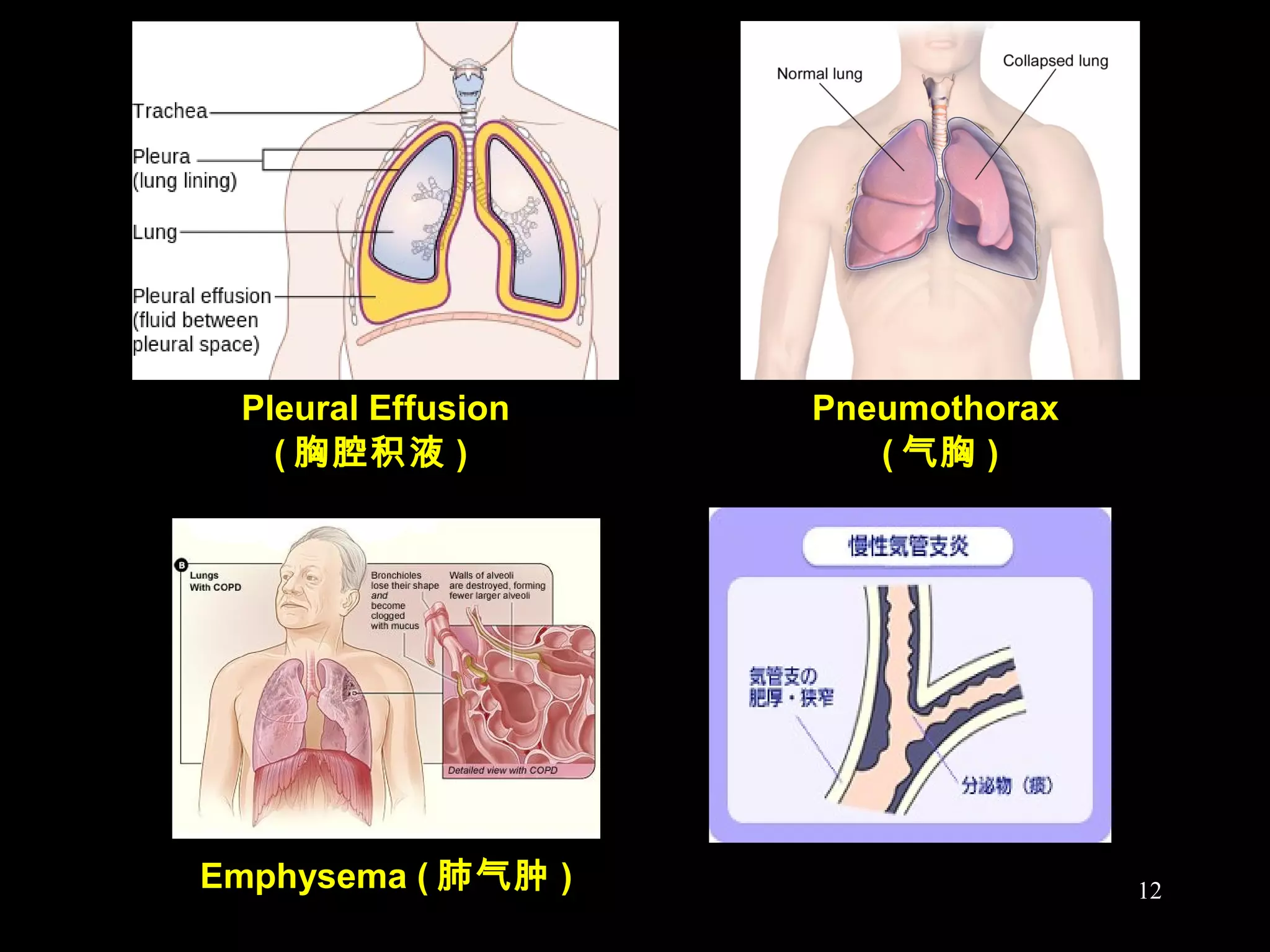
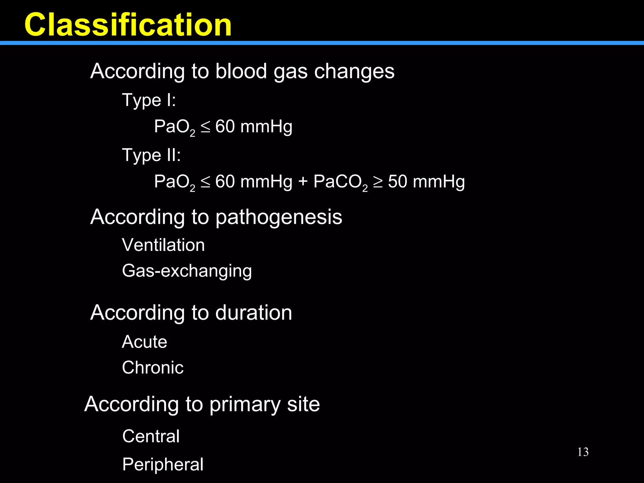
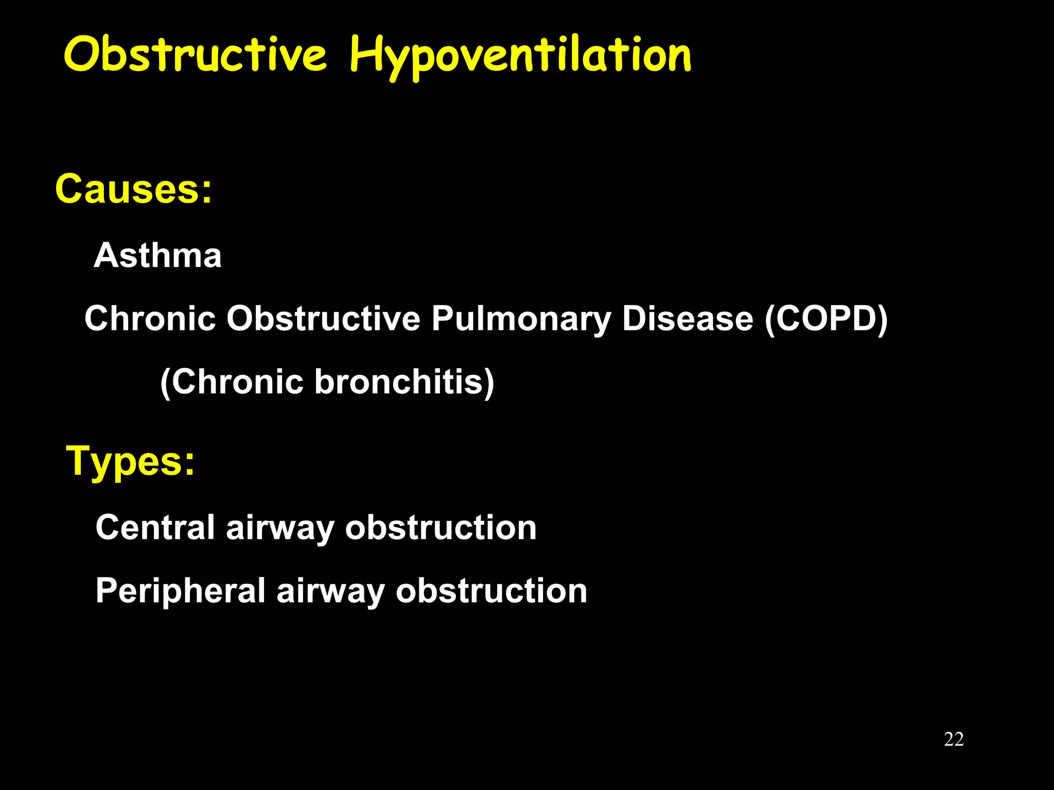
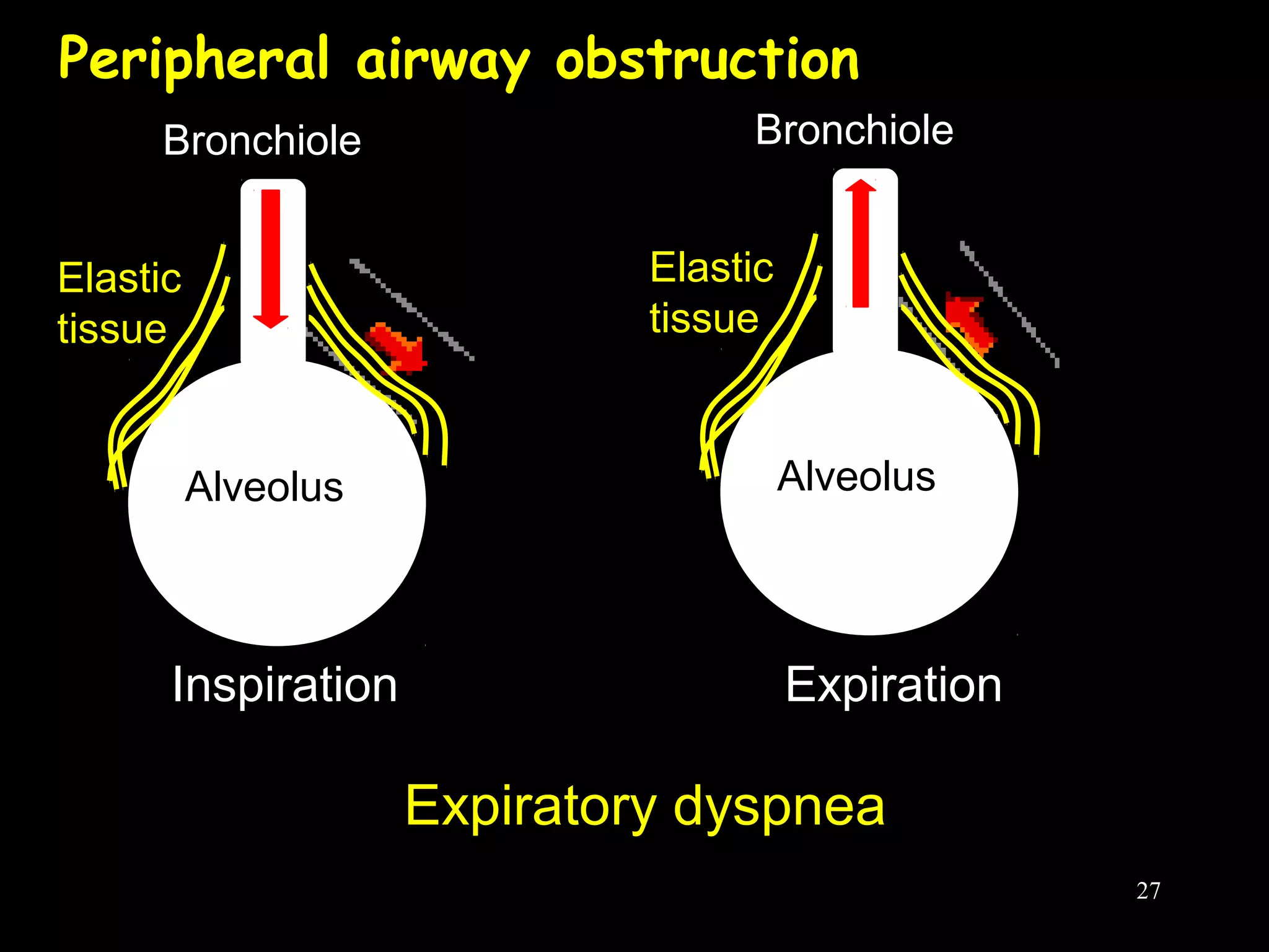
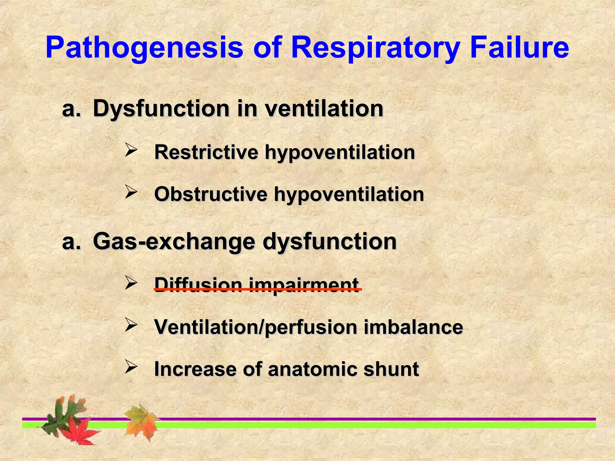
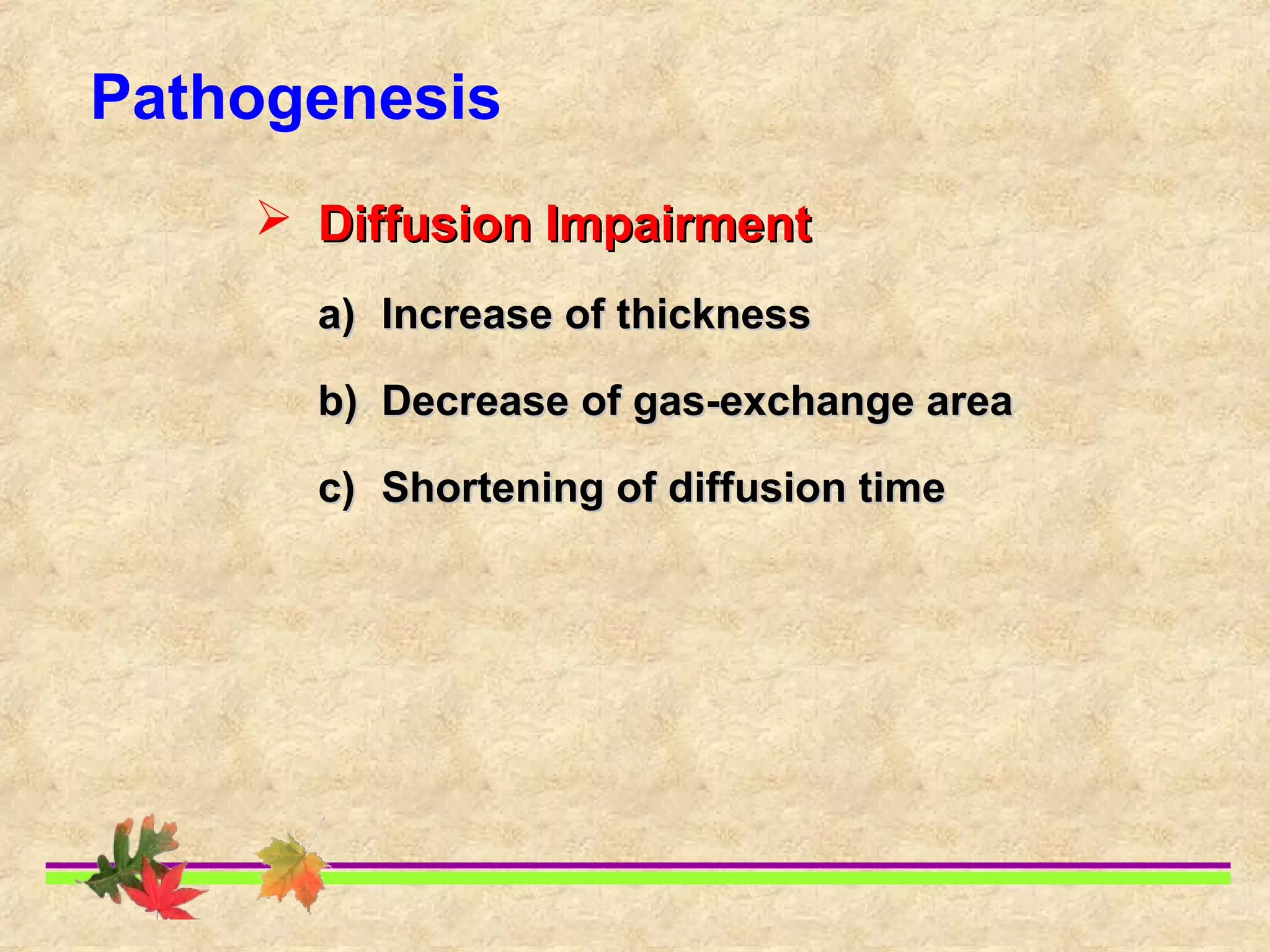
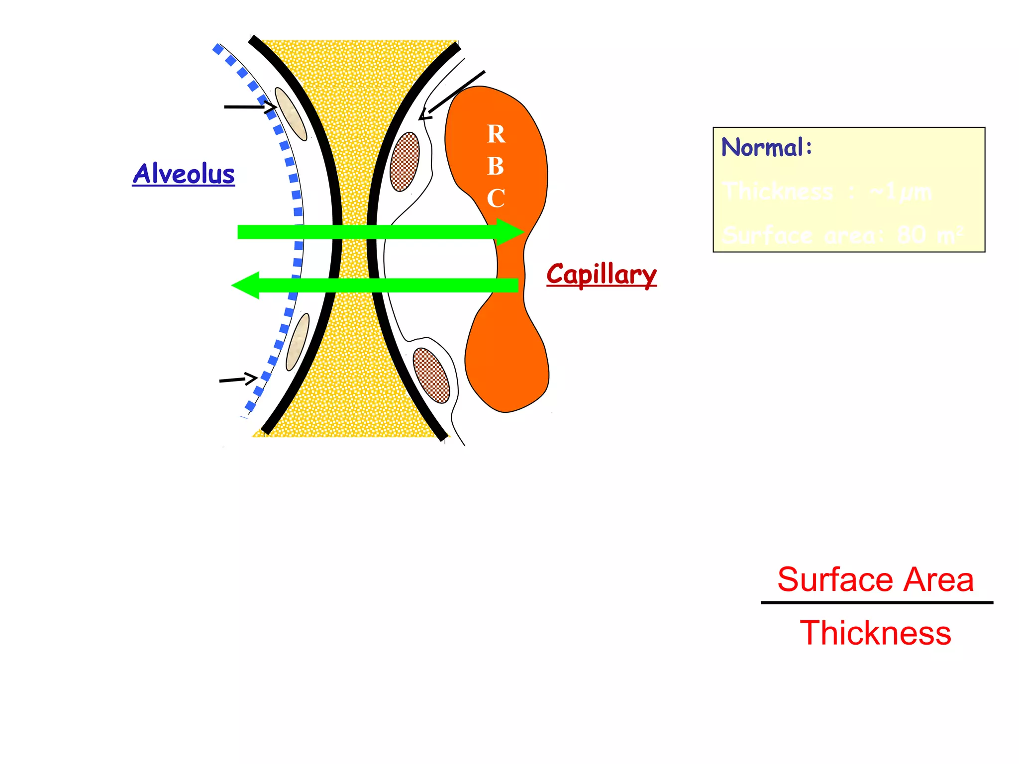
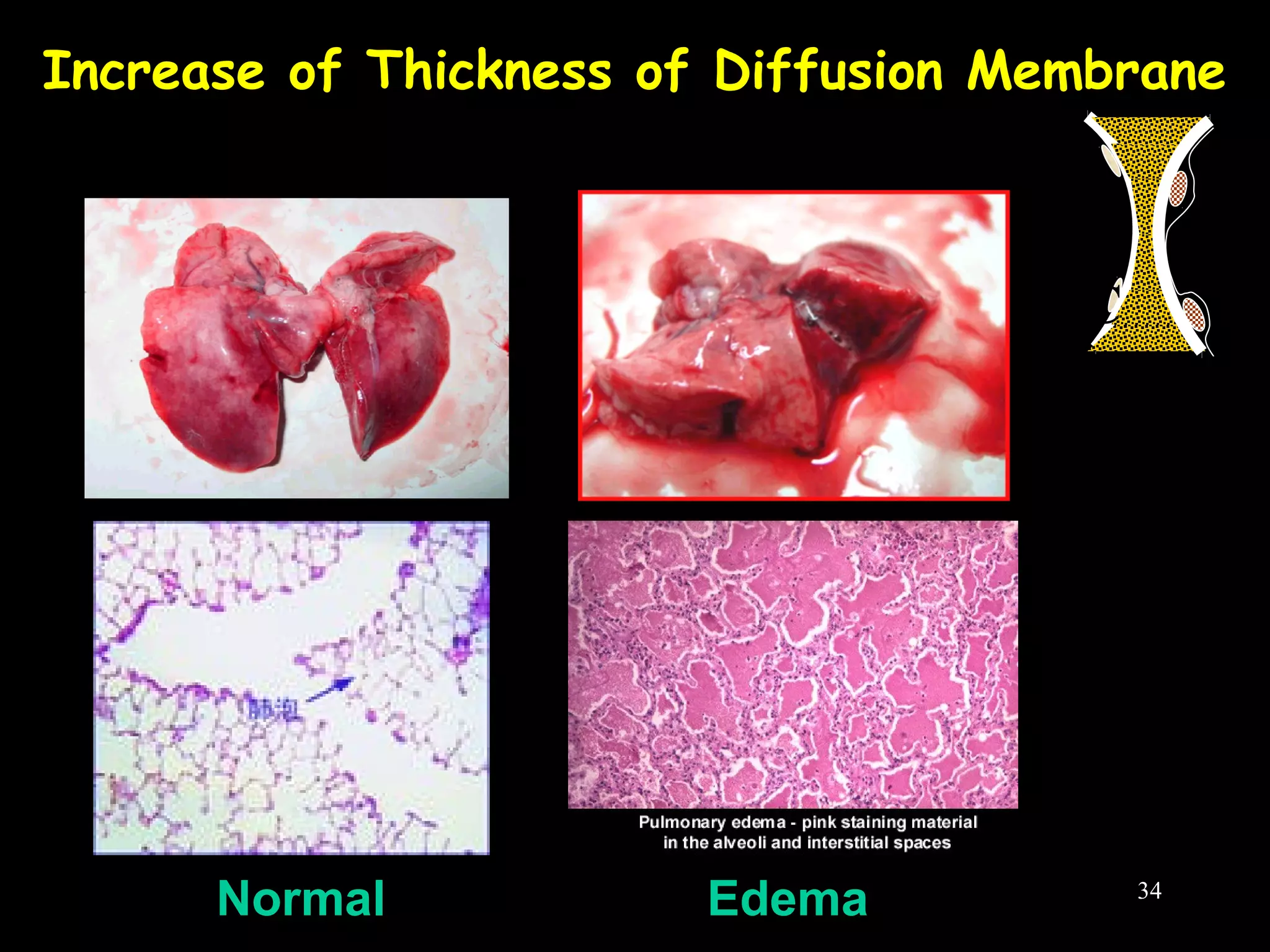
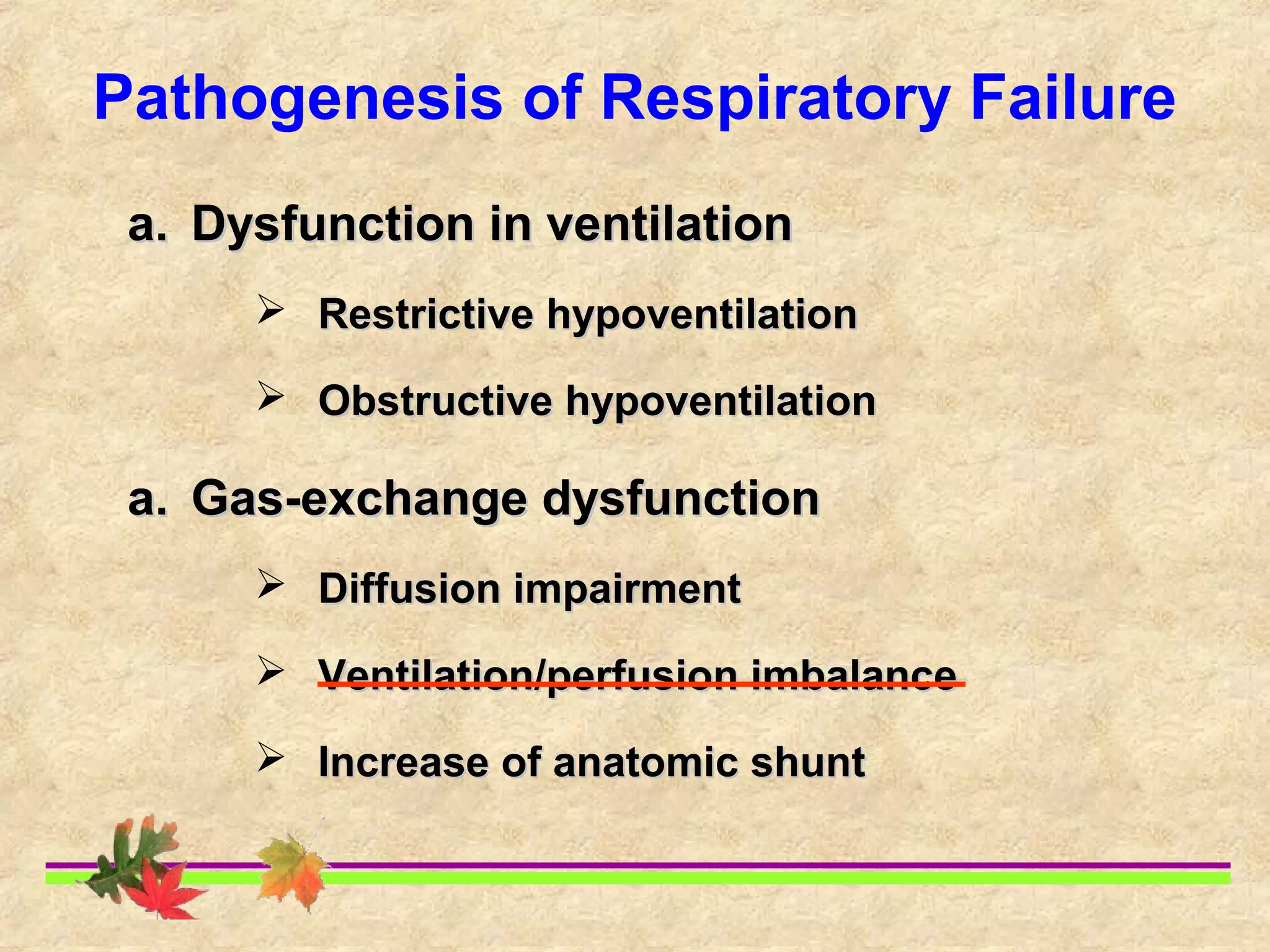
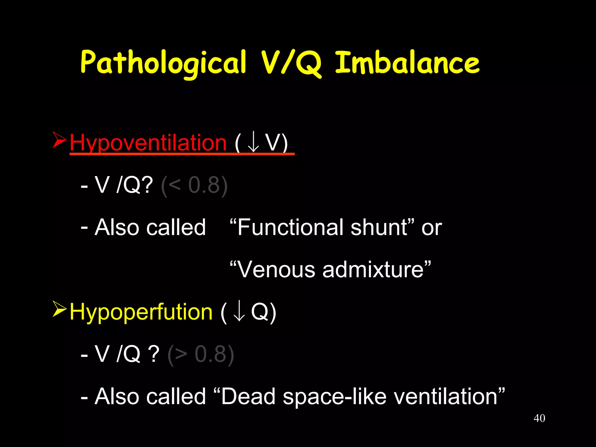
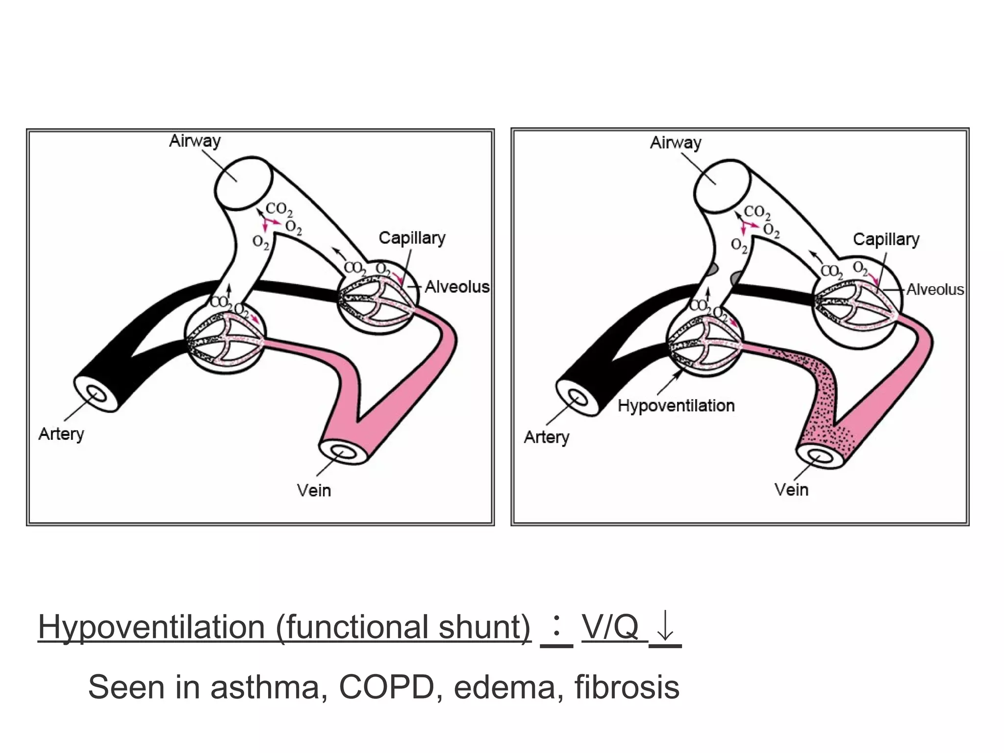
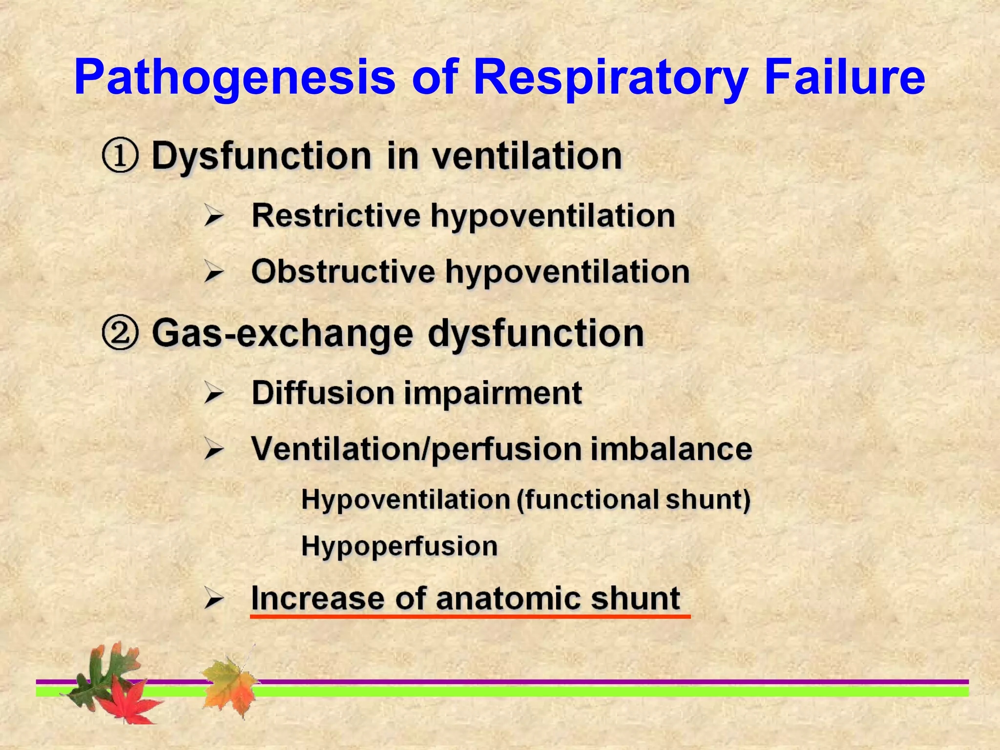
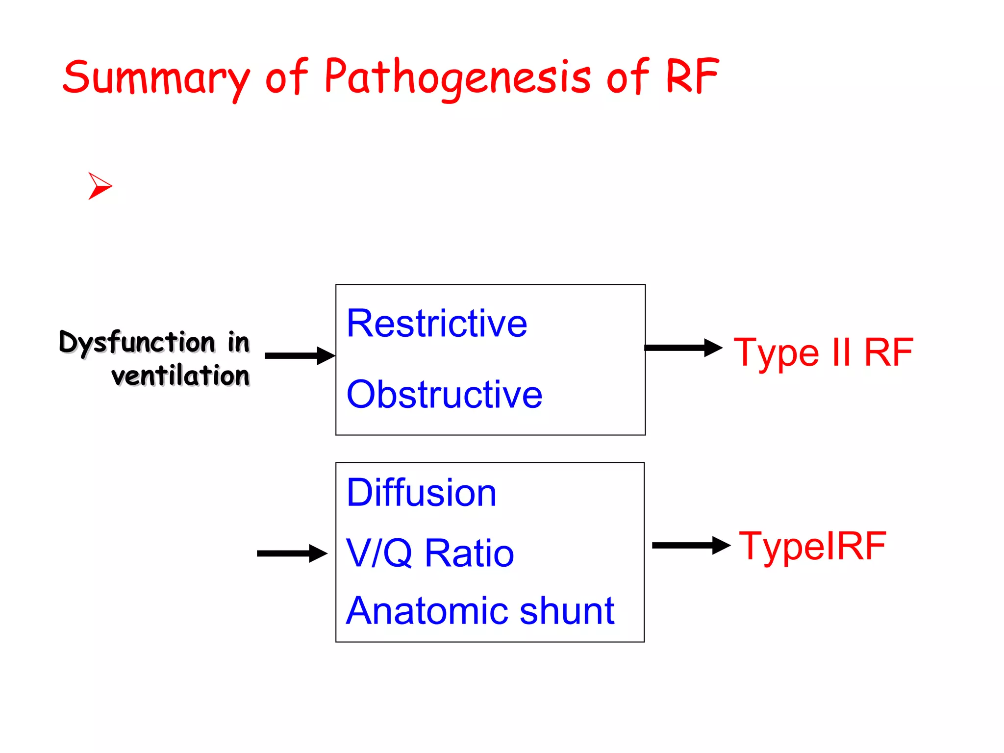
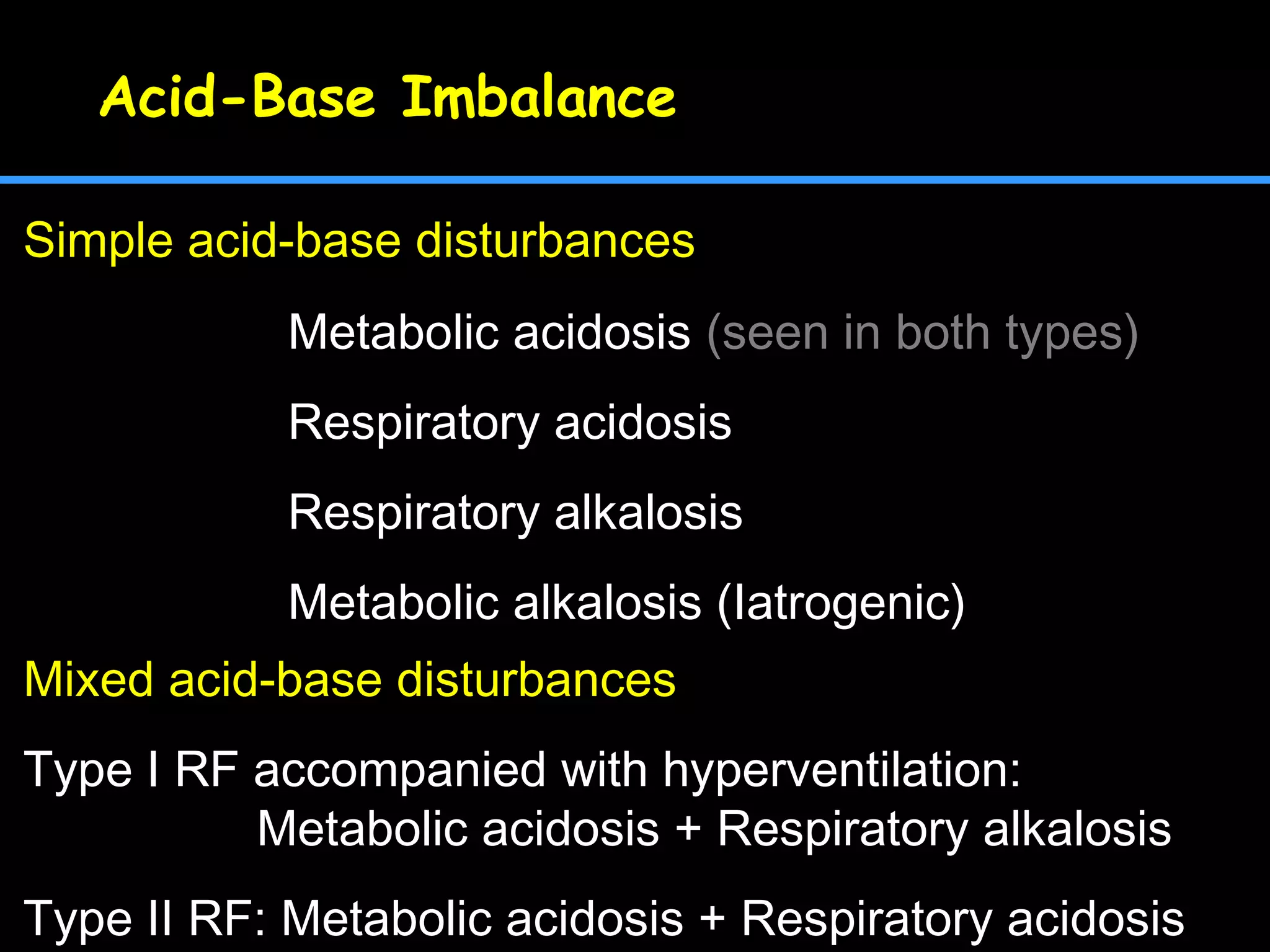
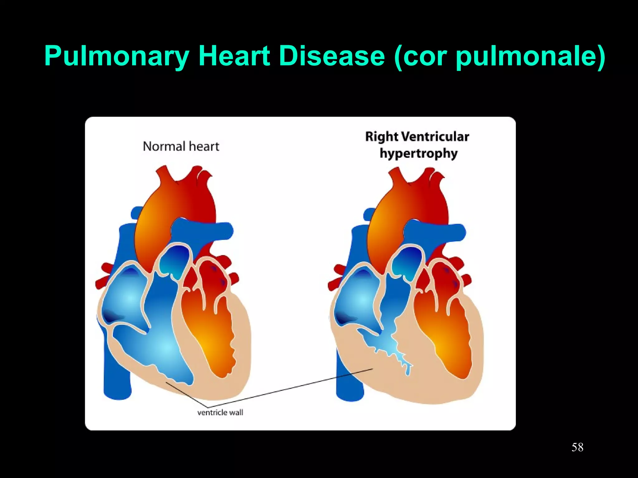
This document discusses respiratory failure, including its definition, causes, classification, pathogenesis, and effects on organ systems. Respiratory failure is defined as inadequate oxygenation of venous blood due to low PaO2 levels and potentially high PaCO2 levels. It can be caused by issues affecting ventilation like restrictive lung disease or obstructive airway diseases. The pathogenesis involves dysfunction of ventilation leading to hypoventilation, as well as gas exchange issues from diffusion impairment, ventilation/perfusion mismatching, or anatomic shunts. Respiratory failure leads to acid-base imbalances, electrolyte disturbances, and dysfunction of organ systems like the lungs, heart, brain, kidneys and gastrointestinal tract.
The document discusses respiratory failure, including its definition, classification, causes, mechanisms, clinical manifestations, diagnosis and treatment. It defines respiratory failure as a clinical syndrome caused by conditions severely affecting the lung's ability to oxygenate blood or eliminate carbon dioxide. Treatment involves identifying and managing the underlying cause, ensuring an open airway, providing oxygen therapy, and mechanical ventilation if needed.
This document discusses portal hypertension, its causes, signs, symptoms, diagnosis, and management. The most common cause is cirrhosis. Portal hypertension develops from increased vascular resistance and blood flow in the portal vein. Signs include weakness, weight loss, jaundice, edema, and gastrointestinal bleeding. Diagnosis involves blood tests, imaging, and endoscopy. Management focuses on treating the underlying cause, preventing variceal bleeding, and treating complications.
The document discusses biliary dyskinesia in children, which is a disturbance in the coordination of contractions in the biliary ducts that can cause pain in the upper right quadrant of the abdomen. It notes that biliary dyskinesia has various potential causes including neuro-circulatory dysfunction, viral hepatitis, genetic factors, and gastrointestinal issues. The document also covers pathogenesis, diagnosis, and potential treatment options which can include laparoscopic cholecystectomy, proton pump inhibitors, osteopathic treatment, and magnesium and enzyme supplements.
This document provides an overview of respiratory failure, including its causes, types, symptoms, diagnosis, and management. It begins by defining respiratory failure as the failure of the respiratory system in gas exchange functions of oxygenation and carbon dioxide elimination. Respiratory failure is then classified based on PaO2 and PaCO2 levels into hypoxemic (Type I) and hypercapnic (Type II) types. Common causes, clinical features, investigations, and general management principles are discussed for respiratory failure. Key aspects of managing hypoxemia and hypercapnia are also summarized.
Respiratory failure occurs when the lungs fail to oxygenate the blood or eliminate carbon dioxide. It can be classified as type I (hypoxemic) or type II (hypercapnic). Common causes include pneumonia, COPD, pulmonary embolism, and cardiac failure. Diagnosis involves blood gas analysis, imaging, and identifying the underlying cause. Management focuses on treating the cause, supporting oxygenation and ventilation, and mechanical ventilation if needed. Type II respiratory failure requires careful oxygen therapy to prevent worsening acidosis.
Peptic ulcers are lesions that occur in areas of the gastrointestinal tract exposed to stomach acid. Risk factors include H. pylori infection and NSAID use. Clinical features include recurrent abdominal pain related to food. Diagnosis involves endoscopy with biopsy or breath/stool tests for H. pylori. Management involves eradicating H. pylori with triple therapy antibiotics and PPIs. Surgery is rarely needed and reserved for complications like perforation or bleeding.
This document discusses respiratory failure, which occurs when the respiratory system fails in gas exchange. It defines two main types - hypoxemic respiratory failure, defined as low blood oxygen, and hypercapnic respiratory failure, defined as high blood carbon dioxide. The document then covers the anatomy and physiology of respiration, diagnostic evaluation of respiratory failure, treatment including mechanical ventilation, and specific causes of respiratory failure like infection, airway obstruction, and cardiac issues.
This document summarizes respiratory failure, including its types, causes, clinical manifestations, diagnosis, treatment, and considerations for older patients. Respiratory failure occurs when the lungs cannot effectively exchange oxygen and carbon dioxide and can be acute or chronic. The two main types are hypoxemic respiratory failure, characterized by low blood oxygen, and hypercapnic respiratory failure, characterized by high blood carbon dioxide. Causes range from lung diseases like COPD to neurological conditions. Treatment involves oxygen therapy, medications, and nutrition management.
This document summarizes COPD (chronic obstructive pulmonary disease). It defines COPD as a disease characterized by irreversible airflow limitation. COPD includes emphysema, chronic bronchitis, and small airways disease. The major risk factor is cigarette smoking. The pathology involves changes in the large airways, small airways (<2mm), and lung parenchyma. Emphysema is classified as centriacinar or panacinar. Treatment focuses on smoking cessation, bronchodilators, inhaled corticosteroids, lung volume reduction surgery, lung transplantation, and managing exacerbations.
This document covers several topics related to respiratory pathophysiology:
1. It describes the anatomy and control of breathing, including the medullary respiratory center and pontine and apneustic areas.
2. Various types of breathing patterns are defined, such as Cheyne-Stokes respirations and Biot's respiration, along with the areas of brain injury that cause each pattern.
3. Common respiratory symptoms like cough, dyspnea, and hemoptysis are discussed alongside their typical causes.
4. Physical exam findings on chest auscultation and percussion are outlined, including vocal fremitus and lung sounds.
5. The calculation of the alveolar-arterial oxygen
1. Hypoxemia, defined as low oxygen levels in arterial blood, can be caused by hypoventilation, low inspired oxygen, right-to-left shunts, ventilation-perfusion mismatching, or diffusion impairment in the lungs.
2. Physical exam and arterial blood gas analysis are used to diagnose hypoxemia and its underlying causes. Treatment focuses on oxygen supplementation, treating the underlying condition, correcting acid-base imbalances, and mechanical ventilation if needed.
3. The causes, mechanisms, diagnosis and management of hypoxemia are complex but critical for treatment of respiratory failure.
This document discusses respiratory failure, including its definition, types, causes, clinical manifestations, diagnostic evaluations, management, and complications. Respiratory failure is when the respiratory system fails to adequately oxygenate the blood or eliminate carbon dioxide. It can be classified as hypoxemic or hypercapnic. Acute respiratory failure develops rapidly over hours while chronic develops over days. Management involves treating the underlying cause, providing oxygen supplementation, monitoring vital signs, and supporting respiratory function. Complications can affect the lungs, heart, gastrointestinal system, and risk of infection.
This document discusses the differences between obstructive and restrictive lung diseases. Obstructive lung diseases involve obstruction in the airways from the trachea to the bronchioles, causing increased resistance to airflow. Restrictive lung diseases involve reduced expansion of the lung parenchyma and decreased total lung capacity. Pulmonary function tests can help distinguish the two, as obstructive diseases typically show decreased FEV1 and FVC with increased TLC, while restrictive diseases show decreased FEV1, FVC and TLC with a normal FEV1/FVC ratio. Common examples of each type of disease are also provided.
Cor pulmonale is a condition where the right ventricle of the heart enlarges and fails due to long-standing increased workload from diseases that affect the lungs like COPD. It is defined as hypertrophy and dilation of the right ventricle resulting from increased pulmonary vascular resistance. The main causes are chronic hypoxemia and pulmonary thromboembolism which lead to remodeling of the pulmonary arteries and increased pressure in the lungs. Over time, this puts strain on the right ventricle and can cause it to fail. Treatment focuses on reducing pulmonary pressures through oxygen therapy and vasodilators while managing symptoms of right heart failure.
this slide focuses on all the acid base disorder pertaining to the respiratory system. it focus on the compensatory mechanism, causes, clinical features and treatment.
The document provides information about Acute Respiratory Distress Syndrome (ARDS) including its definition, pathophysiology, diagnosis, management, and prognosis. ARDS is defined as rapid onset hypoxemia and diffuse pulmonary infiltrates leading to respiratory failure. It is caused by direct lung injury from conditions like pneumonia or indirect injury from sepsis or trauma. Diagnosis involves criteria of acute onset, hypoxemia with PaO2/FiO2 ≤200, and no heart failure. Management focuses on treating the underlying cause and providing ventilator support using low tidal volumes per the ARDSNet protocol to reduce ventilator-induced lung injury. Prognosis depends on risk factors and mortality ranges from 26-44%.
The document discusses pathophysiology of respiratory failure, defining it as failure of oxygenation and/or carbon dioxide elimination. It describes four types of respiratory failure: type 1 (hypoxemic) with low PaO2, type 2 (hypercapnic/ventilatory) with high PaCO2, type 3 (peri-operative) common after surgery, and type 4 (shock) secondary to cardiovascular instability. Causes of types 1 and 2 are explored in depth, including V/Q mismatch, shunts, increased dead space, and factors reducing ventilatory capacity or increasing demand leading to hypercapnia.
This document discusses respiratory failure, defined as inadequate oxygenation, ventilation, or both to meet metabolic demands. It can be classified as type 1 (hypoxemic) or type 2 (hypercapnic) respiratory failure. Risk factors include age, smoking, lung disease, and neurological or muscular disorders. Pathophysiology involves ventilation-perfusion mismatching, right-to-left shunting, or hypoventilation. Causes include pneumonia, pulmonary embolism, neuromuscular disorders, and acute respiratory distress syndrome. The control of breathing and gas exchange physiology are also summarized.
Chronic Obstructive Pulmonary Disease (COPD) is an umbrella term used to describe progressive lung diseases including emphysema, chronic bronchitis, and refractory (non-reversible) asthma. This disease is characterized by increasing breathlessness
Bronchial Asthma: Definition,Pathophysiology and ManagementMarko Makram
Definition and Pathophysiology of Asthma in addition to classification and recent updates in the management of asthma based on GINA-2019 Guidelines, by Dr. Marco Makram.
Small group presentation which was done during our physiology days under the guidance of Prof. Sampath Gunawardena senior lecturer in department of Physiology, Faculty of Medicine University of Ruhuna.
Esophagitis is inflammation of the esophagus caused by injury to the esophageal mucosa. Common causes include prolonged gastric intubation, uremia, ingestion of corrosive substances, radiation, and chemotherapy. Gastroesophageal reflux disease, where stomach acid refluxes into the esophagus, is the most frequent cause in Western countries. Symptoms include dysphagia and heartburn. Treatment involves proton pump inhibitors or H2 blockers to reduce gastric acidity and provide relief. Complications can include esophageal ulceration, bleeding, stricture formation, and Barrett's esophagus.
Pleurisy is inflammation of the pleura covering the lungs and chest wall. It is commonly caused by pneumonia, tuberculosis, pulmonary embolism, or trauma. The inflammation irritates sensory fibers and causes sharp, knifelike pain during inspiration that may radiate to the shoulder or abdomen. Diagnosis involves chest x-ray, sputum examination, or thoracentesis. Treatment focuses on the underlying cause and pain relief through analgesics, heat/cold, or nerve blocks. Complications can include pleural effusions or respiratory issues from shallow breathing.
Helicobacter pylori infection and use of non-steroidal anti-inflammatory drugs (NSAIDs) are the two most common causes of peptic ulcer disease (PUD). H. pylori infection, found in 70-80% of PUD cases, causes chronic gastritis and impairs the stomach's defenses, allowing acid to damage the lining. NSAIDs also impair defenses and can cause ulcers even in the absence of H. pylori. Together these factors disrupt the stomach's balance between aggressive acid and pepsin secretions and protective mucosal defenses, leading to ulcer formation.
Atelectasis/Lung Collapse Part-1 by Dr Bashir Ahmed Dar Associate Professor M...Prof Dr Bashir Ahmed Dar
The term atelectasis is derived from the Greek words ateles and ektasis, which mean incomplete expansion.The incomplete expansion of lung may involve part of lung or entire lung.Most symptoms and signs are determined by the rapidity with which the collapse of lung occurs,the size of the lung area affected, and the presence or absence of complicating infection.
Rapid bronchial occlusion with a large area of lung collapse causes pain on the affected side, sudden onset of dyspnea, and cyanosis. Hypotension, tachycardia, fever, and shock may also occur.
Slowly developing atelectasis may be asymptomatic or may cause only minor symptoms. Middle lobe syndrome often is asymptomatic, although irritation in the right middle and right lower lobe bronchi may cause a severe, hacking, nonproductive cough.
RBCs, also known as red blood cells, are biconcave and flexible cells that lack a nucleus. Their main function is to carry oxygen and carbon dioxide throughout the body. RBCs have an average lifespan of 120 days. They contain hemoglobin, which is made up of protein chains that bind to oxygen and carbon dioxide. When RBCs reach the end of their lifespan, they are broken down by macrophages and the heme portion is recycled to form new RBCs, while the globin portion joins the protein pool.
The document summarizes acid-base balance in the body. It discusses metabolic processes that produce acids and buffer systems that regulate pH. The lungs and kidneys help control acid-base balance by removing carbon dioxide and regulating bicarbonate levels respectively. Chemoreceptors in the body sense changes in acidity and signal the respiratory system to adjust ventilation and maintain pH within a narrow range.
Respiratory failure &ards منتدى تمريض مستشفى غزة الاوروبيegh-nsg
This document provides an overview of respiratory failure (RF) and acute respiratory distress syndrome (ARDS). It defines RF and classifies it as hypoxemic or hypercapnic. ARDS is introduced as a diffuse lung injury causing hypoxemia that cannot be explained by heart failure or fluid overload. The pathophysiology of ARDS involves damage to the alveolar-capillary membrane leading to pulmonary edema. Treatment of ARDS focuses on mechanical ventilation, positive end-expiratory pressure, positioning, and treating the underlying cause.
Slideshow is from the University of Michigan Medical School's M1 Cardiovascular / Respiratory sequence
View additional course materials on Open.Michigan:
openmi.ch/med-M1Cardio
This document discusses the four types of respiratory failure:
1) Type 1 (hypoxemic) is characterized by low oxygen levels in the blood but normal or low carbon dioxide levels, usually due to issues with ventilation/perfusion matching.
2) Type 2 (hypercapnic/ventilatory) involves low oxygen and high carbon dioxide levels due to inadequate alveolar ventilation.
3) Type 3 (peri-operative) commonly occurs after surgery due to effects of anesthesia and abdominal issues.
4) Type 4 (shock) involves intubation during resuscitation for conditions like cardiogenic, hypovolemic, or septic shock. The document then provides details on the causes, characteristics
Chronic bronchitis is defined as a persistent cough with mucus production for at least three months in a year for two consecutive years. It is primarily caused by long-term irritation and inflammation of the airways due to cigarette smoke or other inhaled chemicals. Chronic bronchitis can progress to more severe lung diseases like COPD or lung cancer and, if left untreated, may result in heart failure or respiratory infections. The pathology involves damage to the airways, thickening of the mucus membranes, and increased mucus production.
This document discusses heart failure and respiratory failure. It begins with an introduction to heart failure, including its definition and signs and symptoms. It then covers the etiology, classification, compensatory responses, pathogenesis, clinical manifestations, and treatment principles of heart failure. For respiratory failure, it similarly discusses the definition, etiology and classification, pathogenesis including dysfunction in ventilation and gas exchange, alterations in metabolism and function, and pathophysiological basis of prevention and treatment. The key causes, types, and mechanisms of both restrictive and obstructive hypoventilation and gas exchange dysfunction are explained in detail.
Acute respiratory distress syndrome (ARDS) is a severe lung condition caused by diffuse damage to the alveoli, which results in reduced oxygen exchange and respiratory failure. It has an annual incidence of 75 per 100,000 people in the US and a high mortality rate of 40-60%. ARDS is managed through lung-protective mechanical ventilation with low tidal volumes, application of PEEP, prone positioning, and conservative fluid management. Outcomes are predicted by factors like chronic liver disease, non-pulmonary organ dysfunction, and sepsis, with long-term survivors often having impaired quality of life. Nursing care focuses on supporting respiratory function and managing complications through techniques like airway clearance and treatment of anxiety.
CYSTIC FIBROSIS AND PHYSIOTHERAPY TREATMENT.pptxShilpasree Saha
Physiotherapy has long been considered a cornerstone of condition management for
people with cystic fibrosis (CF). The presentation of CF has changed over time with an increased
life expectancy and increased expectations of people with CF to have a complete lifestyle.
During one lung ventilation (OLV), blood flow is shunted to the non-ventilated lung causing hypoxemia. Anesthetics mildly inhibit hypoxic pulmonary vasoconstriction (HPV), increasing shunt by around 4%. Positioning and techniques like selective lung ventilation and PEEP can optimize ventilation and perfusion matching to minimize hypoxemia during OLV.
This document discusses various hypoventilation disorders including obstructive and restrictive types. It covers topics like upper vs lower airway obstruction, patterns of hypoventilation, obstructive lung diseases like asthma, chronic bronchitis and emphysema. It also discusses tests to evaluate lung function and restrictive vs obstructive patterns. Central sleep apnea and exercise induced bronchoconstriction are also summarized.
This document provides information on acute respiratory distress syndrome (ARDS). It defines ARDS as severe acute lung injury involving diffuse alveolar damage and increased permeability. It notes the criteria for diagnosing ARDS including acute onset of respiratory failure, low blood pressure, low oxygen levels and bilateral lung infiltrates. Common causes include viral or bacterial pneumonia and chest trauma. The management of ARDS focuses on respiratory support through mechanical ventilation with low tidal volumes, application of positive end expiratory pressure and prone positioning. Other treatments aim to correct fluid and electrolyte imbalances while preventing complications like infection. Nursing care centers around monitoring the patient's respiratory status, managing oxygen therapy and supporting ventilation.
This document summarizes various adjunctive treatment strategies for acute respiratory distress syndrome (ARDS). It discusses ventilatory strategies beyond lung protective ventilation including prone positioning, liquid ventilation, high frequency ventilation, and extracorporeal gas exchange. It also covers hemodynamic management, pulmonary vasodilators, surfactant replacement therapy, anti-inflammatory strategies, and adjunctive therapies.
This document summarizes various adjunctive treatment strategies for acute respiratory distress syndrome (ARDS) including ventilatory strategies beyond lung protective ventilation, hemodynamic management, pulmonary vasodilators, surfactant replacement therapy, anti-inflammatory strategies, and corticosteroids. Prone positioning, inhaled nitric oxide, and prolonged corticosteroid treatment in refractory ARDS have shown benefits in oxygenation and survival in clinical trials. However, other strategies such as high frequency ventilation, liquid ventilation, and surfactant replacement have not demonstrated clear clinical benefits.
This document provides a summary of key concepts in gastrointestinal and respiratory physiology for medical students studying for the FRCEM Primary exam. It includes tables outlining the cell types and secretions in the stomach, factors that stimulate and inhibit gastrin release, classifications of respiratory failure and lung volumes. Physiology concepts covered include gastric acid secretion, actions of gastrin, surfactant function, dead space, and respiratory changes in pregnancy. The document is intended to help medical students efficiently review and understand essential physiology for the exam in a short period of time.
This document discusses shock in pediatric patients. It defines shock and describes the different types including hypovolemic, distributive, cardiogenic, obstructive, and dissociative shock. It explains how oxygen delivery works and the factors that affect it. The goals of shock management are to increase oxygen delivery and decrease demand. Treatment involves optimizing preload, contractility, afterload, oxygen content and cardiac output. Initial management follows an ABC approach of securing the airway, providing adequate breathing and circulation. Fluid resuscitation is a key part of treatment, and choices between crystalloids and colloids are discussed. Inotropic support may be needed to optimize cardiac function in shock.
This document discusses acute respiratory distress syndrome (ARDS). It begins by defining ARDS and describing its signs and symptoms. It then discusses the history of ARDS definitions and criteria. It outlines the pathophysiology and three phases of ARDS. Treatment strategies covered include mechanical ventilation, monitoring, infection control, and specific therapies. Prognosis and risk factors are also summarized.
This document discusses several common alterations in respiratory function including pulmonary edema, lower respiratory tract infections, traumatic injuries, neurological diseases, adult respiratory distress syndrome, chronic obstructive pulmonary diseases, upper respiratory tract infections, atelectasis, industrial diseases, and obesity. It outlines the clinical manifestations and critical care management of patients with these respiratory conditions.
This document discusses various adjunctive treatments for acute respiratory distress syndrome (ARDS). It covers ventilatory strategies beyond lung protective ventilation including prone positioning, liquid ventilation, high frequency ventilation, and extracorporeal membrane oxygenation. It also discusses hemodynamic management including fluids and vasopressors. Selective pulmonary vasodilators, surfactant replacement therapy, anti-inflammatory strategies, antioxidants, and anticoagulants are mentioned as potential adjunct treatments for ARDS. Prone positioning is described in more detail as one strategy that can improve oxygenation in ARDS patients.
This document provides guidance on clinically examining the respiratory system. It describes how to inspect, palpate, percuss, and auscultate the chest. Inspection involves examining chest shape, symmetry, movement, and veins. Palpation checks the trachea position, apex beat, chest expansion, vocal fremitus, and tenderness. Percussion distinguishes lung from liver dullness. Auscultation assesses breath sounds and vocal resonance. The examination evaluates the lungs, pleura, chest wall, and underlying bony structures in a systematic manner to detect abnormalities.
Brief Presentation on clinical examination of Respiratory System with Report of Normal case
references:
macleod's clinical examination 13th edition
hutchinson clinical methods
R Alagappan - Manual of Practical Medicine, 4th Edition
This document provides an overview of Acute Lung Injury (ALI), including:
1. The definition, history, epidemiology, and pathogenesis of ALI. It results from direct or indirect lung injury and involves endothelial and epithelial injury, neutrophil infiltration, and cytokine release.
2. The clinical presentation and stages of ALI from the acute/exudative stage with hyaline membrane formation to the proliferative/organizing stage and resolving/fibrotic stage.
3. Investigations for ALI including clinical criteria based on oxygenation and chest imaging findings typically showing bilateral infiltrates. Histopathology often reveals diffuse alveolar damage.
This document provides guidance on evaluating patients presenting with dyspnea (shortness of breath). It defines dyspnea and lists some specific types based on position. Common causes are outlined for pulmonary, cardiac, mixed, and non-cardiopulmonary origins. A clinical approach is described beginning with vital signs and history, followed by physical exam focusing on respiratory, cardiac, and fluid status findings. Initial investigations include chest X-ray, blood gases, ECG, and blood tests. Further tests may include lung function, exercise testing, and biomarkers to differentiate cardiac from pulmonary causes when the chest X-ray is normal. Careful history taking and physical exam remain important to identify underlying conditions.
This document provides information on chronic obstructive pulmonary disease (COPD) including its definition, risk factors, pathophysiology, clinical manifestations, medical management, nursing management, and surgical options. COPD is characterized by persistent airflow limitation that is not fully reversible. It is mostly caused by smoking and results in changes like thickening of the airways and inflammation that narrow the lungs over time. Management involves treatments to improve ventilation and remove secretions, as well as strategies to prevent complications and promote overall health.
COPD is a chronic lung disease characterized by airflow obstruction. It includes emphysema and chronic bronchitis. Cigarette smoking is the leading cause of COPD. Symptoms include dyspnea, cough, sputum production, and wheezing. Diagnosis is confirmed by pulmonary function tests showing reduced FEV1 and FEV1/FVC ratio. Treatment involves smoking cessation, bronchodilators, corticosteroids, oxygen therapy, pulmonary rehabilitation, and surgery for severe cases.
Chronic Obstructive Pulmonary Disease (COPD) is a progressive lung disease characterized by persistent airflow limitation that is usually caused by significant exposure to noxious particles or gases, most commonly from cigarette smoke. [1] COPD is underdiagnosed and undertreated globally despite being a leading cause of mortality. [2] The pathophysiology of COPD involves chronic inflammation in the lungs in response to toxins, resulting in structural changes and impaired airflow over time. [3]
Mycoplasmas are the smallest free-living organisms that lack cell walls. They colonize humans and can cause diseases. The two medically important genera that infect humans are Mycoplasma and Ureaplasma. Mycoplasma pneumoniae is a major cause of atypical pneumonia while Ureaplasma urealyticum and Mycoplasma hominis can cause nongonococcal urethritis. Mycoplasmas are difficult to culture and require complex media with sterols. They are treated with antibiotics like tetracycline and erythromycin.
1. Spirochetes are spiral-shaped, motile bacteria that include pathogens causing syphilis, Lyme disease, and leptospirosis.
2. Treponema pallidum causes syphilis, which has primary, secondary, and tertiary stages and can be transmitted congenitally. Darkfield microscopy and serological tests are used for diagnosis.
3. Other human pathogenic spirochetes include Borrelia burgdorferi, which causes Lyme disease transmitted through tick bites, and Leptospira interrogans, which causes leptospirosis transmitted through contact with infected animal urine.
Here are brief descriptions of the three forms of anthrax and the mechanism of anthrax toxin:
1. The three forms of anthrax are cutaneous (skin), inhalation (lungs), and gastrointestinal (ingestion).
2. Anthrax toxin is composed of three proteins - protective antigen (PA), lethal factor (LF), and edema factor (EF). PA binds to receptors on cells and helps internalize LF and EF. LF is a metalloprotease that cleaves MAPKK, disrupting cell signaling. EF is an adenylate cyclase that increases cyclic AMP levels, impairing immune function. Together they cause edema, tissue necrosis, and sepsis.
Zoonoses are infectious diseases that can be transmitted between animals and humans. A majority (61%) of known human pathogens are zoonotic. Significant zoonotic pandemics in history include the Black Death plague pandemic in the 14th century spread by Mongol invaders, and the 1918 Spanish flu pandemic. Today, zoonotic diseases pose a threat through increased human-animal contact and potential use in biological terrorism. Bacillus anthracis, the cause of anthrax, is a large, spore-forming, gram-positive rod-shaped bacterium. Its virulence depends on its polysaccharide capsule and lethal and edema toxins. Anthrax is typically transmitted through contact with infected animals or their products and
This document provides information on Corynebacterium diphtheriae, which causes diphtheria. It describes the morphology, cultivation, biochemical characteristics, pathogenicity, clinical manifestations, diagnosis, treatment and prevention of diphtheria. C. diphtheriae produces an exotoxin that inhibits protein synthesis and causes pseudomembrane formation in the throat and other areas. Laboratory diagnosis involves microscopy, culture and toxin production testing. Immunization with diphtheria, tetanus and pertussis vaccine (DTP) provides active immunity against the disease.
Mycobacterium is the only genus in the family Mycobacteriaceae. It is classified into four groups: M. tuberculosis complex, non-tuberculous mycobacteria group I, non-tuberculous mycobacteria group II, and rapidly growing non-tuberculous mycobacteria. M. tuberculosis is the pathogen that causes tuberculosis in humans. It is transmitted through the air and infects the lungs, causing symptoms like cough, fever, and weight loss. Inside the body, it is engulfed by macrophages but can survive and multiply, triggering the formation of granulomas.
Clostridium tetani is an obligate anaerobic, gram-positive bacterium that causes the disease tetanus. It forms terminal spores that give it a distinctive drumstick appearance. Though found worldwide in soil, C. tetani enters the body through wounds and causes tetanus by producing a potent neurotoxin. It is a major cause of mortality in developing countries, with neonatal tetanus accounting for about half of worldwide cases and having a mortality rate of 85%.
This document provides information on Campylobacter and Helicobacter, which are Gram-negative, microaerophilic bacteria. Campylobacter causes foodborne illness in humans and is transmitted through contaminated poultry or water. Its main virulence factors include flagella for motility and adhesins for attachment. Helicobacter pylori colonizes the stomach and is associated with gastritis, ulcers, and gastric cancer. It produces urease and adheres tightly to gastric epithelial cells using flagella and adhesins. Both bacteria are transmitted orally between humans or from animal reservoirs and cause disease by invading and damaging intestinal or gastric mucosa.
This document discusses Vibrio cholerae, the bacteria that causes cholera. It provides details on the history, pathogenesis, clinical manifestations, diagnosis, and immunity aspects of cholera. Some key points include:
- V. cholerae causes the severe, acute diarrheal disease cholera and is transmitted through contaminated food or water.
- There have been several global pandemics of cholera since the 1800s. The causative agent was discovered by Pacini and Koch in the 1800s.
- V. cholerae secretes a toxin that increases cyclic AMP levels in intestinal cells, causing a massive outpouring of fluid in the stool. Colonization factors like pili and flagella
1. Enterobacteriaceae are a family of Gram-negative, non-spore forming rods that are normal flora of the gastrointestinal tract. They include many important pathogens like Escherichia, Salmonella, Shigella.
2. They are facultative anaerobes that ferment glucose and reduce nitrates. Identification is based on lactose fermentation and reactions on selective media.
3. Shigella causes bacillary dysentery. It is non-motile and invades the colonic epithelium, causing severe diarrhea, fever and tenesmus. Virulence factors include invasion plasmid antigens.
This document summarizes Gram positive cocci, focusing on Staphylococcus and Streptococcus. Staphylococcus is classified based on coagulase production. It is a facultative anaerobe that can cause skin infections and food poisoning through toxins like enterotoxins. Streptococcus is classified by hemolytic activity and cell wall antigens. It attaches to host cells using M protein and hyaluronidase. It produces invasive enzymes and exotoxins like pyrogenic toxins that allow it to spread. Both bacteria cause disease through various virulence factors including toxins, enzymes, and structural components.
This document discusses drug resistance and nosocomial infections. It begins by describing the discovery of antibiotics by Alexander Fleming in 1928 and how antibiotics work by either killing bacteria or preventing their growth. While antibiotics were initially a "miracle cure", overuse and misuse has led to the development of drug-resistant bacteria. Resistance can arise through genetic mutations that make bacteria less susceptible to antibiotics or through horizontal gene transfer between bacteria. The document examines several antibiotic targets and mechanisms of resistance, such as beta-lactamase enzymes providing resistance to penicillins and altered cell walls conferring vancomycin resistance. It stresses the importance of properly using and prescribing antibiotics to slow the development and spread of drug-resistant bacteria.
There are two methods of artificial immunity: active immunization and passive immunization. Active immunization involves administering antigens to induce an immune response in the recipient. Passive immunization involves transferring antibodies from an immune individual. Vaccines provide active immunization by exposing the immune system to antigens from pathogens in order to stimulate antibody production and develop immunological memory without causing illness. DNA vaccines are a type of vaccine that attempt to make vaccines more effective, cheaper and safer by using recombinant gene technology.
1. Proper specimen collection is essential for accurate laboratory diagnosis of bacterial infections, as the wrong sample, delay in transport, or contamination can limit test usefulness.
2. Common examination methods for diagnosing bacterial infections include morphological analysis, isolation and culture of pathogens, biochemical reactions, antibiotic susceptibility testing, and detection of antigens or nucleic acids.
3. Antibiotic susceptibility testing determines the sensitivity of isolated bacteria to different antibiotics, which helps clinicians select the proper treatment. Methods include minimum inhibitory concentration and disk diffusion tests.
1. Innate immunity provides the first line of defense against pathogens and includes anatomical barriers, inflammation, phagocytosis, and antimicrobial proteins/peptides.
2. Adaptive immunity develops over time upon exposure to specific pathogens and provides enhanced protection through antibody production and immunological memory.
3. The major categories of innate immunity defenses are anatomical barriers, inflammation, phagocytosis, microbial antagonism by normal flora, and antimicrobial substances in tissues. Adaptive immunity involves B cells, T cells, and production of antibodies.
This document discusses bacterial pathogenesis and infection. It covers several key topics:
1) Normal flora are microorganisms that normally live in or on the human body without causing disease. Opportunistic pathogens are normal flora that can cause disease under certain conditions if the host's immunity is compromised.
2) Bacterial infection is determined by factors of both the bacterium and host. The number and virulence of bacteria as well as the host's innate and acquired immunity impact whether infection occurs.
3) Bacterial pathogenicity is influenced by virulence factors like toxins, invasiveness, and the portal of entry. Virulence refers to an organism's ability to cause disease and is determined by its inv
Bacterial heredity and variation can occur through several mechanisms. Genetic variation arises from mutations in bacterial chromosomes and genetic elements like plasmids, bacteriophages, and transposable elements. Gene transfer and recombination can also introduce variation as bacteria can undergo transformation, conjugation, and transduction to exchange genetic material. This allows bacteria to evolve new traits like antibiotic resistance or changes in virulence over multiple generations.
This document discusses biosafety and the safe handling of biological materials. It defines key terms like sterilization, disinfection, and biosafety. Biosafety aims to ensure safe handling, transport, and disposal of biological materials. Biological risks are classified by levels of containment needed, from BSL-1 to BSL-4. Personal protective equipment and disposal of hazardous waste are important biosafety protocols. Biosafety also addresses bioterrorism threats and ways to prevent the spread of dangerous pathogens.
1. The document discusses the field of medical microbiology, including the definition as the study of microorganisms too small to see with the naked eye, such as viruses, bacteria, and fungi.
2. It describes the key research techniques in medical microbiology including microtechnique, aseptic technique, culture technique, and staining technique.
3. The status and developments of medical microbiology are summarized, such as the discovery of new pathogens like HIV and hepatitis viruses, and the direction of further research into pathogenic mechanisms and new treatments.
Bacteria require certain environmental conditions to grow and multiply, including temperature, pH, oxygen, water, and nutrients. Bacterial metabolism allows bacteria to obtain energy and synthesize cellular components through catabolic and anabolic processes. The products of bacterial anabolism like toxins, enzymes, antibiotics, and pigments have medical significance related to pathogenicity, treatment of disease, and identification of bacteria.
A Visual Guide to 1 Samuel | A Tale of Two HeartsSteve Thomason
These slides walk through the story of 1 Samuel. Samuel is the last judge of Israel. The people reject God and want a king. Saul is anointed as the first king, but he is not a good king. David, the shepherd boy is anointed and Saul is envious of him. David shows honor while Saul continues to self destruct.
Level 3 NCEA - NZ: A Nation In the Making 1872 - 1900 SML.pptHenry Hollis
The History of NZ 1870-1900.
Making of a Nation.
From the NZ Wars to Liberals,
Richard Seddon, George Grey,
Social Laboratory, New Zealand,
Confiscations, Kotahitanga, Kingitanga, Parliament, Suffrage, Repudiation, Economic Change, Agriculture, Gold Mining, Timber, Flax, Sheep, Dairying,
THE SACRIFICE HOW PRO-PALESTINE PROTESTS STUDENTS ARE SACRIFICING TO CHANGE T...indexPub
The recent surge in pro-Palestine student activism has prompted significant responses from universities, ranging from negotiations and divestment commitments to increased transparency about investments in companies supporting the war on Gaza. This activism has led to the cessation of student encampments but also highlighted the substantial sacrifices made by students, including academic disruptions and personal risks. The primary drivers of these protests are poor university administration, lack of transparency, and inadequate communication between officials and students. This study examines the profound emotional, psychological, and professional impacts on students engaged in pro-Palestine protests, focusing on Generation Z's (Gen-Z) activism dynamics. This paper explores the significant sacrifices made by these students and even the professors supporting the pro-Palestine movement, with a focus on recent global movements. Through an in-depth analysis of printed and electronic media, the study examines the impacts of these sacrifices on the academic and personal lives of those involved. The paper highlights examples from various universities, demonstrating student activism's long-term and short-term effects, including disciplinary actions, social backlash, and career implications. The researchers also explore the broader implications of student sacrifices. The findings reveal that these sacrifices are driven by a profound commitment to justice and human rights, and are influenced by the increasing availability of information, peer interactions, and personal convictions. The study also discusses the broader implications of this activism, comparing it to historical precedents and assessing its potential to influence policy and public opinion. The emotional and psychological toll on student activists is significant, but their sense of purpose and community support mitigates some of these challenges. However, the researchers call for acknowledging the broader Impact of these sacrifices on the future global movement of FreePalestine.
Leveraging Generative AI to Drive Nonprofit InnovationTechSoup
In this webinar, participants learned how to utilize Generative AI to streamline operations and elevate member engagement. Amazon Web Service experts provided a customer specific use cases and dived into low/no-code tools that are quick and easy to deploy through Amazon Web Service (AWS.)
Gender and Mental Health - Counselling and Family Therapy Applications and In...PsychoTech Services
A proprietary approach developed by bringing together the best of learning theories from Psychology, design principles from the world of visualization, and pedagogical methods from over a decade of training experience, that enables you to: Learn better, faster!
Temple of Asclepius in Thrace. Excavation resultsKrassimira Luka
The temple and the sanctuary around were dedicated to Asklepios Zmidrenus. This name has been known since 1875 when an inscription dedicated to him was discovered in Rome. The inscription is dated in 227 AD and was left by soldiers originating from the city of Philippopolis (modern Plovdiv).
Beyond Degrees - Empowering the Workforce in the Context of Skills-First.pptxEduSkills OECD
Iván Bornacelly, Policy Analyst at the OECD Centre for Skills, OECD, presents at the webinar 'Tackling job market gaps with a skills-first approach' on 12 June 2024
This presentation was provided by Racquel Jemison, Ph.D., Christina MacLaughlin, Ph.D., and Paulomi Majumder. Ph.D., all of the American Chemical Society, for the second session of NISO's 2024 Training Series "DEIA in the Scholarly Landscape." Session Two: 'Expanding Pathways to Publishing Careers,' was held June 13, 2024.
Jemison, MacLaughlin, and Majumder "Broadening Pathways for Editors and Authors"
11 respiratory failure
1. Dept. of PathologyDept. of Pathology
Medical CollegeMedical College
Hunan Normal UniversityHunan Normal University
(( 湖南 范大学医学院病理学教研室师湖南 范大学医学院病理学教研室师 )) 1
Chapter 11Chapter 11
Respiratory FailureRespiratory Failure
(呼吸衰竭)(呼吸衰竭)
2. 22
Respiratory FailureRespiratory Failure
a.a. IntroductionIntroduction
b.b. Etiology and ClassificationEtiology and Classification
c.c. PathogenesisPathogenesis
d.d. Alterations of Metabolism andAlterations of Metabolism and
FunctionFunction
e.e. Pathophysiological Basis ofPathophysiological Basis of
Prevention and TreatmentPrevention and Treatment
3. Normal Process of Respiration
Air Lungs Blood Tissue
External respiration
Internal respiration
Transportation
Ventilation Diffusion Perfusion
3
4. Respiratory Failure: Definition
Respiratory failure (RF) is a syndrome in which the
respiratory system fails to adequately oxygenate the
venous blood w/ or w/o retention of carbon dioxide.
PaO2: ≤ 60 mmHg (when breathing room air)
PaCO2: Normal (type I) or ≥ 50 mmHg (type II)
7
5. Running a race at 12,000 feet
Is This Respiratory Failure?
8
6. 99
Respiratory FailureRespiratory Failure
a.a. IntroductionIntroduction
b.b. Etiology and ClassificationEtiology and Classification
c.c. PathogenesisPathogenesis
d.d. Alterations of Metabolism andAlterations of Metabolism and
FunctionFunction
e.e. Pathophysiological Basis ofPathophysiological Basis of
Prevention and TreatmentPrevention and Treatment
10. Classification
According to blood gas changes
Type I:
PaO2 ≤ 60 mmHg
Type II:
PaO2 ≤ 60 mmHg + PaCO2 ≥ 50 mmHg
According to pathogenesis
Ventilation
Gas-exchanging
According to duration
Acute
Chronic
According to primary site
Central
Peripheral
13
11. 1414
Respiratory FailureRespiratory Failure
a.a. IntroductionIntroduction
b.b. Etiology and ClassificationEtiology and Classification
c.c. PathogenesisPathogenesis
d.d. Alterations of Metabolism andAlterations of Metabolism and
FunctionFunction
e.e. Pathophysiological Basis ofPathophysiological Basis of
Prevention and TreatmentPrevention and Treatment
12. Pathogenesis of Respiratory Failure
a.a. Dysfunction in ventilationDysfunction in ventilation
Restrictive hypoventilationRestrictive hypoventilation
Obstructive hypoventilationObstructive hypoventilation
a.a. Gas-exchange dysfunctionGas-exchange dysfunction
Diffusion impairmentDiffusion impairment
Ventilation/perfusion imbalanceVentilation/perfusion imbalance
Increase of anatomic shuntIncrease of anatomic shunt
13. a.a. Dysfunction in ventilationDysfunction in ventilation
Restrictive hypoventilationRestrictive hypoventilation
a)a) Dysfunction of respiratory pump activityDysfunction of respiratory pump activity
b)b) Decrease of lung compliance (solid lesions)Decrease of lung compliance (solid lesions)
Pathogenesis
14. Pathogenesis of Respiratory Failure
a.a. Dysfunction in ventilationDysfunction in ventilation
Restrictive hypoventilationRestrictive hypoventilation
Obstructive hypoventilationObstructive hypoventilation
a.a. Gas-exchange dysfunctionGas-exchange dysfunction
Diffusion impairmentDiffusion impairment
Ventilation/perfusion imbalanceVentilation/perfusion imbalance
Increase of anatomic shuntIncrease of anatomic shunt
20. Pathogenesis of Respiratory Failure
a.a. Dysfunction in ventilationDysfunction in ventilation
Restrictive hypoventilationRestrictive hypoventilation
Obstructive hypoventilationObstructive hypoventilation
a.a. Gas-exchange dysfunctionGas-exchange dysfunction
Diffusion impairmentDiffusion impairment
Ventilation/perfusion imbalanceVentilation/perfusion imbalance
Increase of anatomic shuntIncrease of anatomic shunt
21. Pathogenesis
Diffusion ImpairmentDiffusion Impairment
a)a) Increase of thicknessIncrease of thickness
b)b) Decrease of gas-exchange areaDecrease of gas-exchange area
c)c) Shortening of diffusion timeShortening of diffusion time
22. Structure of Alveolar-Capillary Membrane
(Diffusion Membrane)
Diffusion Speed∝
Surface Area
Thickness
Alveolus
R
B
C
Capillary
Normal:
Thickness : ~1µm
Surface area: 80 m2
O2
CO2
Epithelium
Surfactant
Endothelium
24. Prolonged Time for Gas Exchange
PO2
100
80
60
40
20
0s 0.25s 0.50s 0.75 s
PCO2
46
PaO2
PaCO2
40
Time of Blood Flow Through Capillary
Dotted lines showing
thickened diffusion
membrane.
C
O
2
Epithelium
Surfactant
33
29. Pathological V/Q Imbalance
Hypoventilation (↓V)
- V /Q? (< 0.8)
- Also called “Functional shunt” or
“Venous admixture”
Hypoperfution (↓Q)
- V /Q ? (> 0.8)
- Also called “Dead space-like ventilation”
40
31. Pathological V/Q Imbalance
Hypoventilation (↓V)
- V /Q? (< 0.8)
- Also called “Functional shunt” or
“Venous admixture”
Hypoperfution (↓Q)
- V /Q ? (> 0.8)
- Also called “Dead space-like ventilation”
42
32. Normal Dead Space-like
Hypoperfusion (dead space-like ventilation) : V/Q ↑
Seen in pulmonary artery embolism, pulmonary
vasoconstriction, pulmonary DIC
34. Anatomic Shunt (True Shunt)
Part of venous blood directly flows into the pulmonary
vein through the bronchial vein or arterio-venous fistula.
Airway
Capillary
Alveolus
vein
Artery
Arterio-venous
fistulas
35. Functional vs. Anatomical Shunt?
Distinquish :
Inspire Pure O2
PaOPaO22 ↑ ↑ ↑↑ ↑ ↑
PaOPaO22 ↑↑ Anatomical
Functional
37. Summary of Pathogenesis of RF
Caused by dysfunction of external respiration.
Dysfunction inDysfunction in
ventilationventilation
Restrictive
Obstructive
Type Ⅱ RF
Dysfunction in
gas-exchange
Diffusion
V/Q Ratio
Anatomic shunt
TypeⅠRF
38. 4949
Respiratory FailureRespiratory Failure
a.a. IntroductionIntroduction
b.b. Etiology and ClassificationEtiology and Classification
c.c. PathogenesisPathogenesis
d.d. Alterations of Metabolism andAlterations of Metabolism and
FunctionFunction
e.e. Pathophysiological Basis ofPathophysiological Basis of
Prevention and TreatmentPrevention and Treatment
39. Alterations of Metabolism and Function
a.a. Acid-base imbalanceAcid-base imbalance
b.b. Electrolyte disturbanceElectrolyte disturbance
c.c. Organ system dysfunctionOrgan system dysfunction
Pulmonary systemPulmonary system
Circulatory systemCirculatory system
Central nervous systemCentral nervous system
Urinary and digestive systemUrinary and digestive system
40. Acid-Base Imbalance
Metabolic acidosis (seen in both types)
Respiratory acidosis
Respiratory alkalosis
Metabolic alkalosis (Iatrogenic)
Mixed acid-base disturbances
Type Ⅰ RF accompanied with hyperventilation:
Metabolic acidosis + Respiratory alkalosis
Type Ⅱ RF: Metabolic acidosis + Respiratory acidosis
Simple acid-base disturbances
43. Alterations of Respiratory System
PaO2
<60 mmHg: ↑ respiratory movement
<30 mmHg:↓respiratory center
PaCO2
>50 mmHg: ↑ respiratory movement
>80 mmHg:↓respiratory center
54
44. Alterations of Circulatory System
Compensatory responses
Hypoxia and hypercapnia → ↑ vasomotor center
Increase HR, CO, myocardial contraction, BP;
blood redistribution
Injurious responses
Hypoxia and hypercapnia → ↓vasomotor center
decrease HR, CO, myocardial contraction, BP;
cor pulmonale
46. Alterations of central nervous system
CNS is the most sensitive organ to hypoxia.
PO2<60 mmHg: gentle impairment of intelligence and vision
PaCO2>80 mmHg: CO2 narcosis
PO2<50 mmHg: appearance of nervous and psychiatric
symptoms
61
47. Alterations of urinary
and digestive system
Functional acute renal insufficiency:
Excitement of sympathetic nerve leads to renal
vessel constriction and RBF and GFR reduction.
Gastro-intestinal insufficiency:
Excitement of sympathetic nerve leads to GI organ
vessel constriction → erosion, necrosis, hemorrhage,
ulcer.
62
48. 6363
Respiratory FailureRespiratory Failure
a.a. IntroductionIntroduction
b.b. Etiology and ClassificationEtiology and Classification
c.c. PathogenesisPathogenesis
d.d. Alterations of Metabolism andAlterations of Metabolism and
FunctionFunction
e.e. Pathophysiological Basis ofPathophysiological Basis of
Prevention and TreatmentPrevention and Treatment
49. Prevention and Treatment
1. Remove the factors that cause RF
2. Raise PaO2 via oxygen therapy
3. Reduce PaCO2 through improving ventilation
4. Others:
Correct acid-base imbalance
Correct electrolyte disturbance
Protect against heart and brain failure
64
50. Type II RF:
Low concentration (30% O2)
Low flow (1 - 2 L/min)
- Avoid too rapid correction of hypoxia
Oxygen Therapy
Type I RF:
High concentration (40% O2)
65
52. Acute Respiratory Distress Syndrome
Definition Clinical concept
defined it as a spectrum of ALI
- Acute onset
- bilateral infiltrates on CXR (“White lung”)
- PCWP =< 18 mmHg
- Hypoxia and PaO2/FiO2 =< 200
( ALI if P/F ratio =< 300 )
-No cardiovascular lesion 67
55. ARDS is a severe lung syndrome (not a
disease) caused by a variety of direct and
indirect issues. It is characterized by
inflammation of the lung parenchyma
leading to impaired gas exchange
Pathophysiological concept
Alveolar-capillary membrane injury
70
60. Causes
Sepsis and Shock
Severe multiple trauma
Aspiration of gastric contents
Inhalation of toxic gases and fumes
etc.
Insults involved in alveolar capillary
membrane injury
76
Respiratory failure relates to external respiration.
Acute respiratory failure can defined as a state in which the pulmonary system is no longer able to meet the metabolic demands of the body. It can be divided into hypoxaemic respiratory failure and hypercapnic respiratory failure. Hypoxaemic respiratory failure is defined as an arterial partial pressure of oxygen of less than or equal to 6.7 kPa when breathing room air and hypercapnic respiratory failure is defined as an arterial partial pressure of carbon dioxide of more than or equal to 6.7 kPa
12,000 feet = 3,780 m.
No, because the external respiratory system functions normally.
Cephalitis: 脑炎
pleural Effusion
胸腔积液; 胸膜渗出(液)
On the left is a diagram of the lungs and airways with an inset showing a detailed cross-section of normal bronchioles and alveoli. On the right is lungs damaged by COPD with an inset showing a cross-section of damaged bronchioles and alveoli
(2) A measure of the distensibility of lung using the following formula: C = ΔV / ΔP, where ΔV is the change in volume and ΔP is the change in pressure, typically expressed in L/cm HOH.
Lung compliance can be influenced by disease states. For instance, fibrosis in lungs makes the lungs stiffer, thereby, decreasing lung compliance. In Emphysema, where many alveolar walls are lost resulting in the lungs becoming loose and floppy that only a small pressure difference is necessary to maintain a large volume., there will be an increase in lung compliance.
The greater surface tension, the greater 弹性回缩力, the lower the lung compliance。
dipalmitoyl lecithin: 二棕榈酰卵磷脂(磷脂酰胆碱)
Pulmonary surfactant decreases surface tension.
The greater surface tension, the greater 弹性回缩力, the lower the lung compliance.
Obstruction mainly comes from big airways (not small airways).
Diffusion impairment = Impairment of diffusion membrane
Normal surface area of respiratory membrane is about 70-80 m^2.
Normally, only 50% (40 m^2) respiratory membrane is used under resting conditions. Respiratory membrane surface area decrease by half, respiratory failure may occur.
&lt;0.25 s is required for O2 and CO2 diffusion.
In case of shortening of diffusion time (as short as 0.25 s), only PaO2 (not PaCO2) will be affected. CO2 diffuses much faster than O2.
Normal case, PaCO2 changes from venous to arterial blood at 0.13 s.
At rest, only about half of alveolus is involved in the respiratory process, while in motion the number will increase.
V/Q at top can be as high as 3.0.
Normal: 3%
COPD: 30-50%
Normal:30%
Lung vessel disease:60%~70%
Normally, anatomic shunt accounts for 2-3% of total cardiac output.
Distinguishment of true or functional shunt by breathing 100% oxygen
Inspire pure O2 for 15-30 min to distinguish.
Patients with anatomical shunt can’t be treated with pure O2, but with mechanical ventilation.
肺泡通气与血流比例失调模式图
Why in gas-exchange dysfunction, PCO2 can be normal or down, just because 1) overventilation stimulated by low O2 level; 2) CO2 diffuses faster than O2.
Why acidosis leads to hyperkalemia? Across cells: H+-K+ interchange; Kidney: H+-Na+ exchange increase, K+-Na+ exchange decrease.
In acidosis, 1) HCO3- goes out from the cell, and Cl- goes in; 2) NH4Cl secretion is increased (therefore lower Cl).
Decrease of Cl- in type II respiratory failure because of respiratory acidosis.
Hypoxia inhibits the central respiratory center and excites peripheral respiratory center. &lt;60 mmHg: Respiration faster and deeper.
This was talked about in Hypoxia.
Cor pulmonale is a condition in which the right ventricle of the heart enlarges (with or without right-sided heart failure) as a result of diseases that affect the structure or function of the lung or its vasculature. Any disease affecting the lungs and accompanied by hypoxemia may result in cor pulmonale.
CNS is activated to control our body, so, CNS need large energy to maintain its activation status.
Too rapid correction of hypoxia will repress respiration (not good for expiration of CO2).
ARDS was defined as the ratio of arterial partial oxygen tension as fraction of inspired oxygen below 200 mmHg in the presence of bilateral infiltrates on the chest x-ray, and PAWP is less than 18 mmHg .