RESPIRATORY
FAILURE
DR. EMMANUEL ALI ADAMU
DEPARTMENT OF
MEDICINE, MODIBBO
ADAMA UNIVERSITY
TEACHING HOSPITAL YOLA,
NIGERIA
Friday, 13th October 2023
OUTLINE
INTRODUCTION
AETIOLOGIES
PATHOPHYSIOLOGY
CLINICAL MANIFESTATION
WORK-UP
TREATMENT
DIFFERENTIAL DIAGNOSIS
CONCLUSION
REFERENCES
11/28/2023 RESPIRATORY FAILURE 2
INTRODUCTION
Respiratory failure is a syndrome in which the respiratory system fails in one
or both of its gas exchange functions: oxygenation and carbon dioxide
elimination.
In practice, it may be classified as either hypoxemic(type I) or
hypercapnic(type II).
Type I respiratory failure is characterized by an arterial oxygen tension
(PaO2) lower than 60 mm Hg with a normal or low arterial carbon dioxide
tension (PaCO2).
This is the most common form of respiratory failure, and it can be
associated with virtually all acute diseases of the lung, which generally
involve fluid filling or collapse of alveolar units.
Some examples of type I respiratory failure are cardiogenic or
11/28/2023 RESPIRATORY FAILURE 3
INTRODUCTION
Type II respiratory failure is characterized by a PaCO2 higher than 50 mm
Hg.
Hypoxemia is common in patients with hypercapnic respiratory failure who
are breathing room air.
The pH depends on the level of bicarbonate, which, in turn, is dependent on
the duration of hypercapnia.
Common etiologies include drug overdose, neuromuscular disease, chest
wall abnormalities, and severe airway disorders (e.g, asthma and chronic
obstructive pulmonary disease (COPD).
11/28/2023 RESPIRATORY FAILURE 4
INTRODUCTION
Respiratory failure may be further classified as either acute or chronic.
Acute respiratory failure is characterized by life-threatening derangements in
arterial blood gases and acid-base status, while the manifestations of
chronic respiratory failure are less dramatic and may not be as readily
apparent.
Acute hypercapnic respiratory failure develops over minutes to hours;
therefore, pH is less than 7.3.
Chronic respiratory failure develops over several days or longer, allowing
time for renal compensation and an increase in bicarbonate concentration.
Therefore, the pH usually is only slightly decreased.
The distinction between acute and chronic hypoxemic respiratory failure
cannot readily be made on the basis of arterial blood gases. The clinical
markers of chronic hypoxemia, such as polycythemia or cor pulmonale,
suggest a long-standing disorder.
11/28/2023 RESPIRATORY FAILURE 5
INTRODUCTION
The act of respiration engages the following three processes:
 Transfer of oxygen across the alveolus
 Transport of oxygen to the tissues
 Removal of carbon dioxide from blood into the alveolus and then into
the environment
Respiratory failure may occur from malfunctioning of any of these
processes. In order to understand the pathophysiologic basis of acute
respiratory failure, an understanding of pulmonary gas exchange is
essential.
11/28/2023 RESPIRATORY FAILURE 6
INTRODUCTION
EPIDEMIOLOGY
Respiratory failure is a syndrome caused by a multitude of pathological states;
therefore, the epidemiology of this disease process is difficult to ascertain.
In 2017 in the United States of America, however, the incidence of respiratory failure
was found to be 1,275 cases per 100,000 adults.
The relationship between acute respiratory failure and race is still debated.
A study by Khan et al suggested that no differences in mortality exist in patients of
Asian and Native Indian descent with acute critical illness after adjusting for
differences in case mix.
Moss and Mannino reported worse outcome for African Americans with ARDS than for
whites after adjustment for case mix.
Future prospective association studies should yield a better knowledge of the impact
of race on the outcome of respiratory failure.
Vincent SM, Eman S, Abdulghani S, Bracken B: Respiratory Failure,Treasure Island (FL): StatPearls Publishing; Jan 2023 accessible at https://www.ncbi.nlm.nih.gov/books/NBK526127/
Khan NA, Palepu A, Norena M, et al. Differences in hospital mortality among critically ill patients of Asian, Native Indian, and European descent. Chest. 2008 Dec. 134(6):1217-22.
Moss M, Mannino DM. Race and gender differences in acute respiratory distress syndrome deaths in the United States: an analysis of multiple-cause mortality data (1979- 1996). Crit Care Med. 2002 Aug. 30(8):1679-85.
11/28/2023 RESPIRATORY FAILURE 7
PATHOPHYSIOLOGY
Respiratory failure can arise from an abnormality in any of the components of the
respiratory system, including the airways, alveoli, central nervous system (CNS),
peripheral nervous system, respiratory muscles, and chest wall.
Patients who have hypoperfusion secondary to cardiogenic, hypovolemic, or septic
shock often present with respiratory failure.
Normally, ventilatory capacity greatly exceeds ventilatory demand.
Respiratory failure may result from either a reduction in ventilatory capacity or an
increase in ventilatory demand (or both).
Ventilatory capacity can be decreased by a disease process involving any of the
functional components of the respiratory system and its controller.
Ventilatory demand is augmented by an increase in minute ventilation and/or an
increase in the work of breathing.
11/28/2023 RESPIRATORY FAILURE 8
PATHOPHYSIOLOGY
HYPOXEMIC RESPIRATORY FAILURE
The pathophysiologic mechanisms that account for the hypoxemia
observed in a wide variety of diseases are V/Q mismatch and shunt.
These 2 mechanisms lead to widening of the alveolar-arterial
PO2 gradient, which normally is less than 15 mm Hg.
They can be differentiated by assessing the response to oxygen
supplementation or calculating the shunt fraction after inhalation of 100%
oxygen.
In most patients with hypoxemic respiratory failure, these 2 mechanisms
coexist.
11/28/2023 RESPIRATORY FAILURE 9
PATHOPHYSIOLOGY
HYPERCAPNIC RESPIRATORY FAILURE
A decrease in alveolar ventilation can result from a reduction in overall (minute)
ventilation or an increase in the proportion of dead space ventilation.
A reduction in minute ventilation is observed primarily in the setting of
neuromuscular disorders and CNS depression.
In pure hypercapnic respiratory failure, the hypoxemia is easily corrected with
oxygen therapy.
Hypoventilation is an uncommon cause of respiratory failure and usually occurs
from depression of the CNS from drugs or neuromuscular diseases affecting
respiratory muscles.
Hypoventilation is characterized by hypercapnia and hypoxemia.
Hypoventilation can be differentiated from other causes of hypoxemia by the
presence of a normal alveolar-arterial PO2 gradient.
11/28/2023 RESPIRATORY FAILURE 10
PATHOPHYSIOLOGY
The underlying mechanisms in type I respiratory failure include:
Alveolar hypoventilation, Low atmospheric pressure/fraction of
inspired oxygen, Diffusion defect, Ventilation/perfusion (V/Q)
mismatch, Right-to-left shunt.
While in type II respiratory failure, Respiratory pump failure,
Increased dead space, Increased CO production are usually observed.
11/28/2023 RESPIRATORY FAILURE 11
RISK FACTORS
Age>60years
Chronic respiratory disorder: COPD, Asthma
Comorbidities such as cardiopulmonary, renal, hepatic, or neurologic
disease.
Poor nutritional status
11/28/2023 RESPIRATORY FAILURE 12
AETIOLOGIES
The aetilogies of respiratory failure can be broadly grouped into the
disorders involving the various components of the respiratory system.
CNS disorders
Peripheral nervous system disorders
Respiratory muscles disorders
Chest wall disorders
Diseases of the Airways, and
Alveolar disorders
11/28/2023 RESPIRATORY FAILURE 13
CNS DISORDERS
Tumors
Vascular abnormalities involving the brain stem
Overdose of a narcotic or sedative
Metabolic disorders such as myxedema
Chronic metabolic alkalosis.
11/28/2023 RESPIRATORY FAILURE 14
PNS, CHEST WALL, AND
RESPIRATORY MUSCLE
DISORDERS
Tetanus
Guillain-Barré syndrome,
Neurotoxic envenomation
Poliomyelitis
Cervical spinal cord injury
Toxins
Muscular dystrophy
Myasthenia gravis
Severe kyphoscoliosis, and
Morbid obesity.
11/28/2023 RESPIRATORY FAILURE 15
DISEASES OF THE AIRWAYS
Upper-airway disorders:
Acute epiglottitis,
Croup,
Diphtheria,
Tumors involving the trachea
Lower-airway disorders:
Bacterial Pneumonia,
Pulmonary TB,
Pneumocystis Jirovecii
Pneumonia,
Aspiration Pneumonitis,
COVID-19,
COPD,
asthma, and
cystic fibrosis.
11/28/2023 RESPIRATORY FAILURE 16
ALVEOLAR DISORDERS
Cardiogenic and non-cardiogenic pulmonary edema
Aspiration pneumonia
Extensive pulmonary hemorrhage.
These disorders are associated with intrapulmonary shunt and an
increased work of breathing
11/28/2023 RESPIRATORY FAILURE 17
COMMON CAUSES OF TYPE I
RESPIRATORY FAILURE
COPD
Pneumonia
Pulmonary edema
Pulmonary fibrosis
Asthma
Pneumothorax
Pulmonary embolism
Bronchiectasis
Acute respiratory distress syndrome
(ARDS)
Fat embolism syndrome
Pulmonary arterial hypertension
Pneumoconiosis
Granulomatous lung diseases
Cyanotic congenital heart disease
Kyphoscoliosis
Obesity
11/28/2023 RESPIRATORY FAILURE 18
COMMON CAUSES OF TYPE II
RESPIRATORY FAILURE
Pulmonary edema
COPD
Severe asthma
Tetanus
Drug overdose
Poisonings
Myasthenia gravis
ARDS
Poliomyelitis
Porphyria
Cervical cordotomy
Head and cervical cord injury
Primary alveolar hypoventilation
Obesity-Hypoventilation
syndrome
Myxedema
Polyneuropathy
Primary muscle disorders
11/28/2023 RESPIRATORY FAILURE 19
CLINICAL FEATURES
HISTORY
 Patients typically present with respiratory symptoms (i.e., dyspnea, cough,
hemoptysis, sputum production, and wheezing); however, symptoms from
other organ systems (i.e., chest pain, decreased appetite, heartburn, fever,
and significant weight loss) are important.
 Loss of smell and/or exposure to sick people or unprotect contact with
individuals with coronavirus infection (COVID-19) is essential in suspecting
COVID-19 illness and associated respiratory failure, particularly in high-risk
patients (older patients, men, and morbidly obese).
 A history of previous cardiac disease, recent symptoms of chest pain,
paroxysmal nocturnal dyspnea, and orthopnea suggest cardiogenic
pulmonary edema.
 Noncardiogenic edema (eg, acute respiratory distress syndrome (ARDS)
occurs in typical clinical contexts, such as sepsis, trauma, aspiration,
pneumonia, pancreatitis, drug toxicity, and multiple transfusions.
11/28/2023 RESPIRATORY FAILURE 20
CLINICAL FEATURES
PHYSICAL EXAMINATION
 General inspection: Accessory muscle use, cachectic, conversational
dyspnea, diaphoresis, fever, pale conjunctiva, respiratory distress, obesity,
and purse-lipped breathing, edema, peripheral cyanosis, central cyanosis,
unilateral swelling, digital clubbing, tobacco staining, and tremor
 Chest: Kussmaul breathing, Cheyne-Stoke breathing, kyphoscoliosis,
Asymmetrical chest expansion, tachypnea, bradypnea, tracheal deviation,
dullness to percussion, hyper-resonance to percussion, bronchial breath
sounds, crackles, decreased breath sounds, pleural rub, rhonchi, stridor,
tactile vocal fremitus, vocal resonance, wheezes, and whispering
pectoriloquy
 Cardiovascular: Jugular venous distention, loud P2,
 CNS: altered mental status, asterixis, Horner's syndrome
 Abdomen: Hepatomegaly, ascites
11/28/2023 RESPIRATORY FAILURE 21
ACUTE RESPIRATORY DISTRESS
SYNDROME(ARDS)
ARDS was recognized as the most severe form of acute lung injury (ALI), a form of
diffuse alveolar injury.
The American European Consensus Conference defined ARDS as an acute condition
characterized by bilateral pulmonary infiltrates and severe hypoxemia in the absence
of evidence for cardiogenic pulmonary edema.
This definition was further refined in 2011 by a panel of experts and is termed the
Berlin definition of ARDS.
ARDS is defined by timing (within 1 week of clinical insult or onset of respiratory
symptoms); radiographic changes (bilateral opacities not fully explained by effusions,
consolidation, or atelectasis); origin of edema (not fully explained by cardiac failure or
fluid overload); and severity based on the PaO2/FiO2 ratio on 5 cm of continuous
positive airway pressure (CPAP).
The 3 categories are mild (PaO2/FiO2 200-300), moderate (PaO2/FiO2 100-200), and
severe (PaO2/FiO2 ≤100).
11/28/2023 RESPIRATORY FAILURE 22
ACUTE RESPIRATORY DISTRESS
SYNDROME(ARDS)
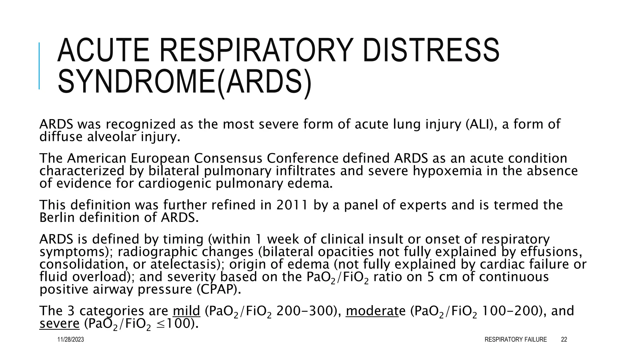
Criteria for the diagnosis of ARDS include the following:
Clinical presentation - Tachypnea and dyspnea; crackles upon auscultation
Clinical setting - Direct insult (aspiration) or systemic process causing lung
injury (sepsis)
Radiologic appearance - 3-quadrant or 4-quadrant alveolar flooding
Lung mechanics - Diminished compliance (< 40 mL/cm water)
Gas exchange - Severe hypoxia refractory to oxygen therapy (ratio of arterial
oxygen tension to fractional concentration of oxygen in inspired gas
[PaO2/FiO2] < 200)
Normal pulmonary vascular properties - Pulmonary capillary wedge pressure
lower than 18 mm Hg
11/28/2023 RESPIRATORY FAILURE 23
WORK-UP
ABGs(PaO2, PaCO2, pH, HCO3) should be evaluated in all patients who are seriously
ill or in whom respiratory failure is suspected.
CXR is essential. ECHO is not routine but is sometimes useful. Pulmonary functions
tests (PFTs) may be helpful. ECG should be performed to assess the possibility of a
cardiovascular cause of respiratory failure; it also may detect dysrhythmias
resulting from severe hypoxemia or acidosis.
Right-sided heart catheterization is controversial.
 Chest CT, Craniocervical CT
 FBC, EUC, LFTs, TFTs, CRP
 Blood Culture, PCR
 Tox Screen(opioids, barbiturates, alcohol)
 Electromyography, Tensilon test
11/28/2023 RESPIRATORY FAILURE 24
TREATMENT
 Hypoxemia is the major immediate threat to organ function.
 After the patient’s hypoxemia is corrected and the ventilatory and
hemodynamic status have stabilized, every attempt should be made to
identify and correct the underlying pathophysiologic process that led to
respiratory failure in the first place.
 The specific treatment depends on the etiology of respiratory failure.
 Consultation with a pulmonologist and an intensivist are often required.
 Patients with acute respiratory failure or exacerbations of chronic
respiratory failure need to be admitted to the intensive care unit for
ventilatory support.
Guidelines on acute respiratory failure (ARF) by the European Respiratory Society/American Thoracic
Society
11/28/2023 RESPIRATORY FAILURE 25
TREATMENT
CORRECTION OF HYPOXEMIA
The first objective in the management of respiratory failure is to reverse and/or
prevent tissue hypoxia.
Hypercapnia unaccompanied by hypoxemia generally is well tolerated and probably is
not a threat to organ function unless accompanied by severe acidosis.
Many experts believe that hypercapnia should be tolerated until the arterial blood pH
falls below 7.2.
Appropriate management of the underlying disease is an important component in the
management of respiratory failure.
A patient with acute respiratory failure generally should be admitted to a respiratory
care unit or intensive care unit (ICU).
Most patients with chronic respiratory failure can be treated at home with oxygen
supplementation and/or ventilatory assist devices along with therapy for their
underlying disease.
Guidelines on acute respiratory failure (ARF) by the European Respiratory Society/American Thoracic Society
11/28/2023 RESPIRATORY FAILURE 26
TREATMENT
CORRECTION OF HYPOXEMIA
Assurance of an adequate airway is vital in a patient with acute respiratory distress.
The most common indication for endotracheal intubation is respiratory failure.
Endotracheal intubation serves as an interface between the patient and the ventilator.
Another indication is airway protection in patients with altered mental status.
Once the airway is secured, attention is turned toward correcting the underlying
hypoxemia, the most life-threatening facet of acute respiratory failure.
The goal is to assure adequate oxygen delivery to tissues, generally achieved with an
arterial oxygen tension (PaO2) of 60 mm Hg or an arterial oxygen saturation (SaO2)
greater than 90%.
Supplemental oxygen is administered via nasal prongs or face mask; however, in
patients with severe hypoxemia, intubation and mechanical ventilation are often
required
Guidelines on acute respiratory failure (ARF) by the European Respiratory Society/American
Thoracic Society
11/28/2023 RESPIRATORY FAILURE 27
TREATMENT
MECHANICAL VENTILATION
Mechanical ventilation is used for two essential reasons: (1) to increase PaO2 and
(2) to lower PaCO2.
Mechanical ventilation also rests the respiratory muscles and is an appropriate
therapy for respiratory muscle fatigue.
The use of mechanical ventilation during the polio epidemics of the 1950s was the
impetus that led to the development of the discipline of critical care medicine.
Before the mid-1950s, negative-pressure ventilation with the use of iron lungs was
the predominant method of ventilatory support.
Currently, virtually all mechanical ventilatory support for acute respiratory failure is
provided by positive-pressure ventilation. Nevertheless, negative-pressure
ventilation still is used occasionally in patients with chronic respiratory failure
Guidelines on acute respiratory failure (ARF) by the European Respiratory Society/American Thoracic Society
11/28/2023 RESPIRATORY FAILURE 28
TREATMENT
VENTILATOR-ASSOCIATED LUNG INJURY
Mechanical ventilation is associated with a variety of insults to the lung.
BAROTRAUMA: pneumothorax, pneumomediastinum, and subcutaneous
and pulmonary interstitial emphysema.
The manifestations of barotrauma probably result from excessive alveolar
wall stress; excessive airway pressure by itself does not appear to cause
barotrauma.
In critically ill patients, the manifestations of barotrauma can be subtle.
For example, the earliest sign of pneumothorax in supine patients may be
the deep-sulcus sign or a collection of air anteriorly along cardiophrenic
angle.
It is now recognized that lung damage indistinguishable from ARDS may
be caused by certain patterns of ventilatory support.
Guidelines on acute respiratory failure (ARF) by the European Respiratory Society/American Thoracic Society
11/28/2023 RESPIRATORY FAILURE 29
TREATMENT
NONINVASIVE VENTILATORY SUPPORT
Ventilatory support via a nasal or full-face mask rather than via an
endotracheal tube is increasingly being employed for patients with
acute or chronic respiratory failure.
Noninvasive ventilation should be considered in patients with mild-
to-moderate acute respiratory failure.
The patient should have an intact airway, airway-protective reflexes,
and be alert enough to follow commands.
Guidelines on acute respiratory failure (ARF) by the European Respiratory Society/American Thoracic Society
11/28/2023 RESPIRATORY FAILURE 30
TREATMENT
WEANING FROM VENTILATOR
Weaning from mechanical ventilation is initiated when the underlying
process that necessitated ventilatory support has improved.
In some patients, such as those recovering from uncomplicated major
surgery or a toxic ingestion, withdrawal of ventilator support may be
done without weaning.
In patients who required more prolonged respiratory therapy, the
process of liberating the patient from ventilatory support may take
much longer.
A patient who has stable underlying respiratory status, adequate
oxygenation (eg, PaO2/FiO2 >200 on PEEP < 10 cm water), intact
respiratory drive, and stable cardiovascular status should be
considered for discontinuance of mechanical ventilation.
Guidelines on acute respiratory failure (ARF) by the European Respiratory Society/American Thoracic Society
11/28/2023 RESPIRATORY FAILURE 31
TREATMENT
MONITORING
Cardiac monitoring, blood pressure, pulse oximetry, SaO2, and
capnometry are recommended.
An arterial blood gas determination should be obtained 15-20 minutes
after the institution of mechanical ventilation.
The pulse oximetry readings direct efforts to reduce FiO2 to a value less
than 0.6, and the PaCO2 guides adjustments of minute ventilation.
Guidelines on acute respiratory failure (ARF) by the European Respiratory Society/American Thoracic Society
11/28/2023 RESPIRATORY FAILURE 32
DIFFERENTIAL DIAGNOSES
Acute Respiratory Distress Syndrome
(ARDS)
Bacterial Pneumonia
Viral Pneumonia
Aspiration Pneumonitis
Asthma
COPD
Cardiogenic Pulmonary Edema
Pulmonary Embolism (PE)
Myocardial Infarction
Pneumothorax
Cor Pulmonale
Bronchiectasis
Diaphragmatic Paralysis
Atelectasis
Shock
Emphysema
Idiopathic Pulmonary Fibrosis (IPF)
Interstitial (Nonidiopathic)
Pulmonary Fibrosis
11/28/2023 RESPIRATORY FAILURE 33
CONCLUSION
Respiratory failure encompasses a spectrum of conditions where the
respiratory system can't adequately oxygenate the blood or remove
excess carbon dioxide.
It can be acute or chronic and has diverse causes, ranging from lung
diseases to neuromuscular disorders.
Patients typically present with symptoms like breathlessness and
altered mental status, but these vary based on the underlying
condition.
Effective management involves treating the root cause, providing
oxygen or mechanical ventilation as necessary, and diligent
monitoring of key parameters.
Understanding these fundamental aspects of respiratory failure is
11/28/2023 RESPIRATORY FAILURE 34
REFERENCES
IJ Clifton, DAB Ellames, Respiratory Failure in Davidson’s principle and practice of Medicine 24th Ed., Chapt. 17, pp.
483-485
Vincent SM, Eman S, Abdulghani S, Bracken B: Respiratory Failure, Treasure Island (FL): StatPearls Publishing; Jan
2023 accessible at https://www.ncbi.nlm.nih.gov/books/NBK526127/
Ata Murat Kaynar, Respiratory Failure, Medscape@emedicine.com
Khan NA, Palepu A, Norena M, et al. Differences in hospital mortality among critically ill patients of Asian, Native
Indian, and European descent. Chest. 2008 Dec. 134(6):1217-22.
Moss M, Mannino DM. Race and gender differences in acute respiratory distress syndrome deaths in the United
States: an analysis of multiple-cause mortality data (1979- 1996). Crit Care Med. 2002 Aug. 30(8):1679-85.
Guideline] Rochwerg B, Brochard L, Elliott MW, Hess D, Hill NS, Nava S, et al. Official ERS/ATS clinical practice
guidelines: noninvasive ventilation for acute respiratory failure. Eur Respir J. 2017 Aug. 50 (2)
[Guideline] Alhazzani W, Møller MH, Arabi YM, et al. Surviving Sepsis Campaign: Guidelines on the Management of
Critically Ill Adults with Coronavirus Disease 2019 (COVID-19). European Society of Intensive Care Medicine.
Available at https://www.esicm.org/wp-content/uploads/2020/03/SSC-COVID19-GUIDELINES.pdf.2020;
11/28/2023 RESPIRATORY FAILURE 35
THANK YOU FOR YOUR
ATTENTION
11/28/2023 RESPIRATORY FAILURE 36

MANAGEMENT OF RESPIRATORY FAILURE

  • 1.
    RESPIRATORY FAILURE DR. EMMANUEL ALIADAMU DEPARTMENT OF MEDICINE, MODIBBO ADAMA UNIVERSITY TEACHING HOSPITAL YOLA, NIGERIA Friday, 13th October 2023
  • 2.
  • 3.
    INTRODUCTION Respiratory failure isa syndrome in which the respiratory system fails in one or both of its gas exchange functions: oxygenation and carbon dioxide elimination. In practice, it may be classified as either hypoxemic(type I) or hypercapnic(type II). Type I respiratory failure is characterized by an arterial oxygen tension (PaO2) lower than 60 mm Hg with a normal or low arterial carbon dioxide tension (PaCO2). This is the most common form of respiratory failure, and it can be associated with virtually all acute diseases of the lung, which generally involve fluid filling or collapse of alveolar units. Some examples of type I respiratory failure are cardiogenic or 11/28/2023 RESPIRATORY FAILURE 3
  • 4.
    INTRODUCTION Type II respiratoryfailure is characterized by a PaCO2 higher than 50 mm Hg. Hypoxemia is common in patients with hypercapnic respiratory failure who are breathing room air. The pH depends on the level of bicarbonate, which, in turn, is dependent on the duration of hypercapnia. Common etiologies include drug overdose, neuromuscular disease, chest wall abnormalities, and severe airway disorders (e.g, asthma and chronic obstructive pulmonary disease (COPD). 11/28/2023 RESPIRATORY FAILURE 4
  • 5.
    INTRODUCTION Respiratory failure maybe further classified as either acute or chronic. Acute respiratory failure is characterized by life-threatening derangements in arterial blood gases and acid-base status, while the manifestations of chronic respiratory failure are less dramatic and may not be as readily apparent. Acute hypercapnic respiratory failure develops over minutes to hours; therefore, pH is less than 7.3. Chronic respiratory failure develops over several days or longer, allowing time for renal compensation and an increase in bicarbonate concentration. Therefore, the pH usually is only slightly decreased. The distinction between acute and chronic hypoxemic respiratory failure cannot readily be made on the basis of arterial blood gases. The clinical markers of chronic hypoxemia, such as polycythemia or cor pulmonale, suggest a long-standing disorder. 11/28/2023 RESPIRATORY FAILURE 5
  • 6.
    INTRODUCTION The act ofrespiration engages the following three processes:  Transfer of oxygen across the alveolus  Transport of oxygen to the tissues  Removal of carbon dioxide from blood into the alveolus and then into the environment Respiratory failure may occur from malfunctioning of any of these processes. In order to understand the pathophysiologic basis of acute respiratory failure, an understanding of pulmonary gas exchange is essential. 11/28/2023 RESPIRATORY FAILURE 6
  • 7.
    INTRODUCTION EPIDEMIOLOGY Respiratory failure isa syndrome caused by a multitude of pathological states; therefore, the epidemiology of this disease process is difficult to ascertain. In 2017 in the United States of America, however, the incidence of respiratory failure was found to be 1,275 cases per 100,000 adults. The relationship between acute respiratory failure and race is still debated. A study by Khan et al suggested that no differences in mortality exist in patients of Asian and Native Indian descent with acute critical illness after adjusting for differences in case mix. Moss and Mannino reported worse outcome for African Americans with ARDS than for whites after adjustment for case mix. Future prospective association studies should yield a better knowledge of the impact of race on the outcome of respiratory failure. Vincent SM, Eman S, Abdulghani S, Bracken B: Respiratory Failure,Treasure Island (FL): StatPearls Publishing; Jan 2023 accessible at https://www.ncbi.nlm.nih.gov/books/NBK526127/ Khan NA, Palepu A, Norena M, et al. Differences in hospital mortality among critically ill patients of Asian, Native Indian, and European descent. Chest. 2008 Dec. 134(6):1217-22. Moss M, Mannino DM. Race and gender differences in acute respiratory distress syndrome deaths in the United States: an analysis of multiple-cause mortality data (1979- 1996). Crit Care Med. 2002 Aug. 30(8):1679-85. 11/28/2023 RESPIRATORY FAILURE 7
  • 8.
    PATHOPHYSIOLOGY Respiratory failure canarise from an abnormality in any of the components of the respiratory system, including the airways, alveoli, central nervous system (CNS), peripheral nervous system, respiratory muscles, and chest wall. Patients who have hypoperfusion secondary to cardiogenic, hypovolemic, or septic shock often present with respiratory failure. Normally, ventilatory capacity greatly exceeds ventilatory demand. Respiratory failure may result from either a reduction in ventilatory capacity or an increase in ventilatory demand (or both). Ventilatory capacity can be decreased by a disease process involving any of the functional components of the respiratory system and its controller. Ventilatory demand is augmented by an increase in minute ventilation and/or an increase in the work of breathing. 11/28/2023 RESPIRATORY FAILURE 8
  • 9.
    PATHOPHYSIOLOGY HYPOXEMIC RESPIRATORY FAILURE Thepathophysiologic mechanisms that account for the hypoxemia observed in a wide variety of diseases are V/Q mismatch and shunt. These 2 mechanisms lead to widening of the alveolar-arterial PO2 gradient, which normally is less than 15 mm Hg. They can be differentiated by assessing the response to oxygen supplementation or calculating the shunt fraction after inhalation of 100% oxygen. In most patients with hypoxemic respiratory failure, these 2 mechanisms coexist. 11/28/2023 RESPIRATORY FAILURE 9
  • 10.
    PATHOPHYSIOLOGY HYPERCAPNIC RESPIRATORY FAILURE Adecrease in alveolar ventilation can result from a reduction in overall (minute) ventilation or an increase in the proportion of dead space ventilation. A reduction in minute ventilation is observed primarily in the setting of neuromuscular disorders and CNS depression. In pure hypercapnic respiratory failure, the hypoxemia is easily corrected with oxygen therapy. Hypoventilation is an uncommon cause of respiratory failure and usually occurs from depression of the CNS from drugs or neuromuscular diseases affecting respiratory muscles. Hypoventilation is characterized by hypercapnia and hypoxemia. Hypoventilation can be differentiated from other causes of hypoxemia by the presence of a normal alveolar-arterial PO2 gradient. 11/28/2023 RESPIRATORY FAILURE 10
  • 11.
    PATHOPHYSIOLOGY The underlying mechanismsin type I respiratory failure include: Alveolar hypoventilation, Low atmospheric pressure/fraction of inspired oxygen, Diffusion defect, Ventilation/perfusion (V/Q) mismatch, Right-to-left shunt. While in type II respiratory failure, Respiratory pump failure, Increased dead space, Increased CO production are usually observed. 11/28/2023 RESPIRATORY FAILURE 11
  • 12.
    RISK FACTORS Age>60years Chronic respiratorydisorder: COPD, Asthma Comorbidities such as cardiopulmonary, renal, hepatic, or neurologic disease. Poor nutritional status 11/28/2023 RESPIRATORY FAILURE 12
  • 13.
    AETIOLOGIES The aetilogies ofrespiratory failure can be broadly grouped into the disorders involving the various components of the respiratory system. CNS disorders Peripheral nervous system disorders Respiratory muscles disorders Chest wall disorders Diseases of the Airways, and Alveolar disorders 11/28/2023 RESPIRATORY FAILURE 13
  • 14.
    CNS DISORDERS Tumors Vascular abnormalitiesinvolving the brain stem Overdose of a narcotic or sedative Metabolic disorders such as myxedema Chronic metabolic alkalosis. 11/28/2023 RESPIRATORY FAILURE 14
  • 15.
    PNS, CHEST WALL,AND RESPIRATORY MUSCLE DISORDERS Tetanus Guillain-Barré syndrome, Neurotoxic envenomation Poliomyelitis Cervical spinal cord injury Toxins Muscular dystrophy Myasthenia gravis Severe kyphoscoliosis, and Morbid obesity. 11/28/2023 RESPIRATORY FAILURE 15
  • 16.
    DISEASES OF THEAIRWAYS Upper-airway disorders: Acute epiglottitis, Croup, Diphtheria, Tumors involving the trachea Lower-airway disorders: Bacterial Pneumonia, Pulmonary TB, Pneumocystis Jirovecii Pneumonia, Aspiration Pneumonitis, COVID-19, COPD, asthma, and cystic fibrosis. 11/28/2023 RESPIRATORY FAILURE 16
  • 17.
    ALVEOLAR DISORDERS Cardiogenic andnon-cardiogenic pulmonary edema Aspiration pneumonia Extensive pulmonary hemorrhage. These disorders are associated with intrapulmonary shunt and an increased work of breathing 11/28/2023 RESPIRATORY FAILURE 17
  • 18.
    COMMON CAUSES OFTYPE I RESPIRATORY FAILURE COPD Pneumonia Pulmonary edema Pulmonary fibrosis Asthma Pneumothorax Pulmonary embolism Bronchiectasis Acute respiratory distress syndrome (ARDS) Fat embolism syndrome Pulmonary arterial hypertension Pneumoconiosis Granulomatous lung diseases Cyanotic congenital heart disease Kyphoscoliosis Obesity 11/28/2023 RESPIRATORY FAILURE 18
  • 19.
    COMMON CAUSES OFTYPE II RESPIRATORY FAILURE Pulmonary edema COPD Severe asthma Tetanus Drug overdose Poisonings Myasthenia gravis ARDS Poliomyelitis Porphyria Cervical cordotomy Head and cervical cord injury Primary alveolar hypoventilation Obesity-Hypoventilation syndrome Myxedema Polyneuropathy Primary muscle disorders 11/28/2023 RESPIRATORY FAILURE 19
  • 20.
    CLINICAL FEATURES HISTORY  Patientstypically present with respiratory symptoms (i.e., dyspnea, cough, hemoptysis, sputum production, and wheezing); however, symptoms from other organ systems (i.e., chest pain, decreased appetite, heartburn, fever, and significant weight loss) are important.  Loss of smell and/or exposure to sick people or unprotect contact with individuals with coronavirus infection (COVID-19) is essential in suspecting COVID-19 illness and associated respiratory failure, particularly in high-risk patients (older patients, men, and morbidly obese).  A history of previous cardiac disease, recent symptoms of chest pain, paroxysmal nocturnal dyspnea, and orthopnea suggest cardiogenic pulmonary edema.  Noncardiogenic edema (eg, acute respiratory distress syndrome (ARDS) occurs in typical clinical contexts, such as sepsis, trauma, aspiration, pneumonia, pancreatitis, drug toxicity, and multiple transfusions. 11/28/2023 RESPIRATORY FAILURE 20
  • 21.
    CLINICAL FEATURES PHYSICAL EXAMINATION General inspection: Accessory muscle use, cachectic, conversational dyspnea, diaphoresis, fever, pale conjunctiva, respiratory distress, obesity, and purse-lipped breathing, edema, peripheral cyanosis, central cyanosis, unilateral swelling, digital clubbing, tobacco staining, and tremor  Chest: Kussmaul breathing, Cheyne-Stoke breathing, kyphoscoliosis, Asymmetrical chest expansion, tachypnea, bradypnea, tracheal deviation, dullness to percussion, hyper-resonance to percussion, bronchial breath sounds, crackles, decreased breath sounds, pleural rub, rhonchi, stridor, tactile vocal fremitus, vocal resonance, wheezes, and whispering pectoriloquy  Cardiovascular: Jugular venous distention, loud P2,  CNS: altered mental status, asterixis, Horner's syndrome  Abdomen: Hepatomegaly, ascites 11/28/2023 RESPIRATORY FAILURE 21
  • 22.
    ACUTE RESPIRATORY DISTRESS SYNDROME(ARDS) ARDSwas recognized as the most severe form of acute lung injury (ALI), a form of diffuse alveolar injury. The American European Consensus Conference defined ARDS as an acute condition characterized by bilateral pulmonary infiltrates and severe hypoxemia in the absence of evidence for cardiogenic pulmonary edema. This definition was further refined in 2011 by a panel of experts and is termed the Berlin definition of ARDS. ARDS is defined by timing (within 1 week of clinical insult or onset of respiratory symptoms); radiographic changes (bilateral opacities not fully explained by effusions, consolidation, or atelectasis); origin of edema (not fully explained by cardiac failure or fluid overload); and severity based on the PaO2/FiO2 ratio on 5 cm of continuous positive airway pressure (CPAP). The 3 categories are mild (PaO2/FiO2 200-300), moderate (PaO2/FiO2 100-200), and severe (PaO2/FiO2 ≤100). 11/28/2023 RESPIRATORY FAILURE 22
  • 23.
    ACUTE RESPIRATORY DISTRESS SYNDROME(ARDS) Criteriafor the diagnosis of ARDS include the following: Clinical presentation - Tachypnea and dyspnea; crackles upon auscultation Clinical setting - Direct insult (aspiration) or systemic process causing lung injury (sepsis) Radiologic appearance - 3-quadrant or 4-quadrant alveolar flooding Lung mechanics - Diminished compliance (< 40 mL/cm water) Gas exchange - Severe hypoxia refractory to oxygen therapy (ratio of arterial oxygen tension to fractional concentration of oxygen in inspired gas [PaO2/FiO2] < 200) Normal pulmonary vascular properties - Pulmonary capillary wedge pressure lower than 18 mm Hg 11/28/2023 RESPIRATORY FAILURE 23
  • 24.
    WORK-UP ABGs(PaO2, PaCO2, pH,HCO3) should be evaluated in all patients who are seriously ill or in whom respiratory failure is suspected. CXR is essential. ECHO is not routine but is sometimes useful. Pulmonary functions tests (PFTs) may be helpful. ECG should be performed to assess the possibility of a cardiovascular cause of respiratory failure; it also may detect dysrhythmias resulting from severe hypoxemia or acidosis. Right-sided heart catheterization is controversial.  Chest CT, Craniocervical CT  FBC, EUC, LFTs, TFTs, CRP  Blood Culture, PCR  Tox Screen(opioids, barbiturates, alcohol)  Electromyography, Tensilon test 11/28/2023 RESPIRATORY FAILURE 24
  • 25.
    TREATMENT  Hypoxemia isthe major immediate threat to organ function.  After the patient’s hypoxemia is corrected and the ventilatory and hemodynamic status have stabilized, every attempt should be made to identify and correct the underlying pathophysiologic process that led to respiratory failure in the first place.  The specific treatment depends on the etiology of respiratory failure.  Consultation with a pulmonologist and an intensivist are often required.  Patients with acute respiratory failure or exacerbations of chronic respiratory failure need to be admitted to the intensive care unit for ventilatory support. Guidelines on acute respiratory failure (ARF) by the European Respiratory Society/American Thoracic Society 11/28/2023 RESPIRATORY FAILURE 25
  • 26.
    TREATMENT CORRECTION OF HYPOXEMIA Thefirst objective in the management of respiratory failure is to reverse and/or prevent tissue hypoxia. Hypercapnia unaccompanied by hypoxemia generally is well tolerated and probably is not a threat to organ function unless accompanied by severe acidosis. Many experts believe that hypercapnia should be tolerated until the arterial blood pH falls below 7.2. Appropriate management of the underlying disease is an important component in the management of respiratory failure. A patient with acute respiratory failure generally should be admitted to a respiratory care unit or intensive care unit (ICU). Most patients with chronic respiratory failure can be treated at home with oxygen supplementation and/or ventilatory assist devices along with therapy for their underlying disease. Guidelines on acute respiratory failure (ARF) by the European Respiratory Society/American Thoracic Society 11/28/2023 RESPIRATORY FAILURE 26
  • 27.
    TREATMENT CORRECTION OF HYPOXEMIA Assuranceof an adequate airway is vital in a patient with acute respiratory distress. The most common indication for endotracheal intubation is respiratory failure. Endotracheal intubation serves as an interface between the patient and the ventilator. Another indication is airway protection in patients with altered mental status. Once the airway is secured, attention is turned toward correcting the underlying hypoxemia, the most life-threatening facet of acute respiratory failure. The goal is to assure adequate oxygen delivery to tissues, generally achieved with an arterial oxygen tension (PaO2) of 60 mm Hg or an arterial oxygen saturation (SaO2) greater than 90%. Supplemental oxygen is administered via nasal prongs or face mask; however, in patients with severe hypoxemia, intubation and mechanical ventilation are often required Guidelines on acute respiratory failure (ARF) by the European Respiratory Society/American Thoracic Society 11/28/2023 RESPIRATORY FAILURE 27
  • 28.
    TREATMENT MECHANICAL VENTILATION Mechanical ventilationis used for two essential reasons: (1) to increase PaO2 and (2) to lower PaCO2. Mechanical ventilation also rests the respiratory muscles and is an appropriate therapy for respiratory muscle fatigue. The use of mechanical ventilation during the polio epidemics of the 1950s was the impetus that led to the development of the discipline of critical care medicine. Before the mid-1950s, negative-pressure ventilation with the use of iron lungs was the predominant method of ventilatory support. Currently, virtually all mechanical ventilatory support for acute respiratory failure is provided by positive-pressure ventilation. Nevertheless, negative-pressure ventilation still is used occasionally in patients with chronic respiratory failure Guidelines on acute respiratory failure (ARF) by the European Respiratory Society/American Thoracic Society 11/28/2023 RESPIRATORY FAILURE 28
  • 29.
    TREATMENT VENTILATOR-ASSOCIATED LUNG INJURY Mechanicalventilation is associated with a variety of insults to the lung. BAROTRAUMA: pneumothorax, pneumomediastinum, and subcutaneous and pulmonary interstitial emphysema. The manifestations of barotrauma probably result from excessive alveolar wall stress; excessive airway pressure by itself does not appear to cause barotrauma. In critically ill patients, the manifestations of barotrauma can be subtle. For example, the earliest sign of pneumothorax in supine patients may be the deep-sulcus sign or a collection of air anteriorly along cardiophrenic angle. It is now recognized that lung damage indistinguishable from ARDS may be caused by certain patterns of ventilatory support. Guidelines on acute respiratory failure (ARF) by the European Respiratory Society/American Thoracic Society 11/28/2023 RESPIRATORY FAILURE 29
  • 30.
    TREATMENT NONINVASIVE VENTILATORY SUPPORT Ventilatorysupport via a nasal or full-face mask rather than via an endotracheal tube is increasingly being employed for patients with acute or chronic respiratory failure. Noninvasive ventilation should be considered in patients with mild- to-moderate acute respiratory failure. The patient should have an intact airway, airway-protective reflexes, and be alert enough to follow commands. Guidelines on acute respiratory failure (ARF) by the European Respiratory Society/American Thoracic Society 11/28/2023 RESPIRATORY FAILURE 30
  • 31.
    TREATMENT WEANING FROM VENTILATOR Weaningfrom mechanical ventilation is initiated when the underlying process that necessitated ventilatory support has improved. In some patients, such as those recovering from uncomplicated major surgery or a toxic ingestion, withdrawal of ventilator support may be done without weaning. In patients who required more prolonged respiratory therapy, the process of liberating the patient from ventilatory support may take much longer. A patient who has stable underlying respiratory status, adequate oxygenation (eg, PaO2/FiO2 >200 on PEEP < 10 cm water), intact respiratory drive, and stable cardiovascular status should be considered for discontinuance of mechanical ventilation. Guidelines on acute respiratory failure (ARF) by the European Respiratory Society/American Thoracic Society 11/28/2023 RESPIRATORY FAILURE 31
  • 32.
    TREATMENT MONITORING Cardiac monitoring, bloodpressure, pulse oximetry, SaO2, and capnometry are recommended. An arterial blood gas determination should be obtained 15-20 minutes after the institution of mechanical ventilation. The pulse oximetry readings direct efforts to reduce FiO2 to a value less than 0.6, and the PaCO2 guides adjustments of minute ventilation. Guidelines on acute respiratory failure (ARF) by the European Respiratory Society/American Thoracic Society 11/28/2023 RESPIRATORY FAILURE 32
  • 33.
    DIFFERENTIAL DIAGNOSES Acute RespiratoryDistress Syndrome (ARDS) Bacterial Pneumonia Viral Pneumonia Aspiration Pneumonitis Asthma COPD Cardiogenic Pulmonary Edema Pulmonary Embolism (PE) Myocardial Infarction Pneumothorax Cor Pulmonale Bronchiectasis Diaphragmatic Paralysis Atelectasis Shock Emphysema Idiopathic Pulmonary Fibrosis (IPF) Interstitial (Nonidiopathic) Pulmonary Fibrosis 11/28/2023 RESPIRATORY FAILURE 33
  • 34.
    CONCLUSION Respiratory failure encompassesa spectrum of conditions where the respiratory system can't adequately oxygenate the blood or remove excess carbon dioxide. It can be acute or chronic and has diverse causes, ranging from lung diseases to neuromuscular disorders. Patients typically present with symptoms like breathlessness and altered mental status, but these vary based on the underlying condition. Effective management involves treating the root cause, providing oxygen or mechanical ventilation as necessary, and diligent monitoring of key parameters. Understanding these fundamental aspects of respiratory failure is 11/28/2023 RESPIRATORY FAILURE 34
  • 35.
    REFERENCES IJ Clifton, DABEllames, Respiratory Failure in Davidson’s principle and practice of Medicine 24th Ed., Chapt. 17, pp. 483-485 Vincent SM, Eman S, Abdulghani S, Bracken B: Respiratory Failure, Treasure Island (FL): StatPearls Publishing; Jan 2023 accessible at https://www.ncbi.nlm.nih.gov/books/NBK526127/ Ata Murat Kaynar, Respiratory Failure, Medscape@emedicine.com Khan NA, Palepu A, Norena M, et al. Differences in hospital mortality among critically ill patients of Asian, Native Indian, and European descent. Chest. 2008 Dec. 134(6):1217-22. Moss M, Mannino DM. Race and gender differences in acute respiratory distress syndrome deaths in the United States: an analysis of multiple-cause mortality data (1979- 1996). Crit Care Med. 2002 Aug. 30(8):1679-85. Guideline] Rochwerg B, Brochard L, Elliott MW, Hess D, Hill NS, Nava S, et al. Official ERS/ATS clinical practice guidelines: noninvasive ventilation for acute respiratory failure. Eur Respir J. 2017 Aug. 50 (2) [Guideline] Alhazzani W, Møller MH, Arabi YM, et al. Surviving Sepsis Campaign: Guidelines on the Management of Critically Ill Adults with Coronavirus Disease 2019 (COVID-19). European Society of Intensive Care Medicine. Available at https://www.esicm.org/wp-content/uploads/2020/03/SSC-COVID19-GUIDELINES.pdf.2020; 11/28/2023 RESPIRATORY FAILURE 35
  • 36.
    THANK YOU FORYOUR ATTENTION 11/28/2023 RESPIRATORY FAILURE 36