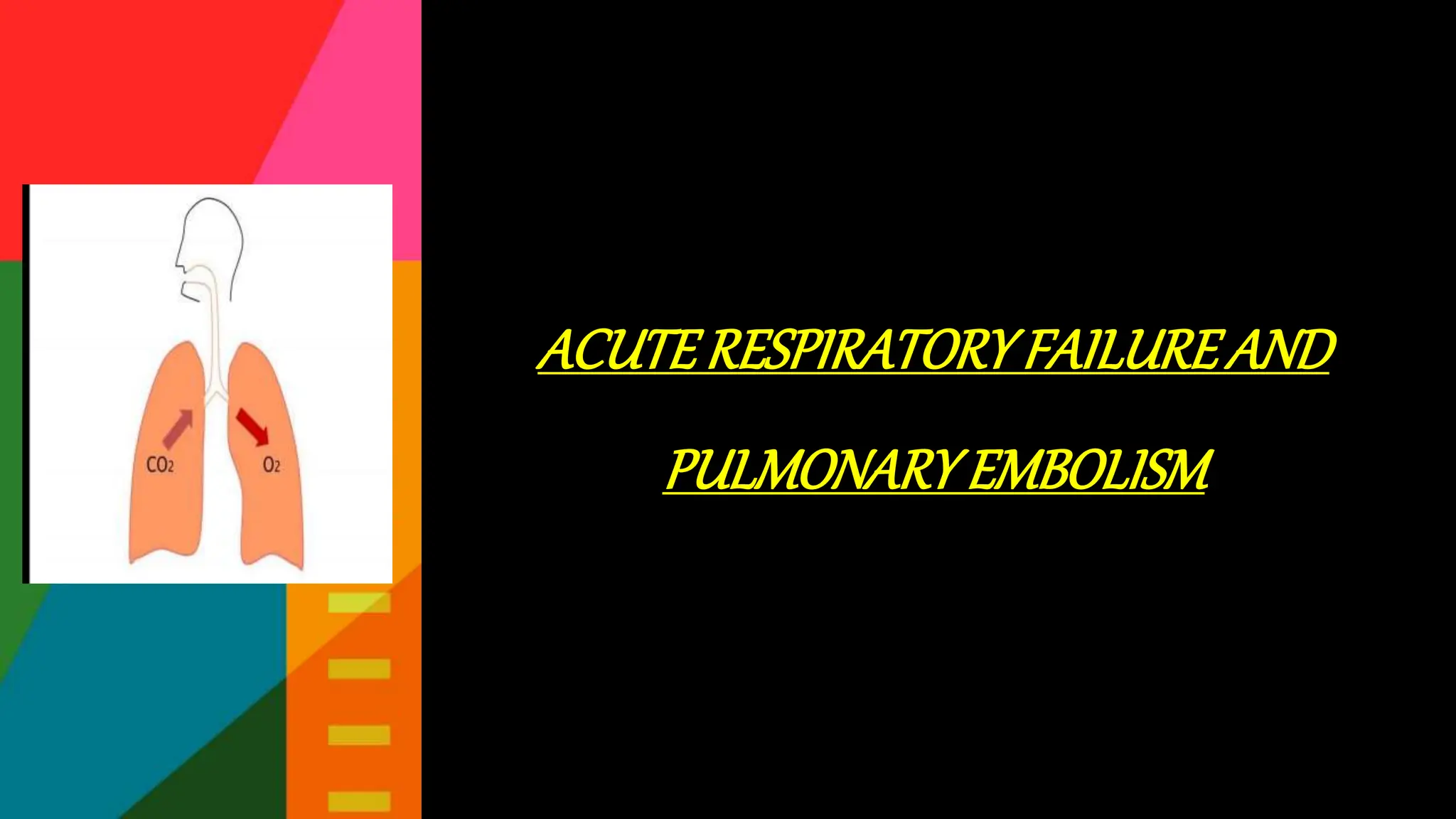
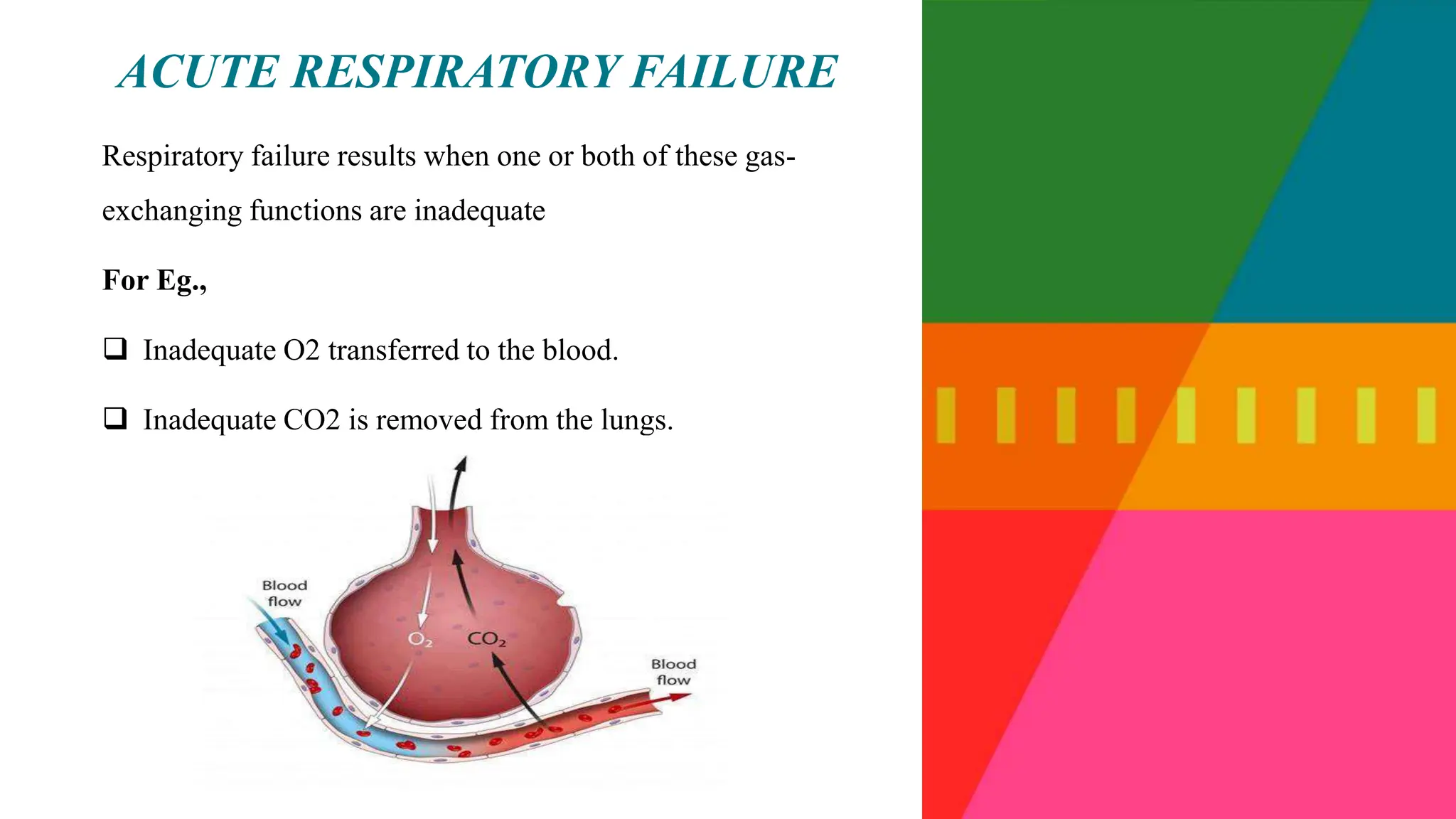
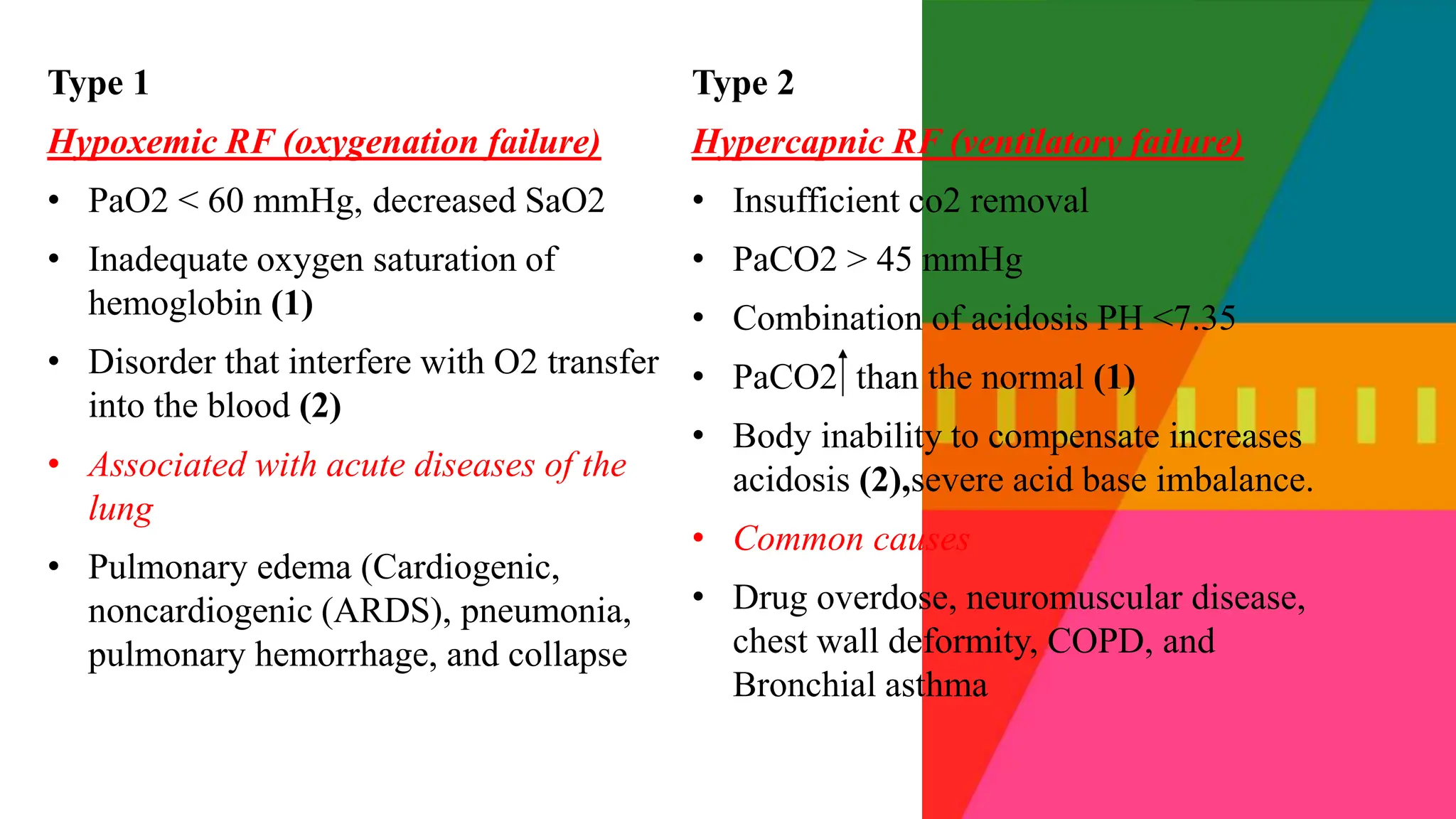
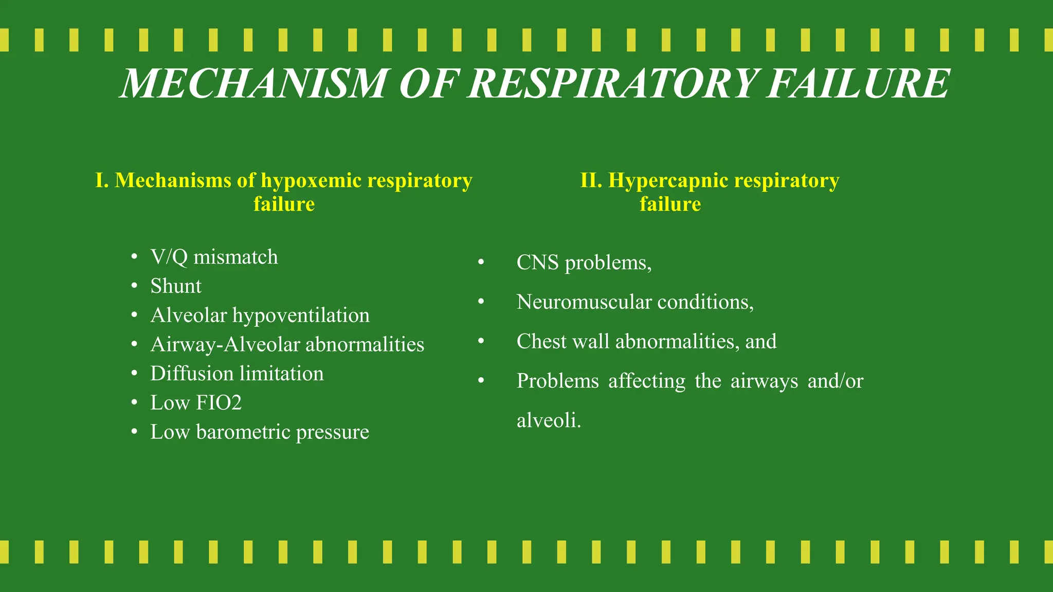
ACUTERESPIRATORYFAILUREAND
PULMONARYEMBOLISM
Respiratory failure results when one or both of these gas-
exchanging functions are inadequate
For Eg.,
 Inadequate O2 transferred to the blood.
 Inadequate CO2 is removed from the lungs.
ACUTE RESPIRATORY FAILURE
Normal gas exchange unit in the lung.
Classification of
respiratory failure
Type 1
Hypoxemic RF (oxygenation failure)
• PaO2 < 60 mmHg, decreased SaO2
• Inadequate oxygen saturation of
hemoglobin (1)
• Disorder that interfere with O2 transfer
into the blood (2)
• Associated with acute diseases of the
lung
• Pulmonary edema (Cardiogenic,
noncardiogenic (ARDS), pneumonia,
pulmonary hemorrhage, and collapse
Type 2
Hypercapnic RF (ventilatory failure)
• Insufficient co2 removal
• PaCO2 > 45 mmHg
• Combination of acidosis PH <7.35
• PaCO2 than the normal (1)
• Body inability to compensate increases
acidosis (2),severe acid base imbalance.
• Common causes
• Drug overdose, neuromuscular disease,
chest wall deformity, COPD, and
Bronchial asthma
Distinction between Acute and Chronic RF
• Acute RF
• Develops over minutes to
hours
• ↓ pH quickly to <7.2
• Example; Pneumonia
Chronic RF
• Develops over days to weeks
• ↑ in HCO3
• ↓ pH slightly
• Polycythemia, Corpulmonale
• Example; COPD
MECHANISM OF RESPIRATORY FAILURE
I. Mechanisms of hypoxemic respiratory
failure
• V/Q mismatch
• Shunt
• Alveolar hypoventilation
• Airway-Alveolar abnormalities
• Diffusion limitation
• Low FIO2
• Low barometric pressure
II. Hypercapnic respiratory
failure
• CNS problems,
• Neuromuscular conditions,
• Chest wall abnormalities, and
• Problems affecting the airways and/or
alveoli.
I . MECHANISMS OF
HYPOXEMIC
RESPIRATORY FAILURE
• V/Q
• Shunt
• Alveolar hypoventilation
• Airway-Alveolar abnormalities
• Diffusion limitation
• Low FIO2
• Low barometric pressure
In normal lungs, the volume of blood perfusing the lungs and the amount of gas reaching
the alveoli are almost identical. So, when you compare normal alveolar ventilation (4 to 6
L/min) to pulmonary blood flow (4 to 6 L/min), V/Q=1 (ventilation-perfusion mismatch )-
1ml of air for 1 ml of blood flow i.e.., 1:1
Condition that alter V/Q (V/Q mismatch )
• Increased secretion in airways-COPD
• Inc secretion in alveoli- pneumonia Limited airflow ventilation
• Bronchospasm- asthma but have no effect on blood flow
• Alveolar collapse- Atelectasis to the gas exchange units .
• Embolus.
PATHOPHYSIOLOGY-V/Q
REGIONAL V/Q DIFFERENCES IN THE NORMAL
LUNG
Range of ventilation to perfusion (V/Q) relationships.
SHUNTS
• Blood exits the heart without participated in gas
exchange
• Two types : Anatomical and intrapulmonary shunts
• Anatomical shunts: e.g. VSD
• intrapulmonary shunts: ARDS , pneumonia, pulmonary
edema (Alveoli filled with fluid prevent gas
exchange)
Intra-pulmonary
• Small airways occluded ( e.g asthma,
chronic bronchitis)
• Alveoli are filled with fluid ( e.g pulm
edema, pneumonia)
• Alveolar collapse ( e.g atelectasis)
Perfusion without ventilation (shunting)
DIFFUSE LIMITATION
Alveolar-capillary membrane
Compromise become thicken or destroys the membrane
Due to severe emphysema and pulmonary emboli that worsen pulmonary vascular bed
Thickening of alveolar capillary membrane
Fibrotic changes
Slow gaseous exchange classic signs: hypoxemia during exercise but not at rest
Alveolar hypoventilation
Decreased in ventilation due to increased PaCo2 and
dec in PaO2
Various conditions that affect hypoventilation such as
restrictive lung disease , CNS disease, chest wall
dysfunction , asthma, neuromuscular failure
Hypoxemia
Hypercarbia
• Hypercarbia is always a reflection of inadequate ventilation & ventilatory failure
• PaCO2 is
• Directly related to CO2 production
• Inversely related to alveolar ventilation PaCO2 = k x VCO2 / VA
When CO2 production increases, ventilation increases rapidly to maintain normal PaCO2
• Alveolar ventilation is only a fraction of total ventilation VA = VE – VD
• Increased dead space or low V/Q areas may adversely effect CO2 removal
II . HYPERCAPNIC RESPIRATORY FAILURE
• ventilatory failure, Many conditions can cause impaired
ventilation. This often occurs from an increase in CO2
production or a decrease in alveolar ventilation. Hypercapnic
respiratory failure can be acute or chronic.
• 4 categories:
• (1) CNS problems,
• (2) neuromuscular conditions,
• (3) chest wall abnormalities, and
• (4) problems affecting the airways and/or alveoli.
1. Abnormalities of airway
2. Central nervous system abnormalities
3. Chest wall abnormalities
4. Neuromuscular conditions
4 categories involved in
Hypercapnic RF
1.AIRWAY AND ALVEOLI ABNORMALITIES
Asthma, COPD, cystic fibrosis, are high risk for hypercapnic RF ,due to
airflow obstruction
Respiratory muscle fatigue
Ventilatory failure
PATHOPHYSIOLOGY
PATHOPHYSIOLOGY
2.CNS Abnormalities
Various CNS may supress the drive to breathe
Overdose of res dep drugs e.g.., opioids, benzodiazepines
It dec O2 reactivity in brain
Arterial CO2 levels rise
Brainstem infarction and head injury may also interfere
Normal function of resp centre in the medulla
PATHOPHYSIOLOGY
3.Chest wall abnormalities
Due to flail chest ,fractures
Difficulty in expanding lungs due to pain , muscle spasm , mechanical restriction
and due to kyphoscoliosis
Change in spinal configuration
Compress the lungs and it prevents normal expansion
Develop risk of respiratory failure
Because of limitation in lung expansion
4.DUE TO NEUROMUSCULAR CONDITION
Due to Guillain-Barre syndrome ,muscular dystrophy , myasthenia
gravis(acute exacerbation) or multiple sclerosis
Respiratory muscles are weakened or paralyzed
Risk for respiratory failure
CAUSES:1-CNS
 Depression of the neural drive to breath
 Brain stem tumors or vascular
abnormality
 Overdose of a narcotic, sedative
Myxedema, chronic metabolic alkalosis
 Acute or chronic hypoventilation and
hypercapnia Causes
2 - Disorders of peripheral nervous system,
• Respiratory dysfunction in multiple sclerosis
• Inability to maintain a level of minute ventilation
appropriate for the rate of CO2 production
• Guillain-Barre syndrome, muscular dystrophy,
myasthenia gravis, Kyphoscoliosis, morbid obesity
• Hypoxemia and hypercapnia
CAUSES
3-Abnormities of the airways
Upper airways
• Acute epiglottitis
• Tracheal tumours
Lower airway
• COPD, Asthma, cystic fibrosis
• Acute and chronic hypercapnia
causes
4 - Abnormities of the alveoli
• Diffuse alveolar filling
• hypoxemic RF
• Cardiogenic and noncardiogenic
pulmonary edema
• Aspiration pneumonia
• Pulmonary hemorrhage
• Associate with Intrapulmonary shunt
and increase work of breathing
causes
CLINICAL MANIFESTATION
Hypoxemia respiratory failure
Respiratory system
 Dyspnoea
 Tachypnoea
 Prolonged expiration (I:E 1:3, 1:4)
 Intercostal muscle retraction
 Use of accessory muscles in respiration
 SpO2, (<80%)
 Paradoxic chest/abdominal wall
movement with respiratory cycle (late)
 Cyanosis (late)
CLINICAL MANIFESTATION
Cerebral
Agitation
Delirium
Confusion.
Coma (late)
Cardiac.
Tachycardia
Skin cool, clammy, and diaphoretic
Other
Fatigue
Unable to speak in complete sentences without
pausing to breathe
CLINICAL MANIFESTATION
Hypercapnia respiratory failure
Respiratory
• Dyspnoea
• Respiratory rate or rapid rate with
shallow respirations
• Tidal volume
• Minute ventilation
Cerebral
• Morning headache
• Progressive somnolence
• Disorientation•
• Coma (late).
Cardiac
• Dysrhythmias.
• Tachycardia
• Hypertension
• Bounding pulse
Neuromuscular
• Muscle weakness
• Tremor, seizures (late)
• Deep tendon reflexes,
OTHER STUDIES
• CBC
• Sr . Electrolytes
• Gram stain
• Culture and sensitivity
• Capnometry
• BLUE PROTOCOLS (bedside
lung ultrasound in emergency)
DIAGNOSIS OF RF
 providing support with oxygenation and ventilation,
 Correction of Hypoxemia.
 Correction of Hypercapnia and Respiratory Acidosis .
 Ventilatory Support.
 HFNC may be as effective as NIV in the management of AHRF,
particularly in improving arterial pH, pCO2, and pO2, as well as
preventing endotracheal intubation and mortality.
Management-goals
Embolus derives from a Greek word meaning “plug” or
“stopper.”
Pulmonary Embolism refers to the obstruction of the
pulmonary artery or one of its branches by a thrombus (or
thrombi) that originates somewhere in the venous system
or in the right side of the heart Most commonly due to
blood clot or thrombus
-HARI PRASATH.
Pulmonary embolism (PE) is the blockage of 1 or more
pulmonary arteries by a thrombus, fat or air embolus, or
tumor tissue
-LEWIS
PULMONARYEMBOLISM
A saddle embolus refers to a large thrombus lodged
at an arterial bifurcation.
Less common causes
• Fat emboli (from fractured long bones),
• Air emboli (from improperly administered IV
therapy),
• Bacterial vegetation on heart valves,
• Amniotic fluid, and
• Cancer.
SADDLE EMBOLUS
Virchow’s Triad criteria: Factors that contributed to
thrombosis.
 Stasis of blood,
 Hypercoagulability, and
 Endothelial vessel wall injury
Virchow’s Triad criteria:
RISK FACTORS
Risk factors for pulmonary embolus:
 Venous stasis
 Prolonged immobilization
 Prolonged periods of sitting/traveling
 Varicose veins
 Spinal cord injury
 Hypercoagulability
 Injury
 Tumour (pancreatic, Gl, breast, lung)
 Increased platelet count (polycythemia,
splenectomy)
 Venous endothelial disease
 Thrombophlebitis
 Vascular disease
 Foreign bodies (IV/central venous
catheters)
 Certain disease States (combination of
stasis, coagulation alterations, and venous
injury)
 Heart disease (especially heart failure).
 Trauma
 Postoperative state/postpartum period
 DM
 COPD
PREDISPOSING FACTORS
 Advanced age
 Obesity
 Pregnancy
 Oral contraceptive use
 History of previous
thrombophlebitis, pulmonary
embolism
 Constrictive clothing
 Smoking
 Hypertension
When a thrombus completely or partially obstructs a pulmonary artery or its branches,
the alveolar dead space is increased
continuing to be ventilated, receives little or no blood flow
gas exchange is impaired or absent
various substances are released from the clot and surrounding area that cause regional blood vessels and
bronchioles to constrict.
increase in pulmonary vascular resistance
V./Q. Mismatch
PATHOPHYSIOLOGY
Increased pulmonary vascular resistance due to the regional vasoconstriction and
reduced size of the pulmonary vascular bed
Increase in pulmonary arterial pressure
Increase in right ventricular work to maintain pulmonary blood flow
Right ventricular failure occurs
Decrease in cardiac output followed by a decrease in systemic blood pressure
Shock develop
PATHOPHYSIOLOGY
Classic triad
⸙ Dyspnea
⸙ Chest pain
⸙ Hemoptysis
Common manifestation
 May be nonspecific, difficult to
diagnose.
 Dyspnea,
 Pleuritic chest pain
 Crackles
 Fever
♠ Accentuation of pulmonic heart sounds
♠ Altered mental status
♠ Hypoxemia
♠ Syncope
♠ Hypotension
♠ Tachycardia
Clinical manifestation
 Collapse of patient with shock , pallor , severe dyspnea, hypoxemia
, crushing chest pain
 Massive-do not have pain
 Pulse rapid and weak
 BP low
 ECG Indicates RT VENT strain
Massive emboli
 Pleuritic chest pain
 Dyspnea
 Slight fever
 Productive cough with blood streaked
sputum
 Tachycardia
 Pleural friction rub
SMALL EMBOLI
 Undetected or produce vague
 Transient symptoms
 Reduction in capillary bed
 pulmonary HT
 Rt vent hypertrophy
Medium sized emboli
 Ventilation perfusion lung scan
 D- dimer testing= measures the amount of cross-linked
fibrin fragments.
 Pulmonary angiography
 Computed tomography
 ABG Analysis
 A spiral (helical) CT scan (also known as CT angiography
or CTA)
Diagnostic studies
The V/Q scan has 2 parts.
1. Perfusion scanning involves IV injection of a
radioisotope. A scanning device images the pulmonary
circulation.
2. Ventilation scanning involves inhalation of a radioactive
gas, such as xenon. Scanning reflects the distribution of
gas through the lung.
CHEST X RAY= R/O Atelectasis, pleural effusion
V/Q SCANNING
 BNP >90 pg/ml
 N-terminal pro BNP >500 pg/ml
 Troponin I >0.4 ng/ml, or troponin T >0.1 ng/ml
 Electrocardiogram (ECG)=RT VENT STRAIN
 Transoesophageal echocardiography- specific
 Ultrasound for lower extremities
Diagnostic studies
European Respiratory Society released the new guidelines for the
diagnosis and management of Pulmonary embolism
 Emergency management (cardiopulmonary
support)=ECMO
 General management
Anticoagulation therapy
Thrombolytic therapy
 Surgical management
 Nursing management
MANAGEMENT
EMERGENCY MANAGEMENT
 Stabilize the cardiopulmonary system
 O2 administration
 Iv lines
 Hemodynamic measurements, perfusion scan , ABG, helical CT , pulmonary angiography
 Treat hypotension-dobutamine infusion
 Monitor ECG-find dysarrthmias , rt vent failure
 Intubation , mechanical ventilation
 Small doses of morphine , sedatives
GENERAL MANAGEMENT- DRUG
THERAPY
Fibrinolytic agent
Unfractionated heparin IV
 Low-molecular-weight heparin (e.g., enoxaparin [Lovenox])
Warfarin (Coumadin) for long-term therapy
Analgesia
ANTICOAGULANT THERAPY (at least 3-6 month)
HEPARIN: 5000 to 10000 units bolus followed by 18U/Kg per
hour not to exceed 1600 U /Hour.
PTT: 1.5 TO 2.5 times normal or 46 to 70 sec
Administered 5 TO 7 DAYS
LMWH: usually cont. 3 to 6 month
maintain INR 2.0 To 3.0
THROMBOLYTIC THERAPY
GENERAL MANAGEMENT- DRUG
THERAPY
 Thrombolytic therapy with recombinant tissue plasminogen activator
(Activase) or other thrombolytic agents like kabikinase (Streptase) are used in
treating massive PE,
 Before thrombolytic therapy is started, INR, partial thromboplastin time
(PTT), hematocrit, and platelet counts are obtained. An anticoagulant is
stopped prior to administration of a thrombolytic agent.
 .During therapy, all but essential invasive procedures are avoided because of
potential bleeding.
 If necessary, fresh whole blood, packed red cells, cryoprecipitate, or frozen
plasma is given to replace blood loss and reverse the bleeding tendency.
 After the thrombolytic infusion is completed (which varies in duration
according to the agent used), maintenance anticoagulation therapy is initiated
Thrombolytic Therapy
Systemic routes of administration of
thrombolytic drugs
Drug name Loading dose Infusion dose Administration time
Streptokinase 250000 IU, 30 min 100000 IU/h 24 h
Urokinase 4400 IU, 10 min 4400 IU/kg/h 12 h
Alteplase (rt-PA) Not needed 50 mg/h
*
2 h
Reteplase Not needed 10 U IV bolus, twice
with 30-min interval
Tenecteplase Not needed 10000 U bolus single
dose in 5–10
seconds
**
 Tissue plasminogen activator (t-pa) infusion of 50–100 mg
intravenously (IV) over 1–2 hours
 Infusing t-pa at a rate of 0.5–1 mg/h under ultrasound or
fluoroscopic guidance
Systemic thrombolysis
catheter-directed thrombolysis(CDT)
• EMBOLECTOMY
• APPLICATION OF TEFLON CLIPS TO THE INFERIOR VENA CAVA
• FILTER INSERTION ( Inferior vena cava filter )
• NURSING MANAGEMENT
• Minimizing the Risk of Pulmonary Embolism
• Preventing Thrombus Formation
• Assessing Potential for Pulmonary Embolism
• Monitoring Thrombolytic Therapy
• Managing Pain
• prophylaxis of VTE
SURGICAL MANAGEMENT
Massive or submissive PE with any of the following:
Contraindication to thrombolytic therapy
- History of intracranial hemorrhage
- - Intracranial malignancy, mass, or aneurysm
- - Cerebrovascular accident with the past 3 months
- - Major surgery within the past 1 month
- - Brain or spinal surgery within the past 2 months
- Failed thrombolytic therapy
- Patent foramen ovale
- Pregnancy
- Right heart failure or cardiogenic shock
- Thrombus-in-transit within the right sided heart chambers
EMBOLECTOMY-INDICATION
 Managing Oxygen Therapy
 Relieving Anxiety
 Monitoring for Complications
 Providing Postoperative Nursing Care
 Promoting Home, Community-Based, and Transitional Care
NURSING MANAGEMENT
 Long-term anticoagulant therapy is essential. Anticoagulant therapy
continues for at least 3 months.
 INR levels are drawn at intervals and warfarin dosage is adjusted.
 Preventing worsening of the condition and avoiding complications and
recurrence.
 Reinforce the need for the patient to return to the hcp for regular follow-
up examinations
PATIENT TEACHING
THANK YOU

Acute_respiratory_failure[1] [Autosaved].pptx

  • 1.
  • 2.
    Respiratory failure resultswhen one or both of these gas- exchanging functions are inadequate For Eg.,  Inadequate O2 transferred to the blood.  Inadequate CO2 is removed from the lungs. ACUTE RESPIRATORY FAILURE
  • 3.
    Normal gas exchangeunit in the lung.
  • 5.
  • 6.
    Type 1 Hypoxemic RF(oxygenation failure) • PaO2 < 60 mmHg, decreased SaO2 • Inadequate oxygen saturation of hemoglobin (1) • Disorder that interfere with O2 transfer into the blood (2) • Associated with acute diseases of the lung • Pulmonary edema (Cardiogenic, noncardiogenic (ARDS), pneumonia, pulmonary hemorrhage, and collapse Type 2 Hypercapnic RF (ventilatory failure) • Insufficient co2 removal • PaCO2 > 45 mmHg • Combination of acidosis PH <7.35 • PaCO2 than the normal (1) • Body inability to compensate increases acidosis (2),severe acid base imbalance. • Common causes • Drug overdose, neuromuscular disease, chest wall deformity, COPD, and Bronchial asthma
  • 8.
    Distinction between Acuteand Chronic RF • Acute RF • Develops over minutes to hours • ↓ pH quickly to <7.2 • Example; Pneumonia Chronic RF • Develops over days to weeks • ↑ in HCO3 • ↓ pH slightly • Polycythemia, Corpulmonale • Example; COPD
  • 9.
    MECHANISM OF RESPIRATORYFAILURE I. Mechanisms of hypoxemic respiratory failure • V/Q mismatch • Shunt • Alveolar hypoventilation • Airway-Alveolar abnormalities • Diffusion limitation • Low FIO2 • Low barometric pressure II. Hypercapnic respiratory failure • CNS problems, • Neuromuscular conditions, • Chest wall abnormalities, and • Problems affecting the airways and/or alveoli.
  • 10.
    I . MECHANISMSOF HYPOXEMIC RESPIRATORY FAILURE • V/Q • Shunt • Alveolar hypoventilation • Airway-Alveolar abnormalities • Diffusion limitation • Low FIO2 • Low barometric pressure
  • 11.
    In normal lungs,the volume of blood perfusing the lungs and the amount of gas reaching the alveoli are almost identical. So, when you compare normal alveolar ventilation (4 to 6 L/min) to pulmonary blood flow (4 to 6 L/min), V/Q=1 (ventilation-perfusion mismatch )- 1ml of air for 1 ml of blood flow i.e.., 1:1 Condition that alter V/Q (V/Q mismatch ) • Increased secretion in airways-COPD • Inc secretion in alveoli- pneumonia Limited airflow ventilation • Bronchospasm- asthma but have no effect on blood flow • Alveolar collapse- Atelectasis to the gas exchange units . • Embolus. PATHOPHYSIOLOGY-V/Q
  • 12.
    REGIONAL V/Q DIFFERENCESIN THE NORMAL LUNG
  • 13.
    Range of ventilationto perfusion (V/Q) relationships.
  • 14.
    SHUNTS • Blood exitsthe heart without participated in gas exchange • Two types : Anatomical and intrapulmonary shunts • Anatomical shunts: e.g. VSD • intrapulmonary shunts: ARDS , pneumonia, pulmonary edema (Alveoli filled with fluid prevent gas exchange)
  • 15.
    Intra-pulmonary • Small airwaysoccluded ( e.g asthma, chronic bronchitis) • Alveoli are filled with fluid ( e.g pulm edema, pneumonia) • Alveolar collapse ( e.g atelectasis) Perfusion without ventilation (shunting)
  • 16.
    DIFFUSE LIMITATION Alveolar-capillary membrane Compromisebecome thicken or destroys the membrane Due to severe emphysema and pulmonary emboli that worsen pulmonary vascular bed Thickening of alveolar capillary membrane Fibrotic changes Slow gaseous exchange classic signs: hypoxemia during exercise but not at rest
  • 18.
    Alveolar hypoventilation Decreased inventilation due to increased PaCo2 and dec in PaO2 Various conditions that affect hypoventilation such as restrictive lung disease , CNS disease, chest wall dysfunction , asthma, neuromuscular failure Hypoxemia
  • 19.
    Hypercarbia • Hypercarbia isalways a reflection of inadequate ventilation & ventilatory failure • PaCO2 is • Directly related to CO2 production • Inversely related to alveolar ventilation PaCO2 = k x VCO2 / VA When CO2 production increases, ventilation increases rapidly to maintain normal PaCO2 • Alveolar ventilation is only a fraction of total ventilation VA = VE – VD • Increased dead space or low V/Q areas may adversely effect CO2 removal
  • 20.
    II . HYPERCAPNICRESPIRATORY FAILURE • ventilatory failure, Many conditions can cause impaired ventilation. This often occurs from an increase in CO2 production or a decrease in alveolar ventilation. Hypercapnic respiratory failure can be acute or chronic. • 4 categories: • (1) CNS problems, • (2) neuromuscular conditions, • (3) chest wall abnormalities, and • (4) problems affecting the airways and/or alveoli.
  • 21.
    1. Abnormalities ofairway 2. Central nervous system abnormalities 3. Chest wall abnormalities 4. Neuromuscular conditions 4 categories involved in Hypercapnic RF
  • 22.
    1.AIRWAY AND ALVEOLIABNORMALITIES Asthma, COPD, cystic fibrosis, are high risk for hypercapnic RF ,due to airflow obstruction Respiratory muscle fatigue Ventilatory failure PATHOPHYSIOLOGY
  • 23.
    PATHOPHYSIOLOGY 2.CNS Abnormalities Various CNSmay supress the drive to breathe Overdose of res dep drugs e.g.., opioids, benzodiazepines It dec O2 reactivity in brain Arterial CO2 levels rise Brainstem infarction and head injury may also interfere Normal function of resp centre in the medulla
  • 24.
    PATHOPHYSIOLOGY 3.Chest wall abnormalities Dueto flail chest ,fractures Difficulty in expanding lungs due to pain , muscle spasm , mechanical restriction and due to kyphoscoliosis Change in spinal configuration Compress the lungs and it prevents normal expansion Develop risk of respiratory failure Because of limitation in lung expansion
  • 25.
    4.DUE TO NEUROMUSCULARCONDITION Due to Guillain-Barre syndrome ,muscular dystrophy , myasthenia gravis(acute exacerbation) or multiple sclerosis Respiratory muscles are weakened or paralyzed Risk for respiratory failure
  • 26.
    CAUSES:1-CNS  Depression ofthe neural drive to breath  Brain stem tumors or vascular abnormality  Overdose of a narcotic, sedative Myxedema, chronic metabolic alkalosis  Acute or chronic hypoventilation and hypercapnia Causes
  • 27.
    2 - Disordersof peripheral nervous system, • Respiratory dysfunction in multiple sclerosis • Inability to maintain a level of minute ventilation appropriate for the rate of CO2 production • Guillain-Barre syndrome, muscular dystrophy, myasthenia gravis, Kyphoscoliosis, morbid obesity • Hypoxemia and hypercapnia CAUSES
  • 28.
    3-Abnormities of theairways Upper airways • Acute epiglottitis • Tracheal tumours Lower airway • COPD, Asthma, cystic fibrosis • Acute and chronic hypercapnia causes
  • 29.
    4 - Abnormitiesof the alveoli • Diffuse alveolar filling • hypoxemic RF • Cardiogenic and noncardiogenic pulmonary edema • Aspiration pneumonia • Pulmonary hemorrhage • Associate with Intrapulmonary shunt and increase work of breathing causes
  • 30.
    CLINICAL MANIFESTATION Hypoxemia respiratoryfailure Respiratory system  Dyspnoea  Tachypnoea  Prolonged expiration (I:E 1:3, 1:4)  Intercostal muscle retraction  Use of accessory muscles in respiration  SpO2, (<80%)  Paradoxic chest/abdominal wall movement with respiratory cycle (late)  Cyanosis (late)
  • 31.
    CLINICAL MANIFESTATION Cerebral Agitation Delirium Confusion. Coma (late) Cardiac. Tachycardia Skincool, clammy, and diaphoretic Other Fatigue Unable to speak in complete sentences without pausing to breathe
  • 32.
    CLINICAL MANIFESTATION Hypercapnia respiratoryfailure Respiratory • Dyspnoea • Respiratory rate or rapid rate with shallow respirations • Tidal volume • Minute ventilation Cerebral • Morning headache • Progressive somnolence • Disorientation• • Coma (late). Cardiac • Dysrhythmias. • Tachycardia • Hypertension • Bounding pulse Neuromuscular • Muscle weakness • Tremor, seizures (late) • Deep tendon reflexes,
  • 35.
    OTHER STUDIES • CBC •Sr . Electrolytes • Gram stain • Culture and sensitivity • Capnometry • BLUE PROTOCOLS (bedside lung ultrasound in emergency) DIAGNOSIS OF RF
  • 36.
     providing supportwith oxygenation and ventilation,  Correction of Hypoxemia.  Correction of Hypercapnia and Respiratory Acidosis .  Ventilatory Support.  HFNC may be as effective as NIV in the management of AHRF, particularly in improving arterial pH, pCO2, and pO2, as well as preventing endotracheal intubation and mortality. Management-goals
  • 52.
    Embolus derives froma Greek word meaning “plug” or “stopper.” Pulmonary Embolism refers to the obstruction of the pulmonary artery or one of its branches by a thrombus (or thrombi) that originates somewhere in the venous system or in the right side of the heart Most commonly due to blood clot or thrombus -HARI PRASATH. Pulmonary embolism (PE) is the blockage of 1 or more pulmonary arteries by a thrombus, fat or air embolus, or tumor tissue -LEWIS PULMONARYEMBOLISM
  • 53.
    A saddle embolusrefers to a large thrombus lodged at an arterial bifurcation. Less common causes • Fat emboli (from fractured long bones), • Air emboli (from improperly administered IV therapy), • Bacterial vegetation on heart valves, • Amniotic fluid, and • Cancer. SADDLE EMBOLUS
  • 54.
    Virchow’s Triad criteria:Factors that contributed to thrombosis.  Stasis of blood,  Hypercoagulability, and  Endothelial vessel wall injury Virchow’s Triad criteria:
  • 55.
    RISK FACTORS Risk factorsfor pulmonary embolus:  Venous stasis  Prolonged immobilization  Prolonged periods of sitting/traveling  Varicose veins  Spinal cord injury  Hypercoagulability  Injury  Tumour (pancreatic, Gl, breast, lung)  Increased platelet count (polycythemia, splenectomy)  Venous endothelial disease  Thrombophlebitis  Vascular disease  Foreign bodies (IV/central venous catheters)  Certain disease States (combination of stasis, coagulation alterations, and venous injury)  Heart disease (especially heart failure).  Trauma  Postoperative state/postpartum period  DM  COPD
  • 56.
    PREDISPOSING FACTORS  Advancedage  Obesity  Pregnancy  Oral contraceptive use  History of previous thrombophlebitis, pulmonary embolism  Constrictive clothing  Smoking  Hypertension
  • 57.
    When a thrombuscompletely or partially obstructs a pulmonary artery or its branches, the alveolar dead space is increased continuing to be ventilated, receives little or no blood flow gas exchange is impaired or absent various substances are released from the clot and surrounding area that cause regional blood vessels and bronchioles to constrict. increase in pulmonary vascular resistance V./Q. Mismatch PATHOPHYSIOLOGY
  • 58.
    Increased pulmonary vascularresistance due to the regional vasoconstriction and reduced size of the pulmonary vascular bed Increase in pulmonary arterial pressure Increase in right ventricular work to maintain pulmonary blood flow Right ventricular failure occurs Decrease in cardiac output followed by a decrease in systemic blood pressure Shock develop PATHOPHYSIOLOGY
  • 59.
    Classic triad ⸙ Dyspnea ⸙Chest pain ⸙ Hemoptysis Common manifestation  May be nonspecific, difficult to diagnose.  Dyspnea,  Pleuritic chest pain  Crackles  Fever ♠ Accentuation of pulmonic heart sounds ♠ Altered mental status ♠ Hypoxemia ♠ Syncope ♠ Hypotension ♠ Tachycardia Clinical manifestation
  • 60.
     Collapse ofpatient with shock , pallor , severe dyspnea, hypoxemia , crushing chest pain  Massive-do not have pain  Pulse rapid and weak  BP low  ECG Indicates RT VENT strain Massive emboli
  • 61.
     Pleuritic chestpain  Dyspnea  Slight fever  Productive cough with blood streaked sputum  Tachycardia  Pleural friction rub SMALL EMBOLI  Undetected or produce vague  Transient symptoms  Reduction in capillary bed  pulmonary HT  Rt vent hypertrophy Medium sized emboli
  • 62.
     Ventilation perfusionlung scan  D- dimer testing= measures the amount of cross-linked fibrin fragments.  Pulmonary angiography  Computed tomography  ABG Analysis  A spiral (helical) CT scan (also known as CT angiography or CTA) Diagnostic studies
  • 63.
    The V/Q scanhas 2 parts. 1. Perfusion scanning involves IV injection of a radioisotope. A scanning device images the pulmonary circulation. 2. Ventilation scanning involves inhalation of a radioactive gas, such as xenon. Scanning reflects the distribution of gas through the lung. CHEST X RAY= R/O Atelectasis, pleural effusion V/Q SCANNING
  • 64.
     BNP >90pg/ml  N-terminal pro BNP >500 pg/ml  Troponin I >0.4 ng/ml, or troponin T >0.1 ng/ml  Electrocardiogram (ECG)=RT VENT STRAIN  Transoesophageal echocardiography- specific  Ultrasound for lower extremities Diagnostic studies
  • 65.
    European Respiratory Societyreleased the new guidelines for the diagnosis and management of Pulmonary embolism
  • 66.
     Emergency management(cardiopulmonary support)=ECMO  General management Anticoagulation therapy Thrombolytic therapy  Surgical management  Nursing management MANAGEMENT
  • 67.
    EMERGENCY MANAGEMENT  Stabilizethe cardiopulmonary system  O2 administration  Iv lines  Hemodynamic measurements, perfusion scan , ABG, helical CT , pulmonary angiography  Treat hypotension-dobutamine infusion  Monitor ECG-find dysarrthmias , rt vent failure  Intubation , mechanical ventilation  Small doses of morphine , sedatives
  • 68.
    GENERAL MANAGEMENT- DRUG THERAPY Fibrinolyticagent Unfractionated heparin IV  Low-molecular-weight heparin (e.g., enoxaparin [Lovenox]) Warfarin (Coumadin) for long-term therapy Analgesia
  • 69.
    ANTICOAGULANT THERAPY (atleast 3-6 month) HEPARIN: 5000 to 10000 units bolus followed by 18U/Kg per hour not to exceed 1600 U /Hour. PTT: 1.5 TO 2.5 times normal or 46 to 70 sec Administered 5 TO 7 DAYS LMWH: usually cont. 3 to 6 month maintain INR 2.0 To 3.0 THROMBOLYTIC THERAPY GENERAL MANAGEMENT- DRUG THERAPY
  • 70.
     Thrombolytic therapywith recombinant tissue plasminogen activator (Activase) or other thrombolytic agents like kabikinase (Streptase) are used in treating massive PE,  Before thrombolytic therapy is started, INR, partial thromboplastin time (PTT), hematocrit, and platelet counts are obtained. An anticoagulant is stopped prior to administration of a thrombolytic agent.  .During therapy, all but essential invasive procedures are avoided because of potential bleeding.  If necessary, fresh whole blood, packed red cells, cryoprecipitate, or frozen plasma is given to replace blood loss and reverse the bleeding tendency.  After the thrombolytic infusion is completed (which varies in duration according to the agent used), maintenance anticoagulation therapy is initiated Thrombolytic Therapy
  • 71.
    Systemic routes ofadministration of thrombolytic drugs Drug name Loading dose Infusion dose Administration time Streptokinase 250000 IU, 30 min 100000 IU/h 24 h Urokinase 4400 IU, 10 min 4400 IU/kg/h 12 h Alteplase (rt-PA) Not needed 50 mg/h * 2 h Reteplase Not needed 10 U IV bolus, twice with 30-min interval Tenecteplase Not needed 10000 U bolus single dose in 5–10 seconds **
  • 72.
     Tissue plasminogenactivator (t-pa) infusion of 50–100 mg intravenously (IV) over 1–2 hours  Infusing t-pa at a rate of 0.5–1 mg/h under ultrasound or fluoroscopic guidance Systemic thrombolysis catheter-directed thrombolysis(CDT)
  • 73.
    • EMBOLECTOMY • APPLICATIONOF TEFLON CLIPS TO THE INFERIOR VENA CAVA • FILTER INSERTION ( Inferior vena cava filter ) • NURSING MANAGEMENT • Minimizing the Risk of Pulmonary Embolism • Preventing Thrombus Formation • Assessing Potential for Pulmonary Embolism • Monitoring Thrombolytic Therapy • Managing Pain • prophylaxis of VTE SURGICAL MANAGEMENT
  • 74.
    Massive or submissivePE with any of the following: Contraindication to thrombolytic therapy - History of intracranial hemorrhage - - Intracranial malignancy, mass, or aneurysm - - Cerebrovascular accident with the past 3 months - - Major surgery within the past 1 month - - Brain or spinal surgery within the past 2 months - Failed thrombolytic therapy - Patent foramen ovale - Pregnancy - Right heart failure or cardiogenic shock - Thrombus-in-transit within the right sided heart chambers EMBOLECTOMY-INDICATION
  • 75.
     Managing OxygenTherapy  Relieving Anxiety  Monitoring for Complications  Providing Postoperative Nursing Care  Promoting Home, Community-Based, and Transitional Care NURSING MANAGEMENT
  • 76.
     Long-term anticoagulanttherapy is essential. Anticoagulant therapy continues for at least 3 months.  INR levels are drawn at intervals and warfarin dosage is adjusted.  Preventing worsening of the condition and avoiding complications and recurrence.  Reinforce the need for the patient to return to the hcp for regular follow- up examinations PATIENT TEACHING
  • 77.