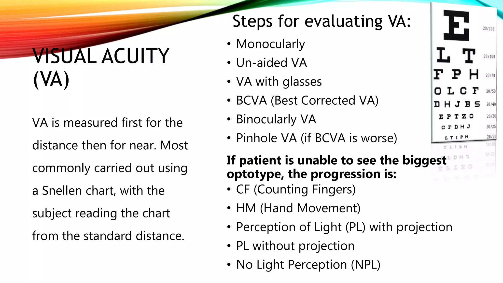
METHODS OF
EYE
EXAMINATION
By: Qurat-ul-ain
Ophthalmic Medical
Technologist/ MBA Health &
Hospital management
PREAMBLE
External Examination
Visual Acuity Assessment
Color-vision Testing
Pupillary Examination
Extraocular Motility and Alignment
Binocular Vision Testing
Amsler Grid
Slit-lamp Bi-microscopy
Indirect Ophthalmoscope
Tonometry
Gonioscopy
Central Corneal Thickness
EXTERNAL EXAMINATION:
• Look for any ptosis.
• Look for lagophthalmos
• Note any unusual growths or lesions that may require a biopsy.
• Measure proptosis or enophthalmos with an exophthalmometer.
• Perform a full cranial nerve exam for patients with diplopia or other
neurologic symptoms.
VISUAL ACUITY
(VA)
• Monocularly
• Un-aided VA
• VA with glasses
• BCVA (Best Corrected VA)
• Binocularly VA
• Pinhole VA (if BCVA is worse)
If patient is unable to see the biggest
optotype, the progression is:
• CF (Counting Fingers)
• HM (Hand Movement)
• Perception of Light (PL) with projection
• PL without projection
• No Light Perception (NPL)
VA is measured first for the
distance then for near. Most
commonly carried out using
a Snellen chart, with the
subject reading the chart
from the standard distance.
Steps for evaluating VA:
• Children who are too young to
use “Allen pictures” employ the
Central Steady Maintain (CSM)
approach.
Near VA:
• Near VA is a sensitive indicator
for the macular disease.
• Near chart held at comfortable
reading distance.
• Patient wear necessary distance
correction together with a
presbyopia correction.
TOOLS:
• Snellen chart
• Log MAR chart
• ETDRS chart
• Computer chart
• Jaeger chart
COLOR VISION TESTING:
• It is useful in the evaluation of optic nerve disease and
in determining the presence of a congenitally
anomalous color defect.
• Color vision depends on 3-populations of retinal cones
with specific peak sensitivity:
Blue (tritan): 414-424nm
Green (deuteron): 522-539
Red (protan): 549-570nm
• If any cone pigment may be deficient (e.g.;
protanomaly-red weakness) or entirely absent
(protanopia- red blindness)
• Acquired macular disease tends to produce blue-yellow
defects, and optic nerve lesions red-green defects.
TOOLS:
• Ishihara test
• City University test
• Hardy-Rand-Rittler test
• Farnsworth-Munsell
100-hue test
PUPILLARY EXAM:
• Look for anisocoria, if present check for the pupil size
• Check the reactivity of each pupil with a pen light or Finoff trans-illuminator.
• Use the swinging flash light test to look for a relative afferent pupillary
defect.
EXTRAOCULAR MOTILITY AND
ALIGNMENT:
• Test with both eyes open to test versions in six cardinal positions of gaze.
• Repeat monocularly to test ductions.
• Use cover/uncover tests to assess for heterotropias.
• Use the alternate cover test to assess the total amount of deviation.
BINOCULAR VISION TESTING:
• Evaluating the sensory status of children is an essential part of the
pediatric eye examination
• Testing stereo acuity is an excellent way of screening for various
conditions that may interfere with the development of binocular
depth perception.
There are two different types of stereo tests:
• Contour stereopsis tests like Titmus test
• Random dot tests like Randot or Lang tests.
AMSLER GRID:
• Evaluates 20 of the visual field
centered on fixation.
• Useful for screening and
monitoring macular disease. It
measures between 5 and 6 mm
in diameter.
• Patients with risk of
CNV(Choroidal
Neovascularization) should
provided with an Amsler grid
for regular use at home.
• Relative scotoma
• Absolute scotoma
• Metamorphopsia
• Micropsia
• Macropsia
SLIT-LAMP BI-MICROSCOPY:
Anterior Segment:
• Direct illumination: use to detect
gross abnormalities
• Scleral scatter: to detect stomal
haze, cellular or lipid infiltration.
• Retro-illumination: use after pupil
dilation to detect fine epithelial and
endothelial changes.
• Specular reflection: shows
abnormalities of the endothelium
Posterior Segment:
Different dioptric power lenses are
used to view fundus and optic nerve.
60D
90D
78D
INDIRECT OPHTHALMOSCOPE:
• Used to refer to the head mounted technique.
• It allows retinal visualization through a greater degree of media
opacity than slit lamp bi-microscopy.
• Lens of various powers are available:
20D
28D
40D
TONOMETRY:
Goldmann Tonometry:
• It states that for an ideal, dry, thin-walled sphere, the pressure inside the
sphere (P) equals the force necessary to flatten its surface (F) divided by the
area of flattening (A) i.e. P=F/A
• It is an accurate variable-force tonometer consisting of a double prism.
Other forms of Tonometry:
• Pneumo-tonometry
• Portable applanation tonometry
• Dynamic contour tonometry
• Electronic indentation/applanation tonometry
GONIOSCOPY:
• It is a method of evaluating the anterior chamber angle (ACA) and
can be used therapeutically for procedures such as laser
trabeculoplasty and goniotomy.
Other means of angle assessment:
• Anterior Segment Optical Coherence Tomography (AS-OCT)
• High frequency ultrasound biomicroscopy (UBM)
CENTRAL CORNEAL THICKNESS
(CCT):
• It can be measured using pachymetry or by Orbscan,
• The normal distribution is 540 ± 30 microns.
• Eyes with a thin cornea have a true IOP that is greater than the
measured IOP.
• Eyes with a thick cornea have a true IOP that is lower than the
measured IOP.
• Patients with NTG tends to have thin CCT measurement.
• It is a vital element when determining the risk of conversion to
glaucoma in individuals with raised IOP.
RESOURCES:
• https://www.aao.org/young-ophthalmologists/yo-info/article/how-to-
conduct-eight-point-ophthalmology-exam
• Harley’s Pediatric Ophthalmology (6th Edition) by Leonard B. Nelson and
Scott E. Olitsky
• Kanski’s Clinical Ophthalmology (9th Edition)
THANK YOU!

1. Methods of eye examination.pptx

  • 1.
    METHODS OF EYE EXAMINATION By: Qurat-ul-ain OphthalmicMedical Technologist/ MBA Health & Hospital management
  • 2.
    PREAMBLE External Examination Visual AcuityAssessment Color-vision Testing Pupillary Examination Extraocular Motility and Alignment Binocular Vision Testing Amsler Grid Slit-lamp Bi-microscopy Indirect Ophthalmoscope Tonometry Gonioscopy Central Corneal Thickness
  • 3.
    EXTERNAL EXAMINATION: • Lookfor any ptosis. • Look for lagophthalmos • Note any unusual growths or lesions that may require a biopsy. • Measure proptosis or enophthalmos with an exophthalmometer. • Perform a full cranial nerve exam for patients with diplopia or other neurologic symptoms.
  • 4.
    VISUAL ACUITY (VA) • Monocularly •Un-aided VA • VA with glasses • BCVA (Best Corrected VA) • Binocularly VA • Pinhole VA (if BCVA is worse) If patient is unable to see the biggest optotype, the progression is: • CF (Counting Fingers) • HM (Hand Movement) • Perception of Light (PL) with projection • PL without projection • No Light Perception (NPL) VA is measured first for the distance then for near. Most commonly carried out using a Snellen chart, with the subject reading the chart from the standard distance. Steps for evaluating VA:
  • 5.
    • Children whoare too young to use “Allen pictures” employ the Central Steady Maintain (CSM) approach. Near VA: • Near VA is a sensitive indicator for the macular disease. • Near chart held at comfortable reading distance. • Patient wear necessary distance correction together with a presbyopia correction. TOOLS: • Snellen chart • Log MAR chart • ETDRS chart • Computer chart • Jaeger chart
  • 6.
    COLOR VISION TESTING: •It is useful in the evaluation of optic nerve disease and in determining the presence of a congenitally anomalous color defect. • Color vision depends on 3-populations of retinal cones with specific peak sensitivity: Blue (tritan): 414-424nm Green (deuteron): 522-539 Red (protan): 549-570nm • If any cone pigment may be deficient (e.g.; protanomaly-red weakness) or entirely absent (protanopia- red blindness) • Acquired macular disease tends to produce blue-yellow defects, and optic nerve lesions red-green defects. TOOLS: • Ishihara test • City University test • Hardy-Rand-Rittler test • Farnsworth-Munsell 100-hue test
  • 7.
    PUPILLARY EXAM: • Lookfor anisocoria, if present check for the pupil size • Check the reactivity of each pupil with a pen light or Finoff trans-illuminator. • Use the swinging flash light test to look for a relative afferent pupillary defect.
  • 8.
    EXTRAOCULAR MOTILITY AND ALIGNMENT: •Test with both eyes open to test versions in six cardinal positions of gaze. • Repeat monocularly to test ductions. • Use cover/uncover tests to assess for heterotropias. • Use the alternate cover test to assess the total amount of deviation.
  • 9.
    BINOCULAR VISION TESTING: •Evaluating the sensory status of children is an essential part of the pediatric eye examination • Testing stereo acuity is an excellent way of screening for various conditions that may interfere with the development of binocular depth perception. There are two different types of stereo tests: • Contour stereopsis tests like Titmus test • Random dot tests like Randot or Lang tests.
  • 10.
    AMSLER GRID: • Evaluates20 of the visual field centered on fixation. • Useful for screening and monitoring macular disease. It measures between 5 and 6 mm in diameter. • Patients with risk of CNV(Choroidal Neovascularization) should provided with an Amsler grid for regular use at home. • Relative scotoma • Absolute scotoma • Metamorphopsia • Micropsia • Macropsia
  • 11.
    SLIT-LAMP BI-MICROSCOPY: Anterior Segment: •Direct illumination: use to detect gross abnormalities • Scleral scatter: to detect stomal haze, cellular or lipid infiltration. • Retro-illumination: use after pupil dilation to detect fine epithelial and endothelial changes. • Specular reflection: shows abnormalities of the endothelium Posterior Segment: Different dioptric power lenses are used to view fundus and optic nerve. 60D 90D 78D
  • 12.
    INDIRECT OPHTHALMOSCOPE: • Usedto refer to the head mounted technique. • It allows retinal visualization through a greater degree of media opacity than slit lamp bi-microscopy. • Lens of various powers are available: 20D 28D 40D
  • 13.
    TONOMETRY: Goldmann Tonometry: • Itstates that for an ideal, dry, thin-walled sphere, the pressure inside the sphere (P) equals the force necessary to flatten its surface (F) divided by the area of flattening (A) i.e. P=F/A • It is an accurate variable-force tonometer consisting of a double prism. Other forms of Tonometry: • Pneumo-tonometry • Portable applanation tonometry • Dynamic contour tonometry • Electronic indentation/applanation tonometry
  • 14.
    GONIOSCOPY: • It isa method of evaluating the anterior chamber angle (ACA) and can be used therapeutically for procedures such as laser trabeculoplasty and goniotomy. Other means of angle assessment: • Anterior Segment Optical Coherence Tomography (AS-OCT) • High frequency ultrasound biomicroscopy (UBM)
  • 15.
    CENTRAL CORNEAL THICKNESS (CCT): •It can be measured using pachymetry or by Orbscan, • The normal distribution is 540 ± 30 microns. • Eyes with a thin cornea have a true IOP that is greater than the measured IOP. • Eyes with a thick cornea have a true IOP that is lower than the measured IOP. • Patients with NTG tends to have thin CCT measurement. • It is a vital element when determining the risk of conversion to glaucoma in individuals with raised IOP.
  • 16.
    RESOURCES: • https://www.aao.org/young-ophthalmologists/yo-info/article/how-to- conduct-eight-point-ophthalmology-exam • Harley’sPediatric Ophthalmology (6th Edition) by Leonard B. Nelson and Scott E. Olitsky • Kanski’s Clinical Ophthalmology (9th Edition)
  • 17.