SlideShare a Scribd company logo
1 of 57
Respiratory
system
Dr. V. Chanukya
Pharm -D
Contents
3
i)
Respiratory
system
a)Anatomy of respiratory organs and
functions
a)Mechanism / physiology of respiration and
regulation of respiration
a)Transport of respiratory gases
a)Respiratory volumes and capacities, and
a)Definition of: Hypoxia, Asphyxia, Dybarism,
Oxygen therapy and resuscitation.
INTRODUCTION
• Respiration is the process by which oxygen is
taken in and carbon dioxide is given out.
• The first breath takes place only after birth.
• Fetal lungs are non-functional.
• So, during intrauterine life the exchange of gases
between fetal blood and mother’s blood occurs
through placenta.
• After the first breath, the respiratory process con-
tinues throughout the life.
• Permanent stoppage of respiration occurs only at
death.
• The respiratory system contributes to homeostasis by providing for the
exchange of gases—oxygen and carbon dioxide—between the atmospheric
air, blood, and tissue cells. It also helps adjust the pH of body fluids.
• Cells of the body continually use oxygen (O2) for the metabolic reactions
that release energy from nutrient molecules and produce ATP. At the same
time, these reactions release carbon dioxide (CO2).
• Because an excessive amount of CO2 produces acidity that can be toxic to
cells, excess CO2 must be eliminated quickly and efficiently.
• The cardiovascular and respiratory systems cooperate to supply O2 and
eliminate CO2. The respiratory system provides.
• Failure of either system disrupts homeostasis by causing rapid death of cells
from oxygen starvation and buildup of waste products.
• The respiratory system provides the route by which the supply of oxygen
present in the atmospheric air gains entry to the body and it provides the
route of excretion of carbon dioxide.
• The condition of the atmospheric air entering the body varies considerably
according to the external environment, e.g., it may be dry, cold and contain
dust particles or it may be moist and hot.
• In addition to functioning in gas exchange, the respiratory system also
participates in regulating blood pH, contains receptors for the sense of smell,
filters inspired air, produces sounds, and rids the body of some water and
heat in exhaled air.
• Exchange of gases between the blood and the lungs is called external
respiration and that between the blood and the cells internal respiration.
Respiratory system can be classified according to
either structure or function
Structurally, the respiratory
system consists of two parts:
The upper respiratory
system includes the nose,
nasal cavity, pharynx, and
associated structures.
The lower respiratory
system includes the larynx,
trachea, bronchi, and lungs.
Functionally, the respiratory
system also consists of two parts:
The conducting zone
consists of a series of
interconnecting cavities and
tubes both outside and within
the lungs.
These include the nose, nasal
cavity, pharynx, larynx,
trachea, bronchi, bronchioles,
and terminal bronchioles;
their function is to filter,
warm, and moisten air and
conduct it into the lungs.
The respiratory zone
consists of tubes and tissues
within the lungs where gas
exchange occurs.
These include the respiratory
bronchioles, alveolar ducts,
alveolar sacs, and alveoli and
are the main sites of gas
exchange between air and
blood.
Anatomy of
respiratory organs
and functions
• The organs of respiratory system are
• Nose
• Pharynx (Throat)
• Larynx (Voice Box)
• Trachea (Windpipe)
• Two bronchi (one bronchus to each
lung)
• Bronchioles and smaller air passages
• T wo Lungs and their coverings
• The pleura
• Muscles of respiration (The intercostal
muscles and the diaphragm)
Nose and Nasal cavity - Anatomy
Position and structure
• The nasal cavity is the first of the respiratory organs
and consists of a large irregular cavity divided into
two equal passages by a septum.
• The posterior bony part of the septum is formed by
the perpendicular plate of the ethmoid bone and the
vomer.
• Anteriorly it consists of hyaline cartilage.
• The roof is formed by the cribriform plate of the
ethmoid bone, and the sphenoid bone, frontal bone
and nasal bones.
• The floor is formed by the roof of the mouth and
consists of the hard palate in front and the soft
palate behind.
• The hard palate is composed of the maxilla and
palatine bones and the soft palate consists of
involuntary muscle.
• The medial wall is formed by the septum.
• The lateral walls are formed by
the maxilla, the ethmoid bone
and the inferior conchae.
• The posterior wall is formed by
the posterior wall of the
pharynx.
Lining of the nose
• The nose is lined with very
vascular ciliated columnar
epithelium (ciliated mucous
membrane) which contains
mucus-secreting goblet cells .
• At the anterior nares this blends
with the skin and posteriorly it
extends into the nasal part of
the pharynx.
Openings into the nasal cavity
• The anterior nares, or nostrils, are the openings from
the exterior into the nasal cavity.
• Hairs are present in this area.
• The posterior nares are the openings from the nasal
cavity into the pharynx.
• The paranasal sinuses are cavities in the bones of
the face and the cranium which contain air.
• There are tiny openings between the paranasal
sinuses and the nasal cavity.
• They are lined with mucous membrane, continuous
with that of the nasal cavity.
• The main sinuses are:
• Maxillary sinuses in the lateral walls
• Frontal and sphenoidal sinuses in the roof
• Ethmoidal sinuses in the upper part of the
lateral walls.
• The sinuses function in speech and also serve to
lighten the skull.
• The nasolacrimal ducts extend from the lateral walls
of the nose to the conjunctival sacs of the eye. They
drain tears from the eyes.
Physiology-Respiratory
function of the nose
• The nose is the first of the respiratory
passages through which the inspired air
passes.
• The function of the nose is to begin the
process by which the air is warmed,
moistened and 'filtered'.
• The projecting conchae increase the
surface area and cause turbulence,
spreading inspired air over the whole
nasal surface.
• The large surface area maximizes
warming, humidification and filtering.
• Warming. This is due to the immense vascularity of the
mucosa.
• Filtering and cleaning of air. This occurs as hairs at the
anterior nares trap larger particles. Smaller particles such
as dust and microbes settle and adhere to the mucus.
Mucus protects the underlying epithelium from irritation
and prevents drying. Synchronous beating of the cilia
wafts the mucus towards the throat where it is swallowed
or expectorated.
• Humidification. This occurs as air travels over the moist
mucosa and becomes saturated with water vapour.
Irritation of the nasal mucosa results in sneezing, a reflex
action that forcibly expels an irritant.
Physiology- Olfactory
function of the nose
• The nose is the organ of the sense
of smell. There are nerve endings
that detect smell, located in the
roof of the nose in the area of the
cribriform plate of the ethmoid
bones and the superior conchae.
These nerve endings are
stimulated by chemical substances
given off by odorous materials.
• The resultant nerve impulses are
conveyed by the olfactory nerves
to the brain where the sensation of
smell is perceived .
PHARYNX
Position The pharynx is a tube 12 to 14 cm long that extends
from the base of the skull to the level of the 6th cervical
vertebra.
It lies behind the nose, mouth and larynx and is wider at
its upper end.
Structures
associated
with
the
pharynx
Superiorly — the inferior surface of the base of the skull
Inferiorly — it is continuous with the oesophagus
Anteriorly — the wall is incomplete because of the
openings into the nose, mouth and larynx
Posteriorly — areolar tissue, involuntary muscle and the
bodies of the first six cervical vertebrae
• The pharynx is divided into three parts:
nasopharynx, oropharynx and
laryngopharynx.
• The nasopharynx.
• The nasal part of the pharynx lies behind
the nose above the level of the soft palate
• On its lateral walls are the two openings of
the auditory tubes, one leading to each
middle ear.
• On the posterior wall there are the
pharyngeal tonsils (adenoids), consisting of
lymphoid tissue.
• They are most prominent in children up to
approximately 7 years of age. Thereafter
they gradually atrophy.
• The oropharynx
• The oral part of the pharynx lies behind the mouth, extending from
below the level of the soft palate to the level of the upper part of the
body of the 3rd cervical vertebra.
• The lateral walls of the pharynx blend with the soft palate to form
two folds on each side.
• Between each pair of folds there is a collection of lymphoid tissue
called the palatine tonsil.
• During swallowing, the nasal and oral parts are separated by the soft
palate and the uvula.
• The laryngopharynx
• The laryngeal part of the pharynx extends from the oropharynx
above and continues as the oesophagus below, i.e. from the level of
the 3rd to the 6th cervical vertebrae.
Structure
• The pharynx is composed of three layers of tissue:
• 1. Mucous membrane lining. The mucosa varies
slightly in the different parts. In the nasopharynx it is
continuous with the lining of the nose and consists of
ciliated columnar epithelium; in the oropharynx and
laryngopharynx it is formed by tougher stratified
squamous epithelium which is continuous with the
lining of the mouth and oesophagus.
• 2. Fibrous tissue. This forms the intermediate layer. It
is thicker in the nasopharynx, where there is little
muscle, and becomes thinner towards the lower end,
where the muscle layer is thicker.
• 3. Muscle tissue. This consists of several involuntary
constrictor muscles that play an important part in the
mechanism of swallowing (deglutition) which, in the
pharynx, is not under voluntary control. The upper
end of the esophagus is closed by the lower
constrictor muscle, except during swallowing.
Blood and nerve
supply
• Blood is supplied to the pharynx by
several branches of the facial artery.
The venous return is into the facial and
internal jugular veins.
• The nerve supply is from the pharyngeal
plexus, formed by parasympathetic and
sympathetic nerves. Parasympathetic
supply is by the vagus and
glossopharyngeal nerves.
• Sympathetic supply is by nerves from
the superior cervical ganglia
Functions
Passageway
for air and
food.
The pharynx is an organ involved in both the respiratory and the
digestive systems: air passes through the nasal and oral parts, and food
through the oral and laryngeal parts.
Warming
and
humidifying.
By the same methods as in the nose, the air is further warmed and
moistened as it passes through the pharynx.
Taste. There are olfactory nerve endings of the sense of taste in the
epithelium of the oral and pharyngeal parts.
Hearing. The auditory tube, extending from the nasal part to each middle ear,
allows air to enter the middle ear. Satisfactory hearing depends on the
presence of air at atmospheric pressure on each side of the tympanic
membrane (ear drum)
Protection. The lymphatic tissue of the pharyngeal and laryngeal tonsils produces
antibodies in response to antigens, e.g. microbes The tonsils are larger
in children and tend to atrophy in adults.
Speech. The pharynx functions in speech; by acting as a resonating chamber for
the sound ascending from the larynx, it helps (together with the
sinuses) to give the voice its individual characteristics.
LARYNX
Position
• The larynx or 'voice box' extends from
the root of the tongue and the hyoid
bone to the trachea.
• It lies in front of the laryngopharynx at
the level of the 3rd, 4th, 5th and 6th
cervical vertebrae.
• Until puberty there is little difference in
the size of the larynx between the sexes.
Thereafter it grows larger in the male,
which explains the prominence of the
'Adam's apple' and the generally deeper
voice.
Structures associated
with the larynx
Superiorly
•the hyoid bone and the root of the
tongue
Inferiorly •it is continuous with the trachea
Anteriorly
•the muscles attached to the hyoid
bone and the muscles of the neck
Posteriorly
• the laryngopharynx and 3rd to 6th
cervical vertebrae
Laterally •the lobes of the thyroid gland
Structure
Cartilages
• The larynx is composed of several
irregularly shaped cartilages attached to
each other by ligaments and
membranes.
The main cartilages are:
1 thyroid cartilage
1 cricoid cartilage
2 arytenoid cartilages
1 epiglottis
hyaline cartilage
elastic fibrocartilage.
The thyroid cartilage
This is the most prominent and consists of two flat pieces
of hyaline cartilage, or laminae, fused anteriorly, forming
the laryngeal prominence (Adam’s apple).
• Immediately above the laryngeal prominence the
laminae are separated, forming a V-shaped notch
known as the thyroid notch.
• The thyroid cartilage is incomplete posteriorly and the
posterior border of each lamina is extended to form
two processes called the superior and inferior cornu.
• The upper part of the thyroid cartilage is lined with
stratified squamous epithelium like the larynx, and the
lower part with ciliated columnar epithelium like the
trachea.
• There are many muscles attached to its outer surface.
The cricoid cartilage
• This lies below the thyroid cartilage and is also
composed of hyaline cartilage.
• It is shaped like a signet ring, completely
encircling the larynx with the narrow part
anteriorly and the broad part posteriorly.
• The broad posterior part articulates with the
arytenoid cartilages above and with the
inferior cornu of the thyroid cartilage below.
• It is lined with ciliated columnar epithelium
and there are muscles and ligaments attached
to its outer surface.
• The lower border of the cricoid cartilage marks
the end of the upper respiratory tract.
Arytenoid cartilages
• These are two roughly pyramid-shaped
hyaline cartilages situated on top of the
broad part of the cricoid cartilage
forming part of the posterior wall of the
larynx.
• They give attachment to the vocal cords
and to muscles and are lined with ciliated
columnar epithelium
The epiglottis
• This is a leaf-shaped fibroelastic cartilage
attached to the inner surface of the anterior
wall of the thyroid cartilage immediately
below the thyroid notch.
• It rises obliquely upwards behind the
tongue and the body of the hyoid bone.
• It is covered with stratified squamous
epithelium.
• If the larynx is assumed as a box, then the
epiglottis acts as the lid; it closes off the
larynx during swallowing, protecting the
lungs from accidental inhalation of foreign
objects.
Ligaments and membranes
• There are several ligaments that
attach the cartilages to each other and
to the hyoid bone
Blood and nerve supply
• Blood is supplied to the larynx by the
superior and inferior laryngeal arteries
and drained by the thyroid veins,
which join the internal jugular vein.
• The parasympathetic nerve supply is
from the superior
• Laryngeal and recurrent laryngeal
nerves, which are branches of the
vagus nerves, and the sympathetic
nerves are from the superior cervical
ganglia, one on each side.
• These provide the motor nerve supply
to the muscles of the larynx and
sensory fibers to the lining membrane
Interior of the larynx
• The vocal cords are two pale folds of mucous membrane with
cord-like free edges which extend from the inner wall of the
thyroid prominence anteriorly to the arytenoid cartilages
posteriorly.
• When the muscles controlling the vocal cords are relaxed, the
vocal cords open and the passageway for air coming up through
the larynx is clear; the vocal cords are said to be abducted. The
pitch of the sound produced by vibrating the vocal cords in this
position is low.
• When the muscles controlling the vocal cords contract, the vocal
cords are stretched out tightly across the larynx —they are said
to be adducted.
• When the vocal cords are stretched to this extent and are
vibrated by air passing through from the lungs, the sound
produced is high pitched.
• The pitch of the voice is therefore determined by the tension
applied to the vocal cords by the appropriate sets of muscles.
When not in use, the vocal cords are adducted.
Functions
Production of sound
• Sound has the properties of pitch, volume and resonance.
• Pitch of the voice depends upon the length and tightness of the cords. At puberty,
the male vocal cords begin to grow longer, hence the lower pitch of the adult
male voice.
• Volume of the voice depends upon the force with which the cords vibrate. The
greater the force of expired air the more the cords vibrate and the louder the
sound emitted.
• Resonance, or tone, is dependent upon the shape of the mouth, the position of
the tongue and the lips, the facial muscles and the air in the paranasal sinuses.
Speech.
• This occurs during expiration when the sounds produced by the vocal cords are manipulated by the tongue,
cheeks and lips.
Protection of the lower respiratory tract
• During swallowing (deglutition) the larynx moves upwards, occluding the opening into it from the pharynx and
the hinged epiglottis closes over the larynx. This ensures that food passes into the oesophagus and not into the
lower respiratory passages
Passageway for air.
• This is between the pharynx and trachea.
Humidifying, filtering and warming.
• These continue as inspired air travels through the larynx.
TRACHEA
Position
• The trachea or windpipe is a
continuation of the larynx and extends
downwards to about the level of the 5th
thoracic vertebra where it divides
(bifurcates) at the carina into the right
and left bronchi, one bronchus going to
each lung.
• It is approximately 10 to 11 cm long and
lies mainly in the median plane in front
of the oesophagus.
Structures associated
with the trachea
• Superiorly — the larynx
• Inferiorly — the right and left bronchi
• Anteriorly — upper part: the isthmus of
the thyroid gland
lower part: the arch of the
aorta and the sternum
• Posteriorly — the oesophagus separates
the trachea from the vertebral column
• Laterally — the lungs and the lobes of the
thyroid gland.
Structure
• The trachea is composed of from 16 to 20
incomplete (C-shaped) rings of hyaline
cartilages lying one above the other.
• The cartilages are incomplete posteriorly.
• Connective tissue and involuntary muscle
join the cartilages and form the posterior
wall where they are incomplete.
• The soft tissue posterior wall is in contact
with the oesophagus
There are three layers of tissue which 'clothe' the
cartilages of the trachea.
The outer
layer.
This consists of fibrous and elastic tissue and encloses the
cartilages.
The
middle
layer.
This consists of cartilages and bands of smooth muscle that
wind round the trachea in a helical arrangement. There is
some areolar tissue, containing blood and lymph vessels and
autonomic nerves.
The inner
lining.
This consists of ciliated columnar epithelium, containing
mucus-secreting goblet cells
Blood and nerve supply,
lymph drainage
The arterial blood supply
• This is mainly by the inferior thyroid and bronchial arteries and
the venous return is by the inferior thyroid veins into the
brachiocephalic veins.
The nerve supply
• This is by parasympathetic and sympathetic fibers.
• Parasympathetic supply is by the recurrent laryngeal nerves and
other branches of the vagi.
• Sympathetic supply is by nerves from the sympathetic ganglia.
Lymph
• Lymph from the respiratory passages passes through lymph nodes
situated round the trachea and in the carina, the area where it
divides into two bronchi
Functions
• The arrangement of cartilage and elastic tissue prevents kinking
and obstruction of the airway as the head and neck move.
• The absence of cartilage posteriorly allows the trachea to dilate
and constrict in response to nerve stimulation, and for
indentation as the esophagus distends during swallowing.
• The cartilages prevent collapse of the tube when the internal
pressure is less than intrathoracic pressure, i.e., at the end of
forced expiration.
Support and patency.
• This is the synchronous and regular beating of the cilia of the
mucous membrane lining that wafts mucus with adherent
particles upwards towards the larynx where it is swallowed or
expectorated.
Mucociliary escalator.
• Nerve endings in the larynx, trachea and bronchi are sensitive to irritation that
generates nerve impulses which are conducted by the vagus nerves to the
respiratory center in the brain stem.
• The reflex motor response is deep inspiration followed by closure of the glottis. The
abdominal and respiratory muscles then contract and suddenly the air is released
under pressure expelling mucus and/or foreign material from the mouth.
Cough reflex
• These continue as in the nose, although air is normally saturated and at body
temperature when it reaches the trachea.
Warming, humidifying and filtering of air
Bronchi
• The two primary bronchi are formed when the trachea divides, i.e.,
about the level of the 5th thoracic vertebra
• The right bronchus.
• This is wider, shorter and more vertical than the left bronchus and
is therefore the more likely of the two to become obstructed by an
inhaled foreign body.
• It is approximately 2.5 cm long. After entering the right lung at the
hilum, it divides into three branches, one to each lobe. Each
branch then subdivides into numerous smaller branches.
• The left bronchus.
• This is about 5 cm long and is narrower than the right. After
entering the lung at the hilum it divides into two branches, one to
each lobe. Each branch then subdivides into progressively smaller
tubes within the lung substance.
Structure
• The bronchi are composed of the same tissues as the
trachea.
• They are lined with ciliated columnar epithelium.
• The bronchi progressively subdivide into bronchioles,
terminal bronchioles, respiratory bronchioles, alveolar
ducts and finally, alveoli.
• Towards the distal end of the bronchi the cartilages
become irregular in shape and are absent at bronchiolar
level.
• In the absence of cartilage, the smooth muscle in the
walls of the bronchioles becomes thicker and is responsive
to autonomic nerve stimulation and irritation.
• Ciliated columnar mucous membrane changes gradually to
non-ciliated cuboidal shaped cells in the distal bronchioles.
Blood and nerve supply,
lymph drainage
• The arterial blood supply.
• The supply to the walls of
the bronchi and smaller air
passages is through
branches of the right and
left bronchial arteries and
the venous return is mainly
through the bronchial
veins.
• On the right side they
empty into the azygos vein
and on the left into the
superior intercostal vein
The nerve supply
• This is by parasympathetic and
sympathetic nerves.
• The vagus nerves
(parasympathetic) stimulate
contraction of smooth muscle
in the bronchial tree, causing
bronchoconstriction, and
sympathetic stimulation
causes bronchodilatation
The lymphatic vessels and
lymph nodes
• Lymph is drained from the
walls of the air passages in a
network of lymph vessels.
• It passes through lymph nodes
situated around the trachea
and bronchial tree then into
the thoracic duct on the left
side and right lymphatic duct
on the other.
Functions of air passages not involved in gaseous exchange
Control of air entry
• The diameter of the respiratory passages may be altered by contraction or relaxation of the
involuntary muscles in their walls, thus regulating the volume of air entering the lungs.
• These changes are controlled by the autonomic nerve supply: parasympathetic stimulation causes
constriction and sympathetic stimulation causes dilatation
warming and humidifying
support and patency
removal of particulate matter
cough reflex where the other functions continue as in the upper airways.
Respiratory bronchioles and alveoli
Structure
• Lobules are the blind ends of the respiratory tract distal to
the terminal bronchioles, consisting of: respiratory
bronchioles, alveolar ducts and alveoli (tiny air sacs) .
• It is in these structures that the process of gas exchange
occurs. The walls gradually become thinner until muscle and
connective tissue fade out leaving a single layer of simple
squamous epithelial cells in the alveolar ducts and alveoli.
• These distal respiratory passages are supported by a loose
network of elastic connective tissue in which macrophages,
fibroblasts, nerves and blood and lymph vessels are
embedded.
• The alveoli are surrounded by a network of capillaries. The
exchange of gases during respiration takes place across two
membranes, the alveolar and capillary membranes.
• Inter spared between the squamous cells are other cells that secrete surfactant, a
phospholipid fluid which prevents the alveoli from drying out.
• In addition, surfactant reduces surface tension and prevents alveolar walls collapsing
during expiration.
• Secretion of surfactant into the distal air passages and alveoli begins about the 35th
week of fetal life.
• Its presence in newborn babies facilitates expansion of the lungs and the establishment
of respiration.
• It may not be present in sufficient amounts in the immature lungs of premature babies,
causing difficulty in establishing respiration.
Functions of respiratory bronchioles and alveoli
• Aid in external respiration
• Defense against microbes.
• At this level, ciliated epithelium, goblet cells and mucus are no longer present
• Defense relies on protective cells present within the lung tissue.
• These include lymphocytes and plasma cells, which produce antibodies in the
presence of antigens, and macrophages and polymorphonuclear lymphocytes, which
are phagocytic.
• These cells are most active in the distal air passages where ciliated epithelium has
been replaced by flattened cells.
• Warming and humidifying
• These continue as in the upper airways.
• Inhalation of dry or inadequately humidified air over a period causes irritation of the
mucosa and facilitates the establishment of pathogenic microbes.
Pleura and pleural cavity
• The pleura consists of a closed sac of serous membrane (one for each lung) which
contains a small amount of serous fluid.
• The lung is invaginated into this sac so that it forms two layers: one adheres to the
lung and the other to the wall of the thoracic cavity.
• The visceral pleura.
• This is adherent to the lung, covering each lobe and passing into the fissures
which separate them.
• The parietal pleura.
• This is adherent to the inside of the chest wall and the thoracic surface of
the diaphragm.
• It remains detached from the adjacent structures in the mediastinum and is
continuous with the visceral pleura round the edges of the hilum.
• The pleural cavity.
• This is only a potential space. In health, the two layers of pleura are
separated by only a thin film of serous fluid which allows them to glide over
each other, preventing friction between them during breathing. The serous
fluid is secreted by the epithelial cells of the membrane
• The two layers of pleura, with serous fluid between them, behave in the same way as
two pieces of glass separated by a thin film of water.
• They glide over each other easily but can be pulled apart only with difficulty, because of
the surface tension between the membranes and the fluid.
• If either layer of pleura is punctured, the underlying lung collapses due to its inherent
property of elastic recoil.
• Interior of the lungs
• The lungs are composed of the bronchi and smaller air passages, alveoli, connective
tissue, blood vessels, lymph vessels and nerves. The left lung is divided into two
lobes and the right, into three.
Lungs
• Position and associated structures
• There are two lungs, one lying on each side of the midline in the thoracic cavity.
• They are cone-shaped and are described as having an apex, a base, costal surface
and medial surface.
• The apex
• This is rounded and rises into the root of the neck, about 25 mm (1 inch)
above the level of the middle third of the clavicle. The structures associated
with it are the first rib and the blood vessels and nerves in the root of the
neck.
• The base
• This is concave and semilunar in shape and is closely associated with the
thoracic surface of the diaphragm.
• The costal surface
• This surface is convex and is closely associated with the costal cartilages, the
ribs and the intercostal muscles.
• The medial surface
• This surface is concave and has a roughly triangular-shaped area, called the
hilum, at the level of the 5th, 6th and 7th thoracic vertebrae.
• Structures which form the root of the lung enter and leave at the hilum. These
include the primary bronchus, the pulmonary artery supplying the lung and
the two pulmonary veins draining it, the bronchial artery and veins, and the
lymphatic and nerve supply .
• The area between the lungs is the mediastinum. It is occupied by the heart,
great vessels, trachea, right and left bronchi, esophagus, lymph nodes, lymph
vessels and nerves.
Organization of the lungs
• The right lung is divided into
three distinct lobes: superior,
middle and inferior.
• The left lung is smaller as the
heart is situated left of the
midline.
• It is divided into only two lobes:
superior and inferior.
Pulmonary blood supply
• The pulmonary artery divides into two, one branch conveying
deoxygenated blood to each lung. Within the lungs each pulmonary
artery divides into many branches which eventually end in a dense
capillary network around the walls of the alveoli.
• The walls of the alveoli and those of the capillaries each consist of only
one layer of flattened epithelial cells. The exchange of gases between air
in the alveoli and blood in the capillaries takes place across these two
very fine membranes.
• The pulmonary capillaries join up, eventually becoming two pulmonary
veins in each lung.
• They leave the lungs at the hilum and convey oxygenated blood to the
left atrium of the heart.
• The innumerable blood capillaries and blood vessels in the lungs are
supported by connective tissue.
• Each bronchopulmonary segment of the lungs has many small
compartments called lobules; each lobule is wrapped in elastic
connective tissue and contains a lymphatic vessel, an arteriole, a
venule, and a branch from a terminal bronchiole.
• Terminal bronchioles subdivide into microscopic branches called
respiratory bronchioles.
• They also have alveoli (described shortly) budding from their walls.
Alveoli participate in gas exchange, and thus respiratory bronchioles
begin the respiratory zone of the respiratory system.
Definition
• Hypoxia : Hypoxia is a condition in which tissues of the body do not receive sufficient oxygen
(O2) supply.
• Asphyxia: It is a condition arising when the body is deprived of oxygen, causing
unconsciousness or death; suffocation.
• Hypoxia is the term used to indicate a deficiency of oxygen. A related term that is often used in
relation to perinatal brain injury is anoxia, meaning without oxygen. Asphyxia refers to the
physiological results of hypoxia or anoxia.
• Dysbarism: it is defined as any adverse medical condition that results from
changes in ambient pressure. These changes in pressure must occur either at a rate
or duration exceeding the capacity of the body to adapt safely.
• Although the most common cause of dysbarism is underwater diving, dysbaric injury
can occur following exposure to any environments with extreme pressure changes.
Other examples include high altitude, aircraft cabin decompression, explosions or
blasts, outer space, caissons, and tunnel-boring operations.
Oxygen therapy
• Oxygen therapy is a treatment that delivers oxygen gas for you to breathe. It
is also called supplemental oxygen.
• You can receive oxygen therapy from tubes resting in your nose, a face mask,
or a tube placed in your trachea, or windpipe. This treatment increases the
amount of oxygen your lungs receive and deliver to your blood.
• Oxygen therapy may be prescribed for you when you have a condition that
causes your blood oxygen levels to be too low. Low blood oxygen may make
you feel short of breath, tired, or confused, and can damage your body.
• Oxygen therapy can be given for a short or long period of time in the hospital,
another medical setting, or at home.
• There are different types of devices that can give you oxygen. Some use tanks of liquid or gas oxygen.
• Others use an oxygen concentrator, which pulls oxygen out of the air. You will get the oxygen through a
nose tube (cannula), a mask, or a tent. The extra oxygen is breathed in along with normal air.
• You may need oxygen therapy if you have a condition that causes low blood oxygen, such as
• COPD (chronic obstructive pulmonary disease)
• Pneumonia
• COVID-19
• A severe asthma attack
• Late-stage heart failure
• Cystic fibrosis
• Sleep apnea
Cardiopulmonary resuscitation (CPR)
• Cardiopulmonary resuscitation (CPR) is a procedure to support and maintain breathing and circulation for an
infant, child, or adolescent who has stopped breathing (respiratory arrest) and/or whose heart has stopped
(cardiac arrest).
• Purpose
• CPR is performed to restore and maintain breathing and circulation and to provide oxygen and blood flow to
the heart, brain, and other vital organs.
• CPR can be performed by trained laypeople or healthcare professionals on infants, children, adolescents, and
adults.
• CPR should be performed if an infant, child, or adolescent is unconscious and not breathing.
• Respiratory and cardiac arrest can be caused by allergic reactions, an ineffective heartbeat, asphyxiation,
breathing passages that are blocked, choking , drowning, drug reactions or overdoses, electric shock, exposure
to cold, severe shock, or trauma.
• In newborns, the most common cause of cardiopulmonary arrest is respiratory failure caused by sudden infant
death syndrome (SIDS), airway obstruction (usually from inhalation of a foreign body), sepsis, neurologic
disease, or drowning.
• Cardiac arrest in children over one year of age is most commonly caused by shock and/or respiratory failure
resulting from an accident or injury.

More Related Content

What's hot

Respiratory System Anatomy & Physiology
Respiratory System Anatomy & Physiology Respiratory System Anatomy & Physiology
Respiratory System Anatomy & Physiology yousaf shah
 
Structure and anatomy of respiratory system
Structure and anatomy of respiratory systemStructure and anatomy of respiratory system
Structure and anatomy of respiratory systemPrudhvi Raj Moganti
 
Respiratory System Powerpoint
Respiratory System PowerpointRespiratory System Powerpoint
Respiratory System Powerpointguestbd819
 
Respiratory System - Anatomy and Physiology
Respiratory System - Anatomy and PhysiologyRespiratory System - Anatomy and Physiology
Respiratory System - Anatomy and PhysiologyNhelia Santos Perez
 
Respiratory system
Respiratory systemRespiratory system
Respiratory systemcbaker745
 
Anatomy & physiology of respiratory system
Anatomy & physiology of respiratory systemAnatomy & physiology of respiratory system
Anatomy & physiology of respiratory systemZeeshan Ahmad Awan
 
Respiratory system, physiology of respiratory system and neural control
Respiratory system, physiology of respiratory system and neural control Respiratory system, physiology of respiratory system and neural control
Respiratory system, physiology of respiratory system and neural control sunil JMI
 
Anatomy and physiology of ear by maghan das
Anatomy and physiology of ear by maghan dasAnatomy and physiology of ear by maghan das
Anatomy and physiology of ear by maghan dasMaghan Das
 
Respiratory System - Human Anatomy and Physiology bPharm
Respiratory System - Human Anatomy and Physiology bPharmRespiratory System - Human Anatomy and Physiology bPharm
Respiratory System - Human Anatomy and Physiology bPharmPankaj Saha
 
Sense organs ear
Sense organs earSense organs ear
Sense organs earneelu2014
 
Anatomy and physiology of respiratory system
Anatomy and physiology of respiratory systemAnatomy and physiology of respiratory system
Anatomy and physiology of respiratory systemPallavi Lokhande
 
1 respiratory system
1 respiratory system1 respiratory system
1 respiratory systemRayan Mzoury
 

What's hot (20)

Respiratory System Anatomy & Physiology
Respiratory System Anatomy & Physiology Respiratory System Anatomy & Physiology
Respiratory System Anatomy & Physiology
 
ANATOMY OF RESPIRATORY SYSTEM
ANATOMY OF RESPIRATORY SYSTEMANATOMY OF RESPIRATORY SYSTEM
ANATOMY OF RESPIRATORY SYSTEM
 
Structure and anatomy of respiratory system
Structure and anatomy of respiratory systemStructure and anatomy of respiratory system
Structure and anatomy of respiratory system
 
Respiratory System Powerpoint
Respiratory System PowerpointRespiratory System Powerpoint
Respiratory System Powerpoint
 
Respiratory System - Anatomy and Physiology
Respiratory System - Anatomy and PhysiologyRespiratory System - Anatomy and Physiology
Respiratory System - Anatomy and Physiology
 
Respiratory system
Respiratory systemRespiratory system
Respiratory system
 
The Respiratory system
The Respiratory system The Respiratory system
The Respiratory system
 
Respiration Physiology-Respiratory System
Respiration Physiology-Respiratory SystemRespiration Physiology-Respiratory System
Respiration Physiology-Respiratory System
 
Anatomy & physiology of respiratory system
Anatomy & physiology of respiratory systemAnatomy & physiology of respiratory system
Anatomy & physiology of respiratory system
 
Respiratory system, physiology of respiratory system and neural control
Respiratory system, physiology of respiratory system and neural control Respiratory system, physiology of respiratory system and neural control
Respiratory system, physiology of respiratory system and neural control
 
Respiratory system
Respiratory systemRespiratory system
Respiratory system
 
Anatomy and physiology of ear by maghan das
Anatomy and physiology of ear by maghan dasAnatomy and physiology of ear by maghan das
Anatomy and physiology of ear by maghan das
 
Respiratory System - Human Anatomy and Physiology bPharm
Respiratory System - Human Anatomy and Physiology bPharmRespiratory System - Human Anatomy and Physiology bPharm
Respiratory System - Human Anatomy and Physiology bPharm
 
Respiratory system physio
Respiratory system physioRespiratory system physio
Respiratory system physio
 
Sense organs ear
Sense organs earSense organs ear
Sense organs ear
 
The respiratory system
The respiratory systemThe respiratory system
The respiratory system
 
Respiratory system
Respiratory systemRespiratory system
Respiratory system
 
Resperattory system
Resperattory systemResperattory system
Resperattory system
 
Anatomy and physiology of respiratory system
Anatomy and physiology of respiratory systemAnatomy and physiology of respiratory system
Anatomy and physiology of respiratory system
 
1 respiratory system
1 respiratory system1 respiratory system
1 respiratory system
 

Similar to Respiratory system

respiratory Allied.HS.ppt
respiratory Allied.HS.pptrespiratory Allied.HS.ppt
respiratory Allied.HS.pptAnees Hasnain
 
RESPIRATORY SYSTEM.pptxv gnm first year students
RESPIRATORY SYSTEM.pptxv gnm first year studentsRESPIRATORY SYSTEM.pptxv gnm first year students
RESPIRATORY SYSTEM.pptxv gnm first year studentsroy456393
 
THE RESPIRATORY SYSTEM-New.pptx
THE RESPIRATORY SYSTEM-New.pptxTHE RESPIRATORY SYSTEM-New.pptx
THE RESPIRATORY SYSTEM-New.pptxgeorginansiah247
 
UNIT 6-respiratory system for Gnm and b.sc. nursing
UNIT 6-respiratory system for Gnm and b.sc. nursingUNIT 6-respiratory system for Gnm and b.sc. nursing
UNIT 6-respiratory system for Gnm and b.sc. nursingDNYANESHWARBIRADAR1
 
Respiratory system B pharmacy 2nd semester
Respiratory system B pharmacy 2nd semester Respiratory system B pharmacy 2nd semester
Respiratory system B pharmacy 2nd semester ZaibaFathima8
 
The respiratory system
The respiratory systemThe respiratory system
The respiratory systemNikita Sharma
 
Respiratory .System. . pdf bsc nursing b
Respiratory .System.  . pdf bsc nursing bRespiratory .System.  . pdf bsc nursing b
Respiratory .System. . pdf bsc nursing brejimariya941
 
Anatomy & physio of Respiratory System.pptx
Anatomy & physio of Respiratory System.pptxAnatomy & physio of Respiratory System.pptx
Anatomy & physio of Respiratory System.pptxMuneebKhanYousafzai3
 
anatomy of rspiratory system, 2022.ppt
anatomy of rspiratory system, 2022.pptanatomy of rspiratory system, 2022.ppt
anatomy of rspiratory system, 2022.pptmomda
 
Respiratory System
Respiratory SystemRespiratory System
Respiratory SystemMayur Gaikar
 
Johny's A&P structure and function of respiratory system
Johny's A&P structure and function of respiratory systemJohny's A&P structure and function of respiratory system
Johny's A&P structure and function of respiratory systemJohny Kutty Joseph
 
The Respiratory System in the Head and Neck
The Respiratory System in the Head and NeckThe Respiratory System in the Head and Neck
The Respiratory System in the Head and NeckHadi Munib
 
Anatomy of respiratory system
Anatomy of respiratory systemAnatomy of respiratory system
Anatomy of respiratory systemSELINA SRAVANTHI
 
Respiratory system.pptx
Respiratory system.pptxRespiratory system.pptx
Respiratory system.pptxahsanashfaq19
 
respiratory system.pptx
respiratory system.pptxrespiratory system.pptx
respiratory system.pptxAsiyaAsi2
 

Similar to Respiratory system (20)

respiratory Allied.HS.ppt
respiratory Allied.HS.pptrespiratory Allied.HS.ppt
respiratory Allied.HS.ppt
 
RESPIRATORY SYSTEM.pptxv gnm first year students
RESPIRATORY SYSTEM.pptxv gnm first year studentsRESPIRATORY SYSTEM.pptxv gnm first year students
RESPIRATORY SYSTEM.pptxv gnm first year students
 
08 respiratory system
08 respiratory system08 respiratory system
08 respiratory system
 
THE RESPIRATORY SYSTEM-New.pptx
THE RESPIRATORY SYSTEM-New.pptxTHE RESPIRATORY SYSTEM-New.pptx
THE RESPIRATORY SYSTEM-New.pptx
 
UNIT 6-respiratory system for Gnm and b.sc. nursing
UNIT 6-respiratory system for Gnm and b.sc. nursingUNIT 6-respiratory system for Gnm and b.sc. nursing
UNIT 6-respiratory system for Gnm and b.sc. nursing
 
Respiratory system B pharmacy 2nd semester
Respiratory system B pharmacy 2nd semester Respiratory system B pharmacy 2nd semester
Respiratory system B pharmacy 2nd semester
 
RESPIRATORY SYSTEM.PDF
RESPIRATORY SYSTEM.PDFRESPIRATORY SYSTEM.PDF
RESPIRATORY SYSTEM.PDF
 
The respiratory system
The respiratory systemThe respiratory system
The respiratory system
 
Respiratory system
Respiratory systemRespiratory system
Respiratory system
 
Respiratory System - Anatomy
Respiratory System - AnatomyRespiratory System - Anatomy
Respiratory System - Anatomy
 
Respiratory .System. . pdf bsc nursing b
Respiratory .System.  . pdf bsc nursing bRespiratory .System.  . pdf bsc nursing b
Respiratory .System. . pdf bsc nursing b
 
Anatomy & physio of Respiratory System.pptx
Anatomy & physio of Respiratory System.pptxAnatomy & physio of Respiratory System.pptx
Anatomy & physio of Respiratory System.pptx
 
Respiratory system
Respiratory systemRespiratory system
Respiratory system
 
anatomy of rspiratory system, 2022.ppt
anatomy of rspiratory system, 2022.pptanatomy of rspiratory system, 2022.ppt
anatomy of rspiratory system, 2022.ppt
 
Respiratory System
Respiratory SystemRespiratory System
Respiratory System
 
Johny's A&P structure and function of respiratory system
Johny's A&P structure and function of respiratory systemJohny's A&P structure and function of respiratory system
Johny's A&P structure and function of respiratory system
 
The Respiratory System in the Head and Neck
The Respiratory System in the Head and NeckThe Respiratory System in the Head and Neck
The Respiratory System in the Head and Neck
 
Anatomy of respiratory system
Anatomy of respiratory systemAnatomy of respiratory system
Anatomy of respiratory system
 
Respiratory system.pptx
Respiratory system.pptxRespiratory system.pptx
Respiratory system.pptx
 
respiratory system.pptx
respiratory system.pptxrespiratory system.pptx
respiratory system.pptx
 

More from Chanukya Vanam . Dr

6d manufacture of total parenteral nutrition
6d manufacture of total parenteral nutrition6d manufacture of total parenteral nutrition
6d manufacture of total parenteral nutritionChanukya Vanam . Dr
 
6c manufacture of powders and granules
6c manufacture of powders and granules6c manufacture of powders and granules
6c manufacture of powders and granulesChanukya Vanam . Dr
 
6b manufacture of ointments and creams
6b manufacture of ointments and creams6b manufacture of ointments and creams
6b manufacture of ointments and creamsChanukya Vanam . Dr
 
6b manufacture of liquid dosageforms
6b manufacture of liquid dosageforms6b manufacture of liquid dosageforms
6b manufacture of liquid dosageformsChanukya Vanam . Dr
 
6a sterile formulations svps and lvps
6a sterile formulations svps and lvps6a sterile formulations svps and lvps
6a sterile formulations svps and lvpsChanukya Vanam . Dr
 
3 the budget – pre paration and imple mentation
3 the budget – pre paration and imple mentation3 the budget – pre paration and imple mentation
3 the budget – pre paration and imple mentationChanukya Vanam . Dr
 
2 hospital pharmacy organisation and manage ment
2 hospital pharmacy organisation and manage ment2 hospital pharmacy organisation and manage ment
2 hospital pharmacy organisation and manage mentChanukya Vanam . Dr
 
1 hospital its organisation and functions
1 hospital its organisation and functions1 hospital its organisation and functions
1 hospital its organisation and functionsChanukya Vanam . Dr
 
7 continuing professional development programs
7 continuing professional development programs7 continuing professional development programs
7 continuing professional development programsChanukya Vanam . Dr
 

More from Chanukya Vanam . Dr (20)

Inventory control
Inventory controlInventory control
Inventory control
 
6d manufacture of total parenteral nutrition
6d manufacture of total parenteral nutrition6d manufacture of total parenteral nutrition
6d manufacture of total parenteral nutrition
 
6c manufacture of tablets
6c manufacture of tablets6c manufacture of tablets
6c manufacture of tablets
 
6c manufacture of powders and granules
6c manufacture of powders and granules6c manufacture of powders and granules
6c manufacture of powders and granules
 
6c manufacture of capsules
6c manufacture of capsules6c manufacture of capsules
6c manufacture of capsules
 
6b manufacture of ointments and creams
6b manufacture of ointments and creams6b manufacture of ointments and creams
6b manufacture of ointments and creams
 
6b manufacture of liquid dosageforms
6b manufacture of liquid dosageforms6b manufacture of liquid dosageforms
6b manufacture of liquid dosageforms
 
6a sterile formulations svps and lvps
6a sterile formulations svps and lvps6a sterile formulations svps and lvps
6a sterile formulations svps and lvps
 
3 the budget – pre paration and imple mentation
3 the budget – pre paration and imple mentation3 the budget – pre paration and imple mentation
3 the budget – pre paration and imple mentation
 
2 hospital pharmacy organisation and manage ment
2 hospital pharmacy organisation and manage ment2 hospital pharmacy organisation and manage ment
2 hospital pharmacy organisation and manage ment
 
1 hospital its organisation and functions
1 hospital its organisation and functions1 hospital its organisation and functions
1 hospital its organisation and functions
 
7 continuing professional development programs
7 continuing professional development programs7 continuing professional development programs
7 continuing professional development programs
 
Pharmacotherapeutics 1 intro
Pharmacotherapeutics  1 introPharmacotherapeutics  1 intro
Pharmacotherapeutics 1 intro
 
6b arrhythmia
6b arrhythmia6b arrhythmia
6b arrhythmia
 
6a electrophysiology of heart
6a electrophysiology of heart6a electrophysiology of heart
6a electrophysiology of heart
 
5 hyperlipidemias
5 hyperlipidemias5 hyperlipidemias
5 hyperlipidemias
 
4 myocardial infraction
4 myocardial infraction4 myocardial infraction
4 myocardial infraction
 
3 angina pectoris
3 angina pectoris3 angina pectoris
3 angina pectoris
 
CONGESTIVE CARDIAC FAILURE
CONGESTIVE CARDIAC FAILURECONGESTIVE CARDIAC FAILURE
CONGESTIVE CARDIAC FAILURE
 
Oral contraception
Oral contraceptionOral contraception
Oral contraception
 

Recently uploaded

Russian Call Girls in Bangalore Manisha 7001305949 Independent Escort Service...
Russian Call Girls in Bangalore Manisha 7001305949 Independent Escort Service...Russian Call Girls in Bangalore Manisha 7001305949 Independent Escort Service...
Russian Call Girls in Bangalore Manisha 7001305949 Independent Escort Service...narwatsonia7
 
VIP Call Girls Tirunelveli Aaradhya 8250192130 Independent Escort Service Tir...
VIP Call Girls Tirunelveli Aaradhya 8250192130 Independent Escort Service Tir...VIP Call Girls Tirunelveli Aaradhya 8250192130 Independent Escort Service Tir...
VIP Call Girls Tirunelveli Aaradhya 8250192130 Independent Escort Service Tir...narwatsonia7
 
Call Girl Chennai Indira 9907093804 Independent Call Girls Service Chennai
Call Girl Chennai Indira 9907093804 Independent Call Girls Service ChennaiCall Girl Chennai Indira 9907093804 Independent Call Girls Service Chennai
Call Girl Chennai Indira 9907093804 Independent Call Girls Service ChennaiNehru place Escorts
 
Call Girls Service In Shyam Nagar Whatsapp 8445551418 Independent Escort Service
Call Girls Service In Shyam Nagar Whatsapp 8445551418 Independent Escort ServiceCall Girls Service In Shyam Nagar Whatsapp 8445551418 Independent Escort Service
Call Girls Service In Shyam Nagar Whatsapp 8445551418 Independent Escort Serviceparulsinha
 
Russian Call Girls in Pune Tanvi 9907093804 Short 1500 Night 6000 Best call g...
Russian Call Girls in Pune Tanvi 9907093804 Short 1500 Night 6000 Best call g...Russian Call Girls in Pune Tanvi 9907093804 Short 1500 Night 6000 Best call g...
Russian Call Girls in Pune Tanvi 9907093804 Short 1500 Night 6000 Best call g...Miss joya
 
Call Girls Doddaballapur Road Just Call 7001305949 Top Class Call Girl Servic...
Call Girls Doddaballapur Road Just Call 7001305949 Top Class Call Girl Servic...Call Girls Doddaballapur Road Just Call 7001305949 Top Class Call Girl Servic...
Call Girls Doddaballapur Road Just Call 7001305949 Top Class Call Girl Servic...narwatsonia7
 
Russian Call Girl Brookfield - 7001305949 Escorts Service 50% Off with Cash O...
Russian Call Girl Brookfield - 7001305949 Escorts Service 50% Off with Cash O...Russian Call Girl Brookfield - 7001305949 Escorts Service 50% Off with Cash O...
Russian Call Girl Brookfield - 7001305949 Escorts Service 50% Off with Cash O...narwatsonia7
 
Call Girls Whitefield Just Call 7001305949 Top Class Call Girl Service Available
Call Girls Whitefield Just Call 7001305949 Top Class Call Girl Service AvailableCall Girls Whitefield Just Call 7001305949 Top Class Call Girl Service Available
Call Girls Whitefield Just Call 7001305949 Top Class Call Girl Service Availablenarwatsonia7
 
Call Girls Service Noida Maya 9711199012 Independent Escort Service Noida
Call Girls Service Noida Maya 9711199012 Independent Escort Service NoidaCall Girls Service Noida Maya 9711199012 Independent Escort Service Noida
Call Girls Service Noida Maya 9711199012 Independent Escort Service NoidaPooja Gupta
 
Russian Call Girls Chennai Madhuri 9907093804 Independent Call Girls Service ...
Russian Call Girls Chennai Madhuri 9907093804 Independent Call Girls Service ...Russian Call Girls Chennai Madhuri 9907093804 Independent Call Girls Service ...
Russian Call Girls Chennai Madhuri 9907093804 Independent Call Girls Service ...Nehru place Escorts
 
Call Girls Horamavu WhatsApp Number 7001035870 Meeting With Bangalore Escorts
Call Girls Horamavu WhatsApp Number 7001035870 Meeting With Bangalore EscortsCall Girls Horamavu WhatsApp Number 7001035870 Meeting With Bangalore Escorts
Call Girls Horamavu WhatsApp Number 7001035870 Meeting With Bangalore Escortsvidya singh
 
Call Girls Service Jaipur Grishma WhatsApp ❤8445551418 VIP Call Girls Jaipur
Call Girls Service Jaipur Grishma WhatsApp ❤8445551418 VIP Call Girls JaipurCall Girls Service Jaipur Grishma WhatsApp ❤8445551418 VIP Call Girls Jaipur
Call Girls Service Jaipur Grishma WhatsApp ❤8445551418 VIP Call Girls Jaipurparulsinha
 
VIP Call Girls Indore Kirti 💚😋 9256729539 🚀 Indore Escorts
VIP Call Girls Indore Kirti 💚😋  9256729539 🚀 Indore EscortsVIP Call Girls Indore Kirti 💚😋  9256729539 🚀 Indore Escorts
VIP Call Girls Indore Kirti 💚😋 9256729539 🚀 Indore Escortsaditipandeya
 
Housewife Call Girls Hoskote | 7001305949 At Low Cost Cash Payment Booking
Housewife Call Girls Hoskote | 7001305949 At Low Cost Cash Payment BookingHousewife Call Girls Hoskote | 7001305949 At Low Cost Cash Payment Booking
Housewife Call Girls Hoskote | 7001305949 At Low Cost Cash Payment Bookingnarwatsonia7
 
Call Girls In Andheri East Call 9920874524 Book Hot And Sexy Girls
Call Girls In Andheri East Call 9920874524 Book Hot And Sexy GirlsCall Girls In Andheri East Call 9920874524 Book Hot And Sexy Girls
Call Girls In Andheri East Call 9920874524 Book Hot And Sexy Girlsnehamumbai
 
Aspirin presentation slides by Dr. Rewas Ali
Aspirin presentation slides by Dr. Rewas AliAspirin presentation slides by Dr. Rewas Ali
Aspirin presentation slides by Dr. Rewas AliRewAs ALI
 
Vip Call Girls Anna Salai Chennai 👉 8250192130 ❣️💯 Top Class Girls Available
Vip Call Girls Anna Salai Chennai 👉 8250192130 ❣️💯 Top Class Girls AvailableVip Call Girls Anna Salai Chennai 👉 8250192130 ❣️💯 Top Class Girls Available
Vip Call Girls Anna Salai Chennai 👉 8250192130 ❣️💯 Top Class Girls AvailableNehru place Escorts
 
Russian Call Girls in Pune Riya 9907093804 Short 1500 Night 6000 Best call gi...
Russian Call Girls in Pune Riya 9907093804 Short 1500 Night 6000 Best call gi...Russian Call Girls in Pune Riya 9907093804 Short 1500 Night 6000 Best call gi...
Russian Call Girls in Pune Riya 9907093804 Short 1500 Night 6000 Best call gi...Miss joya
 
VIP Call Girls Pune Vani 9907093804 Short 1500 Night 6000 Best call girls Ser...
VIP Call Girls Pune Vani 9907093804 Short 1500 Night 6000 Best call girls Ser...VIP Call Girls Pune Vani 9907093804 Short 1500 Night 6000 Best call girls Ser...
VIP Call Girls Pune Vani 9907093804 Short 1500 Night 6000 Best call girls Ser...Miss joya
 

Recently uploaded (20)
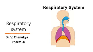
Russian Call Girls in Bangalore Manisha 7001305949 Independent Escort Service...
Russian Call Girls in Bangalore Manisha 7001305949 Independent Escort Service...Russian Call Girls in Bangalore Manisha 7001305949 Independent Escort Service...
Russian Call Girls in Bangalore Manisha 7001305949 Independent Escort Service...
 
Escort Service Call Girls In Sarita Vihar,, 99530°56974 Delhi NCR
Escort Service Call Girls In Sarita Vihar,, 99530°56974 Delhi NCREscort Service Call Girls In Sarita Vihar,, 99530°56974 Delhi NCR
Escort Service Call Girls In Sarita Vihar,, 99530°56974 Delhi NCR
 
VIP Call Girls Tirunelveli Aaradhya 8250192130 Independent Escort Service Tir...
VIP Call Girls Tirunelveli Aaradhya 8250192130 Independent Escort Service Tir...VIP Call Girls Tirunelveli Aaradhya 8250192130 Independent Escort Service Tir...
VIP Call Girls Tirunelveli Aaradhya 8250192130 Independent Escort Service Tir...
 
Call Girl Chennai Indira 9907093804 Independent Call Girls Service Chennai
Call Girl Chennai Indira 9907093804 Independent Call Girls Service ChennaiCall Girl Chennai Indira 9907093804 Independent Call Girls Service Chennai
Call Girl Chennai Indira 9907093804 Independent Call Girls Service Chennai
 
Call Girls Service In Shyam Nagar Whatsapp 8445551418 Independent Escort Service
Call Girls Service In Shyam Nagar Whatsapp 8445551418 Independent Escort ServiceCall Girls Service In Shyam Nagar Whatsapp 8445551418 Independent Escort Service
Call Girls Service In Shyam Nagar Whatsapp 8445551418 Independent Escort Service
 
Russian Call Girls in Pune Tanvi 9907093804 Short 1500 Night 6000 Best call g...
Russian Call Girls in Pune Tanvi 9907093804 Short 1500 Night 6000 Best call g...Russian Call Girls in Pune Tanvi 9907093804 Short 1500 Night 6000 Best call g...
Russian Call Girls in Pune Tanvi 9907093804 Short 1500 Night 6000 Best call g...
 
Call Girls Doddaballapur Road Just Call 7001305949 Top Class Call Girl Servic...
Call Girls Doddaballapur Road Just Call 7001305949 Top Class Call Girl Servic...Call Girls Doddaballapur Road Just Call 7001305949 Top Class Call Girl Servic...
Call Girls Doddaballapur Road Just Call 7001305949 Top Class Call Girl Servic...
 
Russian Call Girl Brookfield - 7001305949 Escorts Service 50% Off with Cash O...
Russian Call Girl Brookfield - 7001305949 Escorts Service 50% Off with Cash O...Russian Call Girl Brookfield - 7001305949 Escorts Service 50% Off with Cash O...
Russian Call Girl Brookfield - 7001305949 Escorts Service 50% Off with Cash O...
 
Call Girls Whitefield Just Call 7001305949 Top Class Call Girl Service Available
Call Girls Whitefield Just Call 7001305949 Top Class Call Girl Service AvailableCall Girls Whitefield Just Call 7001305949 Top Class Call Girl Service Available
Call Girls Whitefield Just Call 7001305949 Top Class Call Girl Service Available
 
Call Girls Service Noida Maya 9711199012 Independent Escort Service Noida
Call Girls Service Noida Maya 9711199012 Independent Escort Service NoidaCall Girls Service Noida Maya 9711199012 Independent Escort Service Noida
Call Girls Service Noida Maya 9711199012 Independent Escort Service Noida
 
Russian Call Girls Chennai Madhuri 9907093804 Independent Call Girls Service ...
Russian Call Girls Chennai Madhuri 9907093804 Independent Call Girls Service ...Russian Call Girls Chennai Madhuri 9907093804 Independent Call Girls Service ...
Russian Call Girls Chennai Madhuri 9907093804 Independent Call Girls Service ...
 
Call Girls Horamavu WhatsApp Number 7001035870 Meeting With Bangalore Escorts
Call Girls Horamavu WhatsApp Number 7001035870 Meeting With Bangalore EscortsCall Girls Horamavu WhatsApp Number 7001035870 Meeting With Bangalore Escorts
Call Girls Horamavu WhatsApp Number 7001035870 Meeting With Bangalore Escorts
 
Call Girls Service Jaipur Grishma WhatsApp ❤8445551418 VIP Call Girls Jaipur
Call Girls Service Jaipur Grishma WhatsApp ❤8445551418 VIP Call Girls JaipurCall Girls Service Jaipur Grishma WhatsApp ❤8445551418 VIP Call Girls Jaipur
Call Girls Service Jaipur Grishma WhatsApp ❤8445551418 VIP Call Girls Jaipur
 
VIP Call Girls Indore Kirti 💚😋 9256729539 🚀 Indore Escorts
VIP Call Girls Indore Kirti 💚😋  9256729539 🚀 Indore EscortsVIP Call Girls Indore Kirti 💚😋  9256729539 🚀 Indore Escorts
VIP Call Girls Indore Kirti 💚😋 9256729539 🚀 Indore Escorts
 
Housewife Call Girls Hoskote | 7001305949 At Low Cost Cash Payment Booking
Housewife Call Girls Hoskote | 7001305949 At Low Cost Cash Payment BookingHousewife Call Girls Hoskote | 7001305949 At Low Cost Cash Payment Booking
Housewife Call Girls Hoskote | 7001305949 At Low Cost Cash Payment Booking
 
Call Girls In Andheri East Call 9920874524 Book Hot And Sexy Girls
Call Girls In Andheri East Call 9920874524 Book Hot And Sexy GirlsCall Girls In Andheri East Call 9920874524 Book Hot And Sexy Girls
Call Girls In Andheri East Call 9920874524 Book Hot And Sexy Girls
 
Aspirin presentation slides by Dr. Rewas Ali
Aspirin presentation slides by Dr. Rewas AliAspirin presentation slides by Dr. Rewas Ali
Aspirin presentation slides by Dr. Rewas Ali
 
Vip Call Girls Anna Salai Chennai 👉 8250192130 ❣️💯 Top Class Girls Available
Vip Call Girls Anna Salai Chennai 👉 8250192130 ❣️💯 Top Class Girls AvailableVip Call Girls Anna Salai Chennai 👉 8250192130 ❣️💯 Top Class Girls Available
Vip Call Girls Anna Salai Chennai 👉 8250192130 ❣️💯 Top Class Girls Available
 
Russian Call Girls in Pune Riya 9907093804 Short 1500 Night 6000 Best call gi...
Russian Call Girls in Pune Riya 9907093804 Short 1500 Night 6000 Best call gi...Russian Call Girls in Pune Riya 9907093804 Short 1500 Night 6000 Best call gi...
Russian Call Girls in Pune Riya 9907093804 Short 1500 Night 6000 Best call gi...
 
VIP Call Girls Pune Vani 9907093804 Short 1500 Night 6000 Best call girls Ser...
VIP Call Girls Pune Vani 9907093804 Short 1500 Night 6000 Best call girls Ser...VIP Call Girls Pune Vani 9907093804 Short 1500 Night 6000 Best call girls Ser...
VIP Call Girls Pune Vani 9907093804 Short 1500 Night 6000 Best call girls Ser...
 

Respiratory system

  • 2. Contents 3 i) Respiratory system a)Anatomy of respiratory organs and functions a)Mechanism / physiology of respiration and regulation of respiration a)Transport of respiratory gases a)Respiratory volumes and capacities, and a)Definition of: Hypoxia, Asphyxia, Dybarism, Oxygen therapy and resuscitation.
  • 3. INTRODUCTION • Respiration is the process by which oxygen is taken in and carbon dioxide is given out. • The first breath takes place only after birth. • Fetal lungs are non-functional. • So, during intrauterine life the exchange of gases between fetal blood and mother’s blood occurs through placenta. • After the first breath, the respiratory process con- tinues throughout the life. • Permanent stoppage of respiration occurs only at death.
  • 4. • The respiratory system contributes to homeostasis by providing for the exchange of gases—oxygen and carbon dioxide—between the atmospheric air, blood, and tissue cells. It also helps adjust the pH of body fluids. • Cells of the body continually use oxygen (O2) for the metabolic reactions that release energy from nutrient molecules and produce ATP. At the same time, these reactions release carbon dioxide (CO2). • Because an excessive amount of CO2 produces acidity that can be toxic to cells, excess CO2 must be eliminated quickly and efficiently. • The cardiovascular and respiratory systems cooperate to supply O2 and eliminate CO2. The respiratory system provides. • Failure of either system disrupts homeostasis by causing rapid death of cells from oxygen starvation and buildup of waste products.
  • 5. • The respiratory system provides the route by which the supply of oxygen present in the atmospheric air gains entry to the body and it provides the route of excretion of carbon dioxide. • The condition of the atmospheric air entering the body varies considerably according to the external environment, e.g., it may be dry, cold and contain dust particles or it may be moist and hot. • In addition to functioning in gas exchange, the respiratory system also participates in regulating blood pH, contains receptors for the sense of smell, filters inspired air, produces sounds, and rids the body of some water and heat in exhaled air. • Exchange of gases between the blood and the lungs is called external respiration and that between the blood and the cells internal respiration.
  • 6. Respiratory system can be classified according to either structure or function Structurally, the respiratory system consists of two parts: The upper respiratory system includes the nose, nasal cavity, pharynx, and associated structures. The lower respiratory system includes the larynx, trachea, bronchi, and lungs. Functionally, the respiratory system also consists of two parts: The conducting zone consists of a series of interconnecting cavities and tubes both outside and within the lungs. These include the nose, nasal cavity, pharynx, larynx, trachea, bronchi, bronchioles, and terminal bronchioles; their function is to filter, warm, and moisten air and conduct it into the lungs. The respiratory zone consists of tubes and tissues within the lungs where gas exchange occurs. These include the respiratory bronchioles, alveolar ducts, alveolar sacs, and alveoli and are the main sites of gas exchange between air and blood.
  • 7.
  • 8. Anatomy of respiratory organs and functions • The organs of respiratory system are • Nose • Pharynx (Throat) • Larynx (Voice Box) • Trachea (Windpipe) • Two bronchi (one bronchus to each lung) • Bronchioles and smaller air passages • T wo Lungs and their coverings • The pleura • Muscles of respiration (The intercostal muscles and the diaphragm)
  • 9.
  • 10. Nose and Nasal cavity - Anatomy Position and structure • The nasal cavity is the first of the respiratory organs and consists of a large irregular cavity divided into two equal passages by a septum. • The posterior bony part of the septum is formed by the perpendicular plate of the ethmoid bone and the vomer. • Anteriorly it consists of hyaline cartilage. • The roof is formed by the cribriform plate of the ethmoid bone, and the sphenoid bone, frontal bone and nasal bones. • The floor is formed by the roof of the mouth and consists of the hard palate in front and the soft palate behind. • The hard palate is composed of the maxilla and palatine bones and the soft palate consists of involuntary muscle. • The medial wall is formed by the septum.
  • 11. • The lateral walls are formed by the maxilla, the ethmoid bone and the inferior conchae. • The posterior wall is formed by the posterior wall of the pharynx. Lining of the nose • The nose is lined with very vascular ciliated columnar epithelium (ciliated mucous membrane) which contains mucus-secreting goblet cells . • At the anterior nares this blends with the skin and posteriorly it extends into the nasal part of the pharynx.
  • 12. Openings into the nasal cavity • The anterior nares, or nostrils, are the openings from the exterior into the nasal cavity. • Hairs are present in this area. • The posterior nares are the openings from the nasal cavity into the pharynx. • The paranasal sinuses are cavities in the bones of the face and the cranium which contain air. • There are tiny openings between the paranasal sinuses and the nasal cavity. • They are lined with mucous membrane, continuous with that of the nasal cavity. • The main sinuses are: • Maxillary sinuses in the lateral walls • Frontal and sphenoidal sinuses in the roof • Ethmoidal sinuses in the upper part of the lateral walls. • The sinuses function in speech and also serve to lighten the skull. • The nasolacrimal ducts extend from the lateral walls of the nose to the conjunctival sacs of the eye. They drain tears from the eyes.
  • 13. Physiology-Respiratory function of the nose • The nose is the first of the respiratory passages through which the inspired air passes. • The function of the nose is to begin the process by which the air is warmed, moistened and 'filtered'. • The projecting conchae increase the surface area and cause turbulence, spreading inspired air over the whole nasal surface. • The large surface area maximizes warming, humidification and filtering.
  • 14. • Warming. This is due to the immense vascularity of the mucosa. • Filtering and cleaning of air. This occurs as hairs at the anterior nares trap larger particles. Smaller particles such as dust and microbes settle and adhere to the mucus. Mucus protects the underlying epithelium from irritation and prevents drying. Synchronous beating of the cilia wafts the mucus towards the throat where it is swallowed or expectorated. • Humidification. This occurs as air travels over the moist mucosa and becomes saturated with water vapour. Irritation of the nasal mucosa results in sneezing, a reflex action that forcibly expels an irritant.
  • 15. Physiology- Olfactory function of the nose • The nose is the organ of the sense of smell. There are nerve endings that detect smell, located in the roof of the nose in the area of the cribriform plate of the ethmoid bones and the superior conchae. These nerve endings are stimulated by chemical substances given off by odorous materials. • The resultant nerve impulses are conveyed by the olfactory nerves to the brain where the sensation of smell is perceived .
  • 16. PHARYNX Position The pharynx is a tube 12 to 14 cm long that extends from the base of the skull to the level of the 6th cervical vertebra. It lies behind the nose, mouth and larynx and is wider at its upper end. Structures associated with the pharynx Superiorly — the inferior surface of the base of the skull Inferiorly — it is continuous with the oesophagus Anteriorly — the wall is incomplete because of the openings into the nose, mouth and larynx Posteriorly — areolar tissue, involuntary muscle and the bodies of the first six cervical vertebrae
  • 17. • The pharynx is divided into three parts: nasopharynx, oropharynx and laryngopharynx. • The nasopharynx. • The nasal part of the pharynx lies behind the nose above the level of the soft palate • On its lateral walls are the two openings of the auditory tubes, one leading to each middle ear. • On the posterior wall there are the pharyngeal tonsils (adenoids), consisting of lymphoid tissue. • They are most prominent in children up to approximately 7 years of age. Thereafter they gradually atrophy.
  • 18. • The oropharynx • The oral part of the pharynx lies behind the mouth, extending from below the level of the soft palate to the level of the upper part of the body of the 3rd cervical vertebra. • The lateral walls of the pharynx blend with the soft palate to form two folds on each side. • Between each pair of folds there is a collection of lymphoid tissue called the palatine tonsil. • During swallowing, the nasal and oral parts are separated by the soft palate and the uvula. • The laryngopharynx • The laryngeal part of the pharynx extends from the oropharynx above and continues as the oesophagus below, i.e. from the level of the 3rd to the 6th cervical vertebrae.
  • 19. Structure • The pharynx is composed of three layers of tissue: • 1. Mucous membrane lining. The mucosa varies slightly in the different parts. In the nasopharynx it is continuous with the lining of the nose and consists of ciliated columnar epithelium; in the oropharynx and laryngopharynx it is formed by tougher stratified squamous epithelium which is continuous with the lining of the mouth and oesophagus. • 2. Fibrous tissue. This forms the intermediate layer. It is thicker in the nasopharynx, where there is little muscle, and becomes thinner towards the lower end, where the muscle layer is thicker. • 3. Muscle tissue. This consists of several involuntary constrictor muscles that play an important part in the mechanism of swallowing (deglutition) which, in the pharynx, is not under voluntary control. The upper end of the esophagus is closed by the lower constrictor muscle, except during swallowing.
  • 20. Blood and nerve supply • Blood is supplied to the pharynx by several branches of the facial artery. The venous return is into the facial and internal jugular veins. • The nerve supply is from the pharyngeal plexus, formed by parasympathetic and sympathetic nerves. Parasympathetic supply is by the vagus and glossopharyngeal nerves. • Sympathetic supply is by nerves from the superior cervical ganglia
  • 21. Functions Passageway for air and food. The pharynx is an organ involved in both the respiratory and the digestive systems: air passes through the nasal and oral parts, and food through the oral and laryngeal parts. Warming and humidifying. By the same methods as in the nose, the air is further warmed and moistened as it passes through the pharynx. Taste. There are olfactory nerve endings of the sense of taste in the epithelium of the oral and pharyngeal parts. Hearing. The auditory tube, extending from the nasal part to each middle ear, allows air to enter the middle ear. Satisfactory hearing depends on the presence of air at atmospheric pressure on each side of the tympanic membrane (ear drum) Protection. The lymphatic tissue of the pharyngeal and laryngeal tonsils produces antibodies in response to antigens, e.g. microbes The tonsils are larger in children and tend to atrophy in adults. Speech. The pharynx functions in speech; by acting as a resonating chamber for the sound ascending from the larynx, it helps (together with the sinuses) to give the voice its individual characteristics.
  • 22. LARYNX Position • The larynx or 'voice box' extends from the root of the tongue and the hyoid bone to the trachea. • It lies in front of the laryngopharynx at the level of the 3rd, 4th, 5th and 6th cervical vertebrae. • Until puberty there is little difference in the size of the larynx between the sexes. Thereafter it grows larger in the male, which explains the prominence of the 'Adam's apple' and the generally deeper voice.
  • 23. Structures associated with the larynx Superiorly •the hyoid bone and the root of the tongue Inferiorly •it is continuous with the trachea Anteriorly •the muscles attached to the hyoid bone and the muscles of the neck Posteriorly • the laryngopharynx and 3rd to 6th cervical vertebrae Laterally •the lobes of the thyroid gland
  • 24. Structure Cartilages • The larynx is composed of several irregularly shaped cartilages attached to each other by ligaments and membranes. The main cartilages are: 1 thyroid cartilage 1 cricoid cartilage 2 arytenoid cartilages 1 epiglottis hyaline cartilage elastic fibrocartilage.
  • 25. The thyroid cartilage This is the most prominent and consists of two flat pieces of hyaline cartilage, or laminae, fused anteriorly, forming the laryngeal prominence (Adam’s apple). • Immediately above the laryngeal prominence the laminae are separated, forming a V-shaped notch known as the thyroid notch. • The thyroid cartilage is incomplete posteriorly and the posterior border of each lamina is extended to form two processes called the superior and inferior cornu. • The upper part of the thyroid cartilage is lined with stratified squamous epithelium like the larynx, and the lower part with ciliated columnar epithelium like the trachea. • There are many muscles attached to its outer surface.
  • 26. The cricoid cartilage • This lies below the thyroid cartilage and is also composed of hyaline cartilage. • It is shaped like a signet ring, completely encircling the larynx with the narrow part anteriorly and the broad part posteriorly. • The broad posterior part articulates with the arytenoid cartilages above and with the inferior cornu of the thyroid cartilage below. • It is lined with ciliated columnar epithelium and there are muscles and ligaments attached to its outer surface. • The lower border of the cricoid cartilage marks the end of the upper respiratory tract.
  • 27. Arytenoid cartilages • These are two roughly pyramid-shaped hyaline cartilages situated on top of the broad part of the cricoid cartilage forming part of the posterior wall of the larynx. • They give attachment to the vocal cords and to muscles and are lined with ciliated columnar epithelium
  • 28. The epiglottis • This is a leaf-shaped fibroelastic cartilage attached to the inner surface of the anterior wall of the thyroid cartilage immediately below the thyroid notch. • It rises obliquely upwards behind the tongue and the body of the hyoid bone. • It is covered with stratified squamous epithelium. • If the larynx is assumed as a box, then the epiglottis acts as the lid; it closes off the larynx during swallowing, protecting the lungs from accidental inhalation of foreign objects.
  • 29. Ligaments and membranes • There are several ligaments that attach the cartilages to each other and to the hyoid bone Blood and nerve supply • Blood is supplied to the larynx by the superior and inferior laryngeal arteries and drained by the thyroid veins, which join the internal jugular vein. • The parasympathetic nerve supply is from the superior • Laryngeal and recurrent laryngeal nerves, which are branches of the vagus nerves, and the sympathetic nerves are from the superior cervical ganglia, one on each side. • These provide the motor nerve supply to the muscles of the larynx and sensory fibers to the lining membrane
  • 30. Interior of the larynx • The vocal cords are two pale folds of mucous membrane with cord-like free edges which extend from the inner wall of the thyroid prominence anteriorly to the arytenoid cartilages posteriorly. • When the muscles controlling the vocal cords are relaxed, the vocal cords open and the passageway for air coming up through the larynx is clear; the vocal cords are said to be abducted. The pitch of the sound produced by vibrating the vocal cords in this position is low. • When the muscles controlling the vocal cords contract, the vocal cords are stretched out tightly across the larynx —they are said to be adducted. • When the vocal cords are stretched to this extent and are vibrated by air passing through from the lungs, the sound produced is high pitched. • The pitch of the voice is therefore determined by the tension applied to the vocal cords by the appropriate sets of muscles. When not in use, the vocal cords are adducted.
  • 31. Functions Production of sound • Sound has the properties of pitch, volume and resonance. • Pitch of the voice depends upon the length and tightness of the cords. At puberty, the male vocal cords begin to grow longer, hence the lower pitch of the adult male voice. • Volume of the voice depends upon the force with which the cords vibrate. The greater the force of expired air the more the cords vibrate and the louder the sound emitted. • Resonance, or tone, is dependent upon the shape of the mouth, the position of the tongue and the lips, the facial muscles and the air in the paranasal sinuses.
  • 32. Speech. • This occurs during expiration when the sounds produced by the vocal cords are manipulated by the tongue, cheeks and lips. Protection of the lower respiratory tract • During swallowing (deglutition) the larynx moves upwards, occluding the opening into it from the pharynx and the hinged epiglottis closes over the larynx. This ensures that food passes into the oesophagus and not into the lower respiratory passages Passageway for air. • This is between the pharynx and trachea. Humidifying, filtering and warming. • These continue as inspired air travels through the larynx.
  • 33. TRACHEA Position • The trachea or windpipe is a continuation of the larynx and extends downwards to about the level of the 5th thoracic vertebra where it divides (bifurcates) at the carina into the right and left bronchi, one bronchus going to each lung. • It is approximately 10 to 11 cm long and lies mainly in the median plane in front of the oesophagus.
  • 34. Structures associated with the trachea • Superiorly — the larynx • Inferiorly — the right and left bronchi • Anteriorly — upper part: the isthmus of the thyroid gland lower part: the arch of the aorta and the sternum • Posteriorly — the oesophagus separates the trachea from the vertebral column • Laterally — the lungs and the lobes of the thyroid gland.
  • 35. Structure • The trachea is composed of from 16 to 20 incomplete (C-shaped) rings of hyaline cartilages lying one above the other. • The cartilages are incomplete posteriorly. • Connective tissue and involuntary muscle join the cartilages and form the posterior wall where they are incomplete. • The soft tissue posterior wall is in contact with the oesophagus
  • 36. There are three layers of tissue which 'clothe' the cartilages of the trachea. The outer layer. This consists of fibrous and elastic tissue and encloses the cartilages. The middle layer. This consists of cartilages and bands of smooth muscle that wind round the trachea in a helical arrangement. There is some areolar tissue, containing blood and lymph vessels and autonomic nerves. The inner lining. This consists of ciliated columnar epithelium, containing mucus-secreting goblet cells
  • 37. Blood and nerve supply, lymph drainage The arterial blood supply • This is mainly by the inferior thyroid and bronchial arteries and the venous return is by the inferior thyroid veins into the brachiocephalic veins. The nerve supply • This is by parasympathetic and sympathetic fibers. • Parasympathetic supply is by the recurrent laryngeal nerves and other branches of the vagi. • Sympathetic supply is by nerves from the sympathetic ganglia. Lymph • Lymph from the respiratory passages passes through lymph nodes situated round the trachea and in the carina, the area where it divides into two bronchi
  • 38. Functions • The arrangement of cartilage and elastic tissue prevents kinking and obstruction of the airway as the head and neck move. • The absence of cartilage posteriorly allows the trachea to dilate and constrict in response to nerve stimulation, and for indentation as the esophagus distends during swallowing. • The cartilages prevent collapse of the tube when the internal pressure is less than intrathoracic pressure, i.e., at the end of forced expiration. Support and patency. • This is the synchronous and regular beating of the cilia of the mucous membrane lining that wafts mucus with adherent particles upwards towards the larynx where it is swallowed or expectorated. Mucociliary escalator.
  • 39. • Nerve endings in the larynx, trachea and bronchi are sensitive to irritation that generates nerve impulses which are conducted by the vagus nerves to the respiratory center in the brain stem. • The reflex motor response is deep inspiration followed by closure of the glottis. The abdominal and respiratory muscles then contract and suddenly the air is released under pressure expelling mucus and/or foreign material from the mouth. Cough reflex • These continue as in the nose, although air is normally saturated and at body temperature when it reaches the trachea. Warming, humidifying and filtering of air
  • 40. Bronchi • The two primary bronchi are formed when the trachea divides, i.e., about the level of the 5th thoracic vertebra • The right bronchus. • This is wider, shorter and more vertical than the left bronchus and is therefore the more likely of the two to become obstructed by an inhaled foreign body. • It is approximately 2.5 cm long. After entering the right lung at the hilum, it divides into three branches, one to each lobe. Each branch then subdivides into numerous smaller branches. • The left bronchus. • This is about 5 cm long and is narrower than the right. After entering the lung at the hilum it divides into two branches, one to each lobe. Each branch then subdivides into progressively smaller tubes within the lung substance.
  • 41. Structure • The bronchi are composed of the same tissues as the trachea. • They are lined with ciliated columnar epithelium. • The bronchi progressively subdivide into bronchioles, terminal bronchioles, respiratory bronchioles, alveolar ducts and finally, alveoli. • Towards the distal end of the bronchi the cartilages become irregular in shape and are absent at bronchiolar level. • In the absence of cartilage, the smooth muscle in the walls of the bronchioles becomes thicker and is responsive to autonomic nerve stimulation and irritation. • Ciliated columnar mucous membrane changes gradually to non-ciliated cuboidal shaped cells in the distal bronchioles.
  • 42. Blood and nerve supply, lymph drainage • The arterial blood supply. • The supply to the walls of the bronchi and smaller air passages is through branches of the right and left bronchial arteries and the venous return is mainly through the bronchial veins. • On the right side they empty into the azygos vein and on the left into the superior intercostal vein
  • 43. The nerve supply • This is by parasympathetic and sympathetic nerves. • The vagus nerves (parasympathetic) stimulate contraction of smooth muscle in the bronchial tree, causing bronchoconstriction, and sympathetic stimulation causes bronchodilatation The lymphatic vessels and lymph nodes • Lymph is drained from the walls of the air passages in a network of lymph vessels. • It passes through lymph nodes situated around the trachea and bronchial tree then into the thoracic duct on the left side and right lymphatic duct on the other.
  • 44. Functions of air passages not involved in gaseous exchange Control of air entry • The diameter of the respiratory passages may be altered by contraction or relaxation of the involuntary muscles in their walls, thus regulating the volume of air entering the lungs. • These changes are controlled by the autonomic nerve supply: parasympathetic stimulation causes constriction and sympathetic stimulation causes dilatation warming and humidifying support and patency removal of particulate matter cough reflex where the other functions continue as in the upper airways.
  • 45. Respiratory bronchioles and alveoli Structure • Lobules are the blind ends of the respiratory tract distal to the terminal bronchioles, consisting of: respiratory bronchioles, alveolar ducts and alveoli (tiny air sacs) . • It is in these structures that the process of gas exchange occurs. The walls gradually become thinner until muscle and connective tissue fade out leaving a single layer of simple squamous epithelial cells in the alveolar ducts and alveoli. • These distal respiratory passages are supported by a loose network of elastic connective tissue in which macrophages, fibroblasts, nerves and blood and lymph vessels are embedded. • The alveoli are surrounded by a network of capillaries. The exchange of gases during respiration takes place across two membranes, the alveolar and capillary membranes.
  • 46. • Inter spared between the squamous cells are other cells that secrete surfactant, a phospholipid fluid which prevents the alveoli from drying out. • In addition, surfactant reduces surface tension and prevents alveolar walls collapsing during expiration. • Secretion of surfactant into the distal air passages and alveoli begins about the 35th week of fetal life. • Its presence in newborn babies facilitates expansion of the lungs and the establishment of respiration. • It may not be present in sufficient amounts in the immature lungs of premature babies, causing difficulty in establishing respiration.
  • 47. Functions of respiratory bronchioles and alveoli • Aid in external respiration • Defense against microbes. • At this level, ciliated epithelium, goblet cells and mucus are no longer present • Defense relies on protective cells present within the lung tissue. • These include lymphocytes and plasma cells, which produce antibodies in the presence of antigens, and macrophages and polymorphonuclear lymphocytes, which are phagocytic. • These cells are most active in the distal air passages where ciliated epithelium has been replaced by flattened cells. • Warming and humidifying • These continue as in the upper airways. • Inhalation of dry or inadequately humidified air over a period causes irritation of the mucosa and facilitates the establishment of pathogenic microbes.
  • 48. Pleura and pleural cavity • The pleura consists of a closed sac of serous membrane (one for each lung) which contains a small amount of serous fluid. • The lung is invaginated into this sac so that it forms two layers: one adheres to the lung and the other to the wall of the thoracic cavity. • The visceral pleura. • This is adherent to the lung, covering each lobe and passing into the fissures which separate them. • The parietal pleura. • This is adherent to the inside of the chest wall and the thoracic surface of the diaphragm. • It remains detached from the adjacent structures in the mediastinum and is continuous with the visceral pleura round the edges of the hilum. • The pleural cavity. • This is only a potential space. In health, the two layers of pleura are separated by only a thin film of serous fluid which allows them to glide over each other, preventing friction between them during breathing. The serous fluid is secreted by the epithelial cells of the membrane
  • 49. • The two layers of pleura, with serous fluid between them, behave in the same way as two pieces of glass separated by a thin film of water. • They glide over each other easily but can be pulled apart only with difficulty, because of the surface tension between the membranes and the fluid. • If either layer of pleura is punctured, the underlying lung collapses due to its inherent property of elastic recoil. • Interior of the lungs • The lungs are composed of the bronchi and smaller air passages, alveoli, connective tissue, blood vessels, lymph vessels and nerves. The left lung is divided into two lobes and the right, into three.
  • 50. Lungs • Position and associated structures • There are two lungs, one lying on each side of the midline in the thoracic cavity. • They are cone-shaped and are described as having an apex, a base, costal surface and medial surface. • The apex • This is rounded and rises into the root of the neck, about 25 mm (1 inch) above the level of the middle third of the clavicle. The structures associated with it are the first rib and the blood vessels and nerves in the root of the neck. • The base • This is concave and semilunar in shape and is closely associated with the thoracic surface of the diaphragm. • The costal surface • This surface is convex and is closely associated with the costal cartilages, the ribs and the intercostal muscles. • The medial surface • This surface is concave and has a roughly triangular-shaped area, called the hilum, at the level of the 5th, 6th and 7th thoracic vertebrae. • Structures which form the root of the lung enter and leave at the hilum. These include the primary bronchus, the pulmonary artery supplying the lung and the two pulmonary veins draining it, the bronchial artery and veins, and the lymphatic and nerve supply . • The area between the lungs is the mediastinum. It is occupied by the heart, great vessels, trachea, right and left bronchi, esophagus, lymph nodes, lymph vessels and nerves.
  • 51. Organization of the lungs • The right lung is divided into three distinct lobes: superior, middle and inferior. • The left lung is smaller as the heart is situated left of the midline. • It is divided into only two lobes: superior and inferior.
  • 52. Pulmonary blood supply • The pulmonary artery divides into two, one branch conveying deoxygenated blood to each lung. Within the lungs each pulmonary artery divides into many branches which eventually end in a dense capillary network around the walls of the alveoli. • The walls of the alveoli and those of the capillaries each consist of only one layer of flattened epithelial cells. The exchange of gases between air in the alveoli and blood in the capillaries takes place across these two very fine membranes. • The pulmonary capillaries join up, eventually becoming two pulmonary veins in each lung. • They leave the lungs at the hilum and convey oxygenated blood to the left atrium of the heart. • The innumerable blood capillaries and blood vessels in the lungs are supported by connective tissue.
  • 53. • Each bronchopulmonary segment of the lungs has many small compartments called lobules; each lobule is wrapped in elastic connective tissue and contains a lymphatic vessel, an arteriole, a venule, and a branch from a terminal bronchiole. • Terminal bronchioles subdivide into microscopic branches called respiratory bronchioles. • They also have alveoli (described shortly) budding from their walls. Alveoli participate in gas exchange, and thus respiratory bronchioles begin the respiratory zone of the respiratory system.
  • 54. Definition • Hypoxia : Hypoxia is a condition in which tissues of the body do not receive sufficient oxygen (O2) supply. • Asphyxia: It is a condition arising when the body is deprived of oxygen, causing unconsciousness or death; suffocation. • Hypoxia is the term used to indicate a deficiency of oxygen. A related term that is often used in relation to perinatal brain injury is anoxia, meaning without oxygen. Asphyxia refers to the physiological results of hypoxia or anoxia. • Dysbarism: it is defined as any adverse medical condition that results from changes in ambient pressure. These changes in pressure must occur either at a rate or duration exceeding the capacity of the body to adapt safely. • Although the most common cause of dysbarism is underwater diving, dysbaric injury can occur following exposure to any environments with extreme pressure changes. Other examples include high altitude, aircraft cabin decompression, explosions or blasts, outer space, caissons, and tunnel-boring operations.
  • 55. Oxygen therapy • Oxygen therapy is a treatment that delivers oxygen gas for you to breathe. It is also called supplemental oxygen. • You can receive oxygen therapy from tubes resting in your nose, a face mask, or a tube placed in your trachea, or windpipe. This treatment increases the amount of oxygen your lungs receive and deliver to your blood. • Oxygen therapy may be prescribed for you when you have a condition that causes your blood oxygen levels to be too low. Low blood oxygen may make you feel short of breath, tired, or confused, and can damage your body. • Oxygen therapy can be given for a short or long period of time in the hospital, another medical setting, or at home.
  • 56. • There are different types of devices that can give you oxygen. Some use tanks of liquid or gas oxygen. • Others use an oxygen concentrator, which pulls oxygen out of the air. You will get the oxygen through a nose tube (cannula), a mask, or a tent. The extra oxygen is breathed in along with normal air. • You may need oxygen therapy if you have a condition that causes low blood oxygen, such as • COPD (chronic obstructive pulmonary disease) • Pneumonia • COVID-19 • A severe asthma attack • Late-stage heart failure • Cystic fibrosis • Sleep apnea
  • 57. Cardiopulmonary resuscitation (CPR) • Cardiopulmonary resuscitation (CPR) is a procedure to support and maintain breathing and circulation for an infant, child, or adolescent who has stopped breathing (respiratory arrest) and/or whose heart has stopped (cardiac arrest). • Purpose • CPR is performed to restore and maintain breathing and circulation and to provide oxygen and blood flow to the heart, brain, and other vital organs. • CPR can be performed by trained laypeople or healthcare professionals on infants, children, adolescents, and adults. • CPR should be performed if an infant, child, or adolescent is unconscious and not breathing. • Respiratory and cardiac arrest can be caused by allergic reactions, an ineffective heartbeat, asphyxiation, breathing passages that are blocked, choking , drowning, drug reactions or overdoses, electric shock, exposure to cold, severe shock, or trauma. • In newborns, the most common cause of cardiopulmonary arrest is respiratory failure caused by sudden infant death syndrome (SIDS), airway obstruction (usually from inhalation of a foreign body), sepsis, neurologic disease, or drowning. • Cardiac arrest in children over one year of age is most commonly caused by shock and/or respiratory failure resulting from an accident or injury.