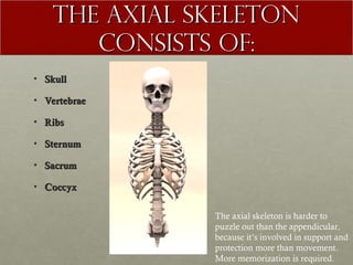
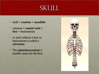
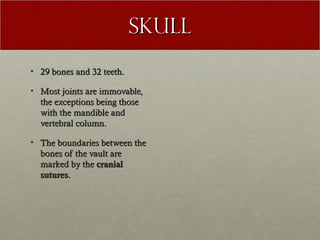
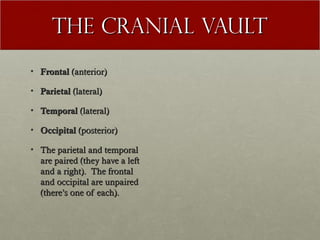
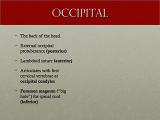
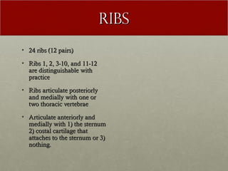
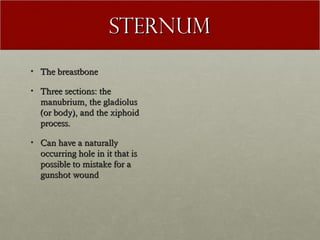
The axial Skeleton
THE AXIAL SKELETON
       CONSISTS OF:
• Skull

• Vertebrae

• Ribs

• Sternum

• Sacrum

• Coccyx

               The axial skeleton is harder to
               puzzle out than the appendicular,
               because it’s involved in support and
               protection more than movement.
               More memorization is required.
SKULL
• skull = cranium + mandible

• cranium = cranial vault +
  face + basicranium

• A vault without a face or
  basicranium is called a
  calvarium.

• The splanchnocranium is
  another name for the face.
Skull
• 29 bones and 32 teeth.

• Most joints are immovable,
  the exceptions being those
  with the mandible and
  vertebral column.

• The boundaries between the
  bones of the vault are
  marked by the cranial
  sutures.
THE CRANIAL VAULT
• Frontal (anterior)

• Parietal (lateral)

• Temporal (lateral)

• Occipital (posterior)

• The parietal and temporal
  are paired (they have a left
  and a right). The frontal
  and occipital are unpaired
  (there’s one of each).
Sutures
• The coronal suture is parallel
  to the coronal plane and
  forms the posterior border of
  the frontal.

• The sagittal suture is along
  the sagittal plane.

• The squamosal suture is
  between the temporal and the
  parietal.

• The lambdoid suture is
  posterior and forms the
  superior border of the
  occipital.
Frontal
• Forehead

• Orbits (anterior and
  inferior)

• Supraorbital torus (brow
  ridge)

• Coronal suture (posterior)

• Zygomatic process (lateral)

• Billowed internal surface

• Frontal crest (internal)
Parietal
• Deeply curved.

• Grooves for middle
  meningeal vessels on the
  inside. They go back and up.
  The deepest is the most
  anterior. It is parallel to the
  coronal suture.

• coronal suture to anterior

• Sagittal suture medially

• Squamosal suture to inferior

• Lambdoid suture posterior
TEMPORAL
• External auditory meatus (ear
  canal)

• Squamosal suture superiorly

• Lambdoid suture posteriorly

• Zygomatic process to anterior

• Mastoid process w/ digastric
  groove to posterior

• External auditory meatus in
  between mastoid and zygomatic

• Petrous process medially and
  internally
Occipital
• The back of the head.

• External occipital
  protuberance (posterior)

• Lambdoid suture (anterior)

• Articulates with first
  cervical vertebrae at
  occipital condyles

• Foramen magnum (“big
  hole”) for spinal cord
  (inferior)
Major bones of the
         Face
• Frontal

• Nasal

• Zygomatic

• Maxilla
zygomatic
• Cheekbone

• Articulates with frontal
  (superiorly), maxilla
  (medially), and temporal
  (posteriorly)

• Contributes to orbits
  superiorly and medially
Maxilla
• Upper jaw

• Articulates with frontal
  (superiorly) and zygomatic
  (laterally/posteriorly)

• Dentition (teeth) inferiorly

• Maxillary sinus internally
Major bones of the
      basicranium
• Occipital (posterior)

• Temporal (lateral)

• Sphenoid (midline)
Sphenoid
• Highly complex bone

• Contributes to vault,
  basicranium, and posterior
  portion of the eye orbit

• Often recovered in
  fragmentary state, usually in
  connection with another
  cranial bone

• To remember its anatomical
  orientation, think of it
  resembling bat, butterfly, or
  owl
Mandible
• Lower jaw

• One of two movable joints
  in the skull

• Articulates with the
  temporal’s glenoid fossa

• Ramus

• Condylar process and
  coronoid process
Vertebrae
• 24 vertebrae: 7 cervical, 12
  thoracic, 5 lumbar

• Cervical vertebrae are
  located in the neck

• Thoracic are located in the
  chest

• Lumbar are located in the
  lower back

• Ribs articulate with the
  thoracic vertebrae.
Ribs
• 24 ribs (12 pairs)

• Ribs 1, 2, 3-10, and 11-12
  are distinguishable with
  practice

• Ribs articulate posteriorly
  and medially with one or
  two thoracic vertebrae

• Articulate anteriorly and
  medially with 1) the sternum
  2) costal cartilage that
  attaches to the sternum or 3)
  nothing.
STERNUM
• The breastbone

• Three sections: the
  manubrium, the gladiolus
  (or body), and the xiphoid
  process.

• Can have a naturally
  occurring hole in it that is
  possible to mistake for a
  gunshot wound
Sacrum + Coccyx
• Sacrum is the posterior,
  medial-most part of the
  pelvis

• Sacrum follows a vertebral
  pattern and ossifies from five
  elements

• coccyx = tailbone. Very
  variable. Not always
  recovered. Sometimes fused
  to sacrum.

05 the axial skeleton english

  • 1.
  • 2.
    THE AXIAL SKELETON CONSISTS OF: • Skull • Vertebrae • Ribs • Sternum • Sacrum • Coccyx The axial skeleton is harder to puzzle out than the appendicular, because it’s involved in support and protection more than movement. More memorization is required.
  • 3.
    SKULL • skull =cranium + mandible • cranium = cranial vault + face + basicranium • A vault without a face or basicranium is called a calvarium. • The splanchnocranium is another name for the face.
  • 4.
    Skull • 29 bonesand 32 teeth. • Most joints are immovable, the exceptions being those with the mandible and vertebral column. • The boundaries between the bones of the vault are marked by the cranial sutures.
  • 5.
    THE CRANIAL VAULT •Frontal (anterior) • Parietal (lateral) • Temporal (lateral) • Occipital (posterior) • The parietal and temporal are paired (they have a left and a right). The frontal and occipital are unpaired (there’s one of each).
  • 6.
    Sutures • The coronalsuture is parallel to the coronal plane and forms the posterior border of the frontal. • The sagittal suture is along the sagittal plane. • The squamosal suture is between the temporal and the parietal. • The lambdoid suture is posterior and forms the superior border of the occipital.
  • 7.
    Frontal • Forehead • Orbits(anterior and inferior) • Supraorbital torus (brow ridge) • Coronal suture (posterior) • Zygomatic process (lateral) • Billowed internal surface • Frontal crest (internal)
  • 8.
    Parietal • Deeply curved. •Grooves for middle meningeal vessels on the inside. They go back and up. The deepest is the most anterior. It is parallel to the coronal suture. • coronal suture to anterior • Sagittal suture medially • Squamosal suture to inferior • Lambdoid suture posterior
  • 9.
    TEMPORAL • External auditorymeatus (ear canal) • Squamosal suture superiorly • Lambdoid suture posteriorly • Zygomatic process to anterior • Mastoid process w/ digastric groove to posterior • External auditory meatus in between mastoid and zygomatic • Petrous process medially and internally
  • 10.
    Occipital • The backof the head. • External occipital protuberance (posterior) • Lambdoid suture (anterior) • Articulates with first cervical vertebrae at occipital condyles • Foramen magnum (“big hole”) for spinal cord (inferior)
  • 11.
    Major bones ofthe Face • Frontal • Nasal • Zygomatic • Maxilla
  • 12.
    zygomatic • Cheekbone • Articulateswith frontal (superiorly), maxilla (medially), and temporal (posteriorly) • Contributes to orbits superiorly and medially
  • 13.
    Maxilla • Upper jaw •Articulates with frontal (superiorly) and zygomatic (laterally/posteriorly) • Dentition (teeth) inferiorly • Maxillary sinus internally
  • 14.
    Major bones ofthe basicranium • Occipital (posterior) • Temporal (lateral) • Sphenoid (midline)
  • 15.
    Sphenoid • Highly complexbone • Contributes to vault, basicranium, and posterior portion of the eye orbit • Often recovered in fragmentary state, usually in connection with another cranial bone • To remember its anatomical orientation, think of it resembling bat, butterfly, or owl
  • 16.
    Mandible • Lower jaw •One of two movable joints in the skull • Articulates with the temporal’s glenoid fossa • Ramus • Condylar process and coronoid process
  • 17.
    Vertebrae • 24 vertebrae:7 cervical, 12 thoracic, 5 lumbar • Cervical vertebrae are located in the neck • Thoracic are located in the chest • Lumbar are located in the lower back • Ribs articulate with the thoracic vertebrae.
  • 18.
    Ribs • 24 ribs(12 pairs) • Ribs 1, 2, 3-10, and 11-12 are distinguishable with practice • Ribs articulate posteriorly and medially with one or two thoracic vertebrae • Articulate anteriorly and medially with 1) the sternum 2) costal cartilage that attaches to the sternum or 3) nothing.
  • 19.
    STERNUM • The breastbone •Three sections: the manubrium, the gladiolus (or body), and the xiphoid process. • Can have a naturally occurring hole in it that is possible to mistake for a gunshot wound
  • 20.
    Sacrum + Coccyx •Sacrum is the posterior, medial-most part of the pelvis • Sacrum follows a vertebral pattern and ossifies from five elements • coccyx = tailbone. Very variable. Not always recovered. Sometimes fused to sacrum.