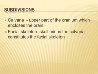
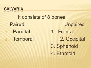
INTRODUCTION TO SKULL
DEFINITION
 Skeleton of head is known as Skull
 It is a highly modified region of axial skeleton
 It is of mesenchymal origin
 It consists of 22 bones.
FUNCTION
 Protection and housing of brain
 Acts as rigid barrier between brain and
impact produced by masticatory and axial
musculature
SUBDIVISIONS
 Calvaria - upper part of the cranium which
encloses the brain
 Facial skeleton- skull minus the calvaria
constitutes the facial skeleton
CALVARIA
It consists of 8 bones
Paired Unpaired
1. Parietal 1. Frontal
2. Temporal 2. Occipital
3. Sphenoid
4. Ethmoid
SUTURES
Most of the joints of skull are immovable and
fibrous in type these are known as Sutures.
 Coronal suture
 Saggital suture
 Lambdoid suture
 Temporoparietal suture
FACIAL SKELETON
It is composed of 14 bones
Paired Unpaired
1. Maxilla 1. Mandible
2. Zygomatic 2. Vomer
3. Nasal
4. Lacrimal
5. Palatine
6. Inferior nasal concha
ANATOMICAL POSITION
Planes for orientation includes:
 Reids’s Base line – horizontal line joining
infraorbital margin to centre of external
acoustic meatus.
 Frankfort Horizontal plane – obtained by
joining the infraorbital margin to upper
margin of external acoustic meatus.
METHODS OF STUDY
1. For the purpose of examination skull maybe viewed
externally :
 From Front- Norma Frontalis
 From Behind – Norma occipitalis
 From the Sides – Norma Lateralis
 From Above – Normal Verticalis
 From Below – Normal Basalis
2. Interior of Skull - it is divided into anterior, middle and
posterior cranial fossa.
3. Study of individual bone.
NORMA VERTICALIS
Shape : oval, wider posteriorly
Bones seen:
Anteriorly - upper part of frontal bone
Posteriorly – uppermost part of occipital bone
Side – parietal bone on each side
Sutures – Coronal
Saggital
Lambdoid
Metopic
NAMED FEATURES
 Vertex – highest point on saggital suture.
 Vault – it is the arched roof for the dome of
the skull.
 Bregma – meeting point between coronal
and saggital sutures.
 Lambda meeting point between saggital and
lambdoid suture.
 Parietal eminence - is the area of maximum
convexity of parietal bone.
 Parietal foramen – one on each side, pierces
parietal bone near its upper border, 2.5cm to
4cm in front of lambda
 Obelion – is the point on saggital suture
between two parietal foramina.
 Temporal lines
NORMA OCCIPITALIS
 Shape – convex upward and on each side,
flattened below
 Bones seen –
Above – posterior parts of parietal bone
below – upper part of squamous part of
occiptal bone below
side – mastoid part of temporal bone on
each side
Sutures : Lambdoid suture
occipitomastoid suture
parietomastoid suture
posterior part of saggital suture
Other feature :
External occipital protuberance
Superior nuchal lines
Highest nuchal lines
Occipital point
Mastoid foramen
Interparietal bone
ATTACHMENTS
1. External occipital protuberance
- Upper part – Trapezius
- Lower part – Ligamentum nuchae
2. Superior nuchal line
Medial 1/3rd – Trapezius
Lateral - Sternocleidomastoid (above)
Splenius capitis (below)
3. Highest Nuchal line
Medially – Epicranial aponeurosis
Laterally – Occipital belly of Occipitofrontalis muscle
NORMA FRONTALIS
Shape : roughly oval, being wider above than
below.
Bones :
1. Frontal bone – forms the forehead
2. rt. & lt. Maxillae – forms the upper jaw
3. rt. & lt. Nasal bone – forms the bridge of nose
4. Zygomatic bone – forms the bony prominence
of cheek
5. Mandible – forms the lower jaw
FRONTAL REGION
 Superciliary arch – rounded, curved
elevation situated just above the medial part
of each orbit.
 Glabella – median elevation connecting two
superciliary arches
 Nasion – median point at root of nose where
internasal suture meets with frontonasal
suture
 Frontal eminence – is a low rounded
elevation above the superciliary arch
ORBITAL OPENING
 Quadrangular in shape
 Bounded by 4 margins :
a) Supraorbital margin – formed by frontal
bone, contain supraorbital notch or foramen
b) Infraorbital margin –
formed laterally by- Zygomatic bone
medially by - Maxilla
Medial orbital margin –
formed Above - by frontal bone
formed Below – by lacrimal crest of
frontal process of maxilla
Lateral orbital margin – formed mostly by
frontal process of zygomatic bone but is
completed above by zygomatic process of
frontal bone
ANTERIOR BONY APERTURE OF NOSE
 It is pear shaped, wide below & narrow
above
 Boundaries –
above – by lower border of nasal bone
below – by nasal notch of body of maxilla
FEATURES
1. Articulation of nasal bone
a. Anteriorly – with opposite bone at internasal
suture
b. Posteriorly – with frontal process of maxilla
c. Superiorly – with frontal bone at frontonasal
suture
d. Inferiorly – upper nasal cartilage is attached to
it
2. Most commonly fractured bone of face
3. Anterior nasal spine – sharp projection in
median plane in lower boundary of piriform
LOWER PART OF FACE
Maxilla
Anterior surface of body of maxilla presents –
nasal notch medially
anterior nasal spine
infraorbital foramen
incisive fossa
canine fossa
Prosesses seen : Frontal process of maxilla
Zygomatic process
Alveolar process
Zygomatic Bone
 Forms the prominence of cheek
 Zygomatic formamen is also seen
Mandible
 Forms the lower jaw
 Upper border or alveolar arch lodges the lower teeth
 Lower border or base is rounded
 Mid point of base is called mental point or anathion
 Anterior surface of mandible presents:
Symphysis menti
Mental protuberance
Mental tubercle
Mental foramen
Oblique line
SUTURES OF NORMA FRONTALIS
 Internasal
 Frontonasal
 Nasomaxillary
 Lacrimomaxillary
 Frontomaxillary
 Intermaxillary
 Zygomaticomaxillary
 Zygomaticofrontal
INTERIOR OF BASE OF SKULL
It is subdivided into :
 Anterior Cranial
Fossa
 Middle Cranial Fossa
 Posterial Cranial
Fossa
FEOTAL SKULL
Dimensions
 Larger in proportion to other parts of skeleton
 The facial skeleton is 1/8th of calvaria whereas it
is ½ of calvaria
 Base of skull is short & narrow
 Internal ear is of same size as in adult
Structure of bones :
 Bones in cranial vault are smooth & unilamellar,
 There is no diploe
 Tables and diploe appear by fourth year of age
Bony Prominences
 Frontal & Parietal tubera are prominent
 Glabella, superciliary arches and mastoid
processes are not developed
Ossification of Bones
 Two halves of frontal bone are separated by
metopic suture.
 Mandible is also present in two halves
 Occipital bone is in 4 parts ( (1)squamous, (2)
condylar, and (1) basilar)
 Unossified membranous gap a total of 6
frontanelle at the angles of parietal bones are
present.
TEMPORAL BONE
 Internal ear, tymphanic cavity, tympanic antrum & ear
ossicles are of adult size
 Mastoid process is absent, appears during later part of
2nd year
 External acoustic meatus is short & straight. Its bony part
is unossified and represented by a fibrocartilaginous
plate.
 Tympanic membrane faces more downwards than
laterally
 Stylomastoid foramen is exposed on lateral surface of
skull
 Mandibular fossa is flat and placed more laterally,and the
auricular tubercle has not developed
 Sub arcuate fossa is deep & prominent
 Facial canal is short.
Orbits
 These are large
 Germs of developing teeth lies close to
orbital floor
Paranasal Air sinuses
 Are rudimentary or absent
POST NATAL GROWTH OF SKULL
 It proceeds at different rate and over different
periods
 Growth of calvaria related to - growth of brain
 Grwoth of facial skeleton related to -
development of dentition, muscle of
mastication & tongue
GROWTH OF VAULT
Rate – rapid during first yr.
slow upto 7th yr.
Growth in breadth : occurs at
saggital suture
sutures bordering greater
wings,
Occipito mastoid suture
Petro- occipital suture at base
Growth in Height - occurs at
Frontozygomatic suture
Pterion
Squamous suture
Asterion
Growth in Anteroposterior Diameter - occurs at
coronal & lambdoid
sutures
GROWTH OF BASE
It grows in antero-posterior diameter at 3
cartilaginous plates situated between –
Occipital & sphenoid bones
Pre & post sphenoid bones
Sphenoid & ethmoid bones
GROWTH OF FACE
 Growth of orbits and ethmoid is complete by
7th yr.
 Growth occurs mostly during first year,
although continues till puberty n even later
CLOSURE OF FRONTANELLES
 Anterior – by 18 months
 Posterior – by 2-3 months
 Mastoid – by 12 months
THICKENING OF BONES
 Two tables and diploe appear by 4th yr.
 Differentiation reaches maximum by about 35
yrs., when diploic vein produce characteristic
arking in radiographs
 Mastoid process appear during 2nd yr, and
mastoid air cells during 6th yr.
OBLITERATION OF SUTURES OF VAULT
 It begins on inner surface between 30-40 yrs. & on
outer surface between 40-50 yrs.
 Occurs first in lower part of coronal suture
Posterior part of saggital suture
Lambdoid suture
AGE RELATED CHANGES
 Skull becomes thinner & lighter
 Reduction in size of maxilla and mandible
 Decrease in vertical height of face
 Angle of mandible becomes more obtuse
SEX DIFFERENCE
CRANIOMETRY
1. Cephalic Index
it expresses shape of head,& is the proportion of
breadth to length of skull
cephalic index = breadth x 100
length
Length/ longest diameter – measured from
glabella to occipital point
Breadth / widest diameter – measured a little
below parietal tubera
Human races may be-
a. Dolichocephalic or long headed ( when C.I
is 75 or less)
b. Mesaticephalic ( when C.I is between 75
and 80)
c. Brachycephalic or Short headed (when C.I
is above 80)
FACIAL ANGLE
 Is the angle between 2 lines drwan from the
nasion to th basion or anterior margin of
foramen magnum and the prosthion or
central point on upper incisor alveolus
 It is a rough index of degree of devlopment of
brain because it is angle between facial
skeleton and calvaria, which are inversely
propotional to each other.
 Angle – smallest- evolved races of man
larger – in lower races
ABNORMAL CRANIA
 Oxycephaly or Acrocephaly – tower like skull
or steeple skull is a abnormally tall skull.
- Occurs due to premature closure of suture
between presphenoid and postsphenoid and
coronal suture
 Scaphocephaly or boat shaped skull is due
to premature synostosis in saggital suture

Introduction to skull[1]

  • 1.
  • 2.
    DEFINITION  Skeleton ofhead is known as Skull  It is a highly modified region of axial skeleton  It is of mesenchymal origin  It consists of 22 bones.
  • 3.
    FUNCTION  Protection andhousing of brain  Acts as rigid barrier between brain and impact produced by masticatory and axial musculature
  • 4.
    SUBDIVISIONS  Calvaria -upper part of the cranium which encloses the brain  Facial skeleton- skull minus the calvaria constitutes the facial skeleton
  • 5.
    CALVARIA It consists of8 bones Paired Unpaired 1. Parietal 1. Frontal 2. Temporal 2. Occipital 3. Sphenoid 4. Ethmoid
  • 6.
    SUTURES Most of thejoints of skull are immovable and fibrous in type these are known as Sutures.  Coronal suture  Saggital suture  Lambdoid suture  Temporoparietal suture
  • 7.
    FACIAL SKELETON It iscomposed of 14 bones Paired Unpaired 1. Maxilla 1. Mandible 2. Zygomatic 2. Vomer 3. Nasal 4. Lacrimal 5. Palatine 6. Inferior nasal concha
  • 8.
    ANATOMICAL POSITION Planes fororientation includes:  Reids’s Base line – horizontal line joining infraorbital margin to centre of external acoustic meatus.  Frankfort Horizontal plane – obtained by joining the infraorbital margin to upper margin of external acoustic meatus.
  • 9.
    METHODS OF STUDY 1.For the purpose of examination skull maybe viewed externally :  From Front- Norma Frontalis  From Behind – Norma occipitalis  From the Sides – Norma Lateralis  From Above – Normal Verticalis  From Below – Normal Basalis 2. Interior of Skull - it is divided into anterior, middle and posterior cranial fossa. 3. Study of individual bone.
  • 10.
    NORMA VERTICALIS Shape :oval, wider posteriorly Bones seen: Anteriorly - upper part of frontal bone Posteriorly – uppermost part of occipital bone Side – parietal bone on each side Sutures – Coronal Saggital Lambdoid Metopic
  • 11.
    NAMED FEATURES  Vertex– highest point on saggital suture.  Vault – it is the arched roof for the dome of the skull.  Bregma – meeting point between coronal and saggital sutures.  Lambda meeting point between saggital and lambdoid suture.
  • 12.
     Parietal eminence- is the area of maximum convexity of parietal bone.  Parietal foramen – one on each side, pierces parietal bone near its upper border, 2.5cm to 4cm in front of lambda  Obelion – is the point on saggital suture between two parietal foramina.  Temporal lines
  • 13.
    NORMA OCCIPITALIS  Shape– convex upward and on each side, flattened below  Bones seen – Above – posterior parts of parietal bone below – upper part of squamous part of occiptal bone below side – mastoid part of temporal bone on each side
  • 14.
    Sutures : Lambdoidsuture occipitomastoid suture parietomastoid suture posterior part of saggital suture Other feature : External occipital protuberance Superior nuchal lines Highest nuchal lines Occipital point Mastoid foramen Interparietal bone
  • 15.
    ATTACHMENTS 1. External occipitalprotuberance - Upper part – Trapezius - Lower part – Ligamentum nuchae 2. Superior nuchal line Medial 1/3rd – Trapezius Lateral - Sternocleidomastoid (above) Splenius capitis (below) 3. Highest Nuchal line Medially – Epicranial aponeurosis Laterally – Occipital belly of Occipitofrontalis muscle
  • 16.
    NORMA FRONTALIS Shape :roughly oval, being wider above than below. Bones : 1. Frontal bone – forms the forehead 2. rt. & lt. Maxillae – forms the upper jaw 3. rt. & lt. Nasal bone – forms the bridge of nose 4. Zygomatic bone – forms the bony prominence of cheek 5. Mandible – forms the lower jaw
  • 17.
    FRONTAL REGION  Superciliaryarch – rounded, curved elevation situated just above the medial part of each orbit.  Glabella – median elevation connecting two superciliary arches  Nasion – median point at root of nose where internasal suture meets with frontonasal suture  Frontal eminence – is a low rounded elevation above the superciliary arch
  • 18.
    ORBITAL OPENING  Quadrangularin shape  Bounded by 4 margins : a) Supraorbital margin – formed by frontal bone, contain supraorbital notch or foramen b) Infraorbital margin – formed laterally by- Zygomatic bone medially by - Maxilla
  • 19.
    Medial orbital margin– formed Above - by frontal bone formed Below – by lacrimal crest of frontal process of maxilla Lateral orbital margin – formed mostly by frontal process of zygomatic bone but is completed above by zygomatic process of frontal bone
  • 20.
    ANTERIOR BONY APERTUREOF NOSE  It is pear shaped, wide below & narrow above  Boundaries – above – by lower border of nasal bone below – by nasal notch of body of maxilla
  • 21.
    FEATURES 1. Articulation ofnasal bone a. Anteriorly – with opposite bone at internasal suture b. Posteriorly – with frontal process of maxilla c. Superiorly – with frontal bone at frontonasal suture d. Inferiorly – upper nasal cartilage is attached to it 2. Most commonly fractured bone of face 3. Anterior nasal spine – sharp projection in median plane in lower boundary of piriform
  • 22.
    LOWER PART OFFACE Maxilla Anterior surface of body of maxilla presents – nasal notch medially anterior nasal spine infraorbital foramen incisive fossa canine fossa Prosesses seen : Frontal process of maxilla Zygomatic process Alveolar process
  • 23.
    Zygomatic Bone  Formsthe prominence of cheek  Zygomatic formamen is also seen Mandible  Forms the lower jaw  Upper border or alveolar arch lodges the lower teeth  Lower border or base is rounded  Mid point of base is called mental point or anathion  Anterior surface of mandible presents: Symphysis menti Mental protuberance Mental tubercle Mental foramen Oblique line
  • 24.
    SUTURES OF NORMAFRONTALIS  Internasal  Frontonasal  Nasomaxillary  Lacrimomaxillary  Frontomaxillary  Intermaxillary  Zygomaticomaxillary  Zygomaticofrontal
  • 33.
    INTERIOR OF BASEOF SKULL It is subdivided into :  Anterior Cranial Fossa  Middle Cranial Fossa  Posterial Cranial Fossa
  • 35.
    FEOTAL SKULL Dimensions  Largerin proportion to other parts of skeleton  The facial skeleton is 1/8th of calvaria whereas it is ½ of calvaria  Base of skull is short & narrow  Internal ear is of same size as in adult Structure of bones :  Bones in cranial vault are smooth & unilamellar,  There is no diploe  Tables and diploe appear by fourth year of age
  • 36.
    Bony Prominences  Frontal& Parietal tubera are prominent  Glabella, superciliary arches and mastoid processes are not developed Ossification of Bones  Two halves of frontal bone are separated by metopic suture.  Mandible is also present in two halves  Occipital bone is in 4 parts ( (1)squamous, (2) condylar, and (1) basilar)  Unossified membranous gap a total of 6 frontanelle at the angles of parietal bones are present.
  • 37.
    TEMPORAL BONE  Internalear, tymphanic cavity, tympanic antrum & ear ossicles are of adult size  Mastoid process is absent, appears during later part of 2nd year  External acoustic meatus is short & straight. Its bony part is unossified and represented by a fibrocartilaginous plate.  Tympanic membrane faces more downwards than laterally  Stylomastoid foramen is exposed on lateral surface of skull  Mandibular fossa is flat and placed more laterally,and the auricular tubercle has not developed  Sub arcuate fossa is deep & prominent  Facial canal is short.
  • 38.
    Orbits  These arelarge  Germs of developing teeth lies close to orbital floor Paranasal Air sinuses  Are rudimentary or absent
  • 39.
    POST NATAL GROWTHOF SKULL  It proceeds at different rate and over different periods  Growth of calvaria related to - growth of brain  Grwoth of facial skeleton related to - development of dentition, muscle of mastication & tongue
  • 40.
    GROWTH OF VAULT Rate– rapid during first yr. slow upto 7th yr. Growth in breadth : occurs at saggital suture sutures bordering greater wings, Occipito mastoid suture Petro- occipital suture at base
  • 41.
    Growth in Height- occurs at Frontozygomatic suture Pterion Squamous suture Asterion Growth in Anteroposterior Diameter - occurs at coronal & lambdoid sutures
  • 42.
    GROWTH OF BASE Itgrows in antero-posterior diameter at 3 cartilaginous plates situated between – Occipital & sphenoid bones Pre & post sphenoid bones Sphenoid & ethmoid bones
  • 43.
    GROWTH OF FACE Growth of orbits and ethmoid is complete by 7th yr.  Growth occurs mostly during first year, although continues till puberty n even later
  • 44.
    CLOSURE OF FRONTANELLES Anterior – by 18 months  Posterior – by 2-3 months  Mastoid – by 12 months
  • 45.
    THICKENING OF BONES Two tables and diploe appear by 4th yr.  Differentiation reaches maximum by about 35 yrs., when diploic vein produce characteristic arking in radiographs  Mastoid process appear during 2nd yr, and mastoid air cells during 6th yr.
  • 46.
    OBLITERATION OF SUTURESOF VAULT  It begins on inner surface between 30-40 yrs. & on outer surface between 40-50 yrs.  Occurs first in lower part of coronal suture Posterior part of saggital suture Lambdoid suture
  • 47.
    AGE RELATED CHANGES Skull becomes thinner & lighter  Reduction in size of maxilla and mandible  Decrease in vertical height of face  Angle of mandible becomes more obtuse
  • 48.
  • 49.
    CRANIOMETRY 1. Cephalic Index itexpresses shape of head,& is the proportion of breadth to length of skull cephalic index = breadth x 100 length Length/ longest diameter – measured from glabella to occipital point Breadth / widest diameter – measured a little below parietal tubera
  • 50.
    Human races maybe- a. Dolichocephalic or long headed ( when C.I is 75 or less) b. Mesaticephalic ( when C.I is between 75 and 80) c. Brachycephalic or Short headed (when C.I is above 80)
  • 51.
    FACIAL ANGLE  Isthe angle between 2 lines drwan from the nasion to th basion or anterior margin of foramen magnum and the prosthion or central point on upper incisor alveolus  It is a rough index of degree of devlopment of brain because it is angle between facial skeleton and calvaria, which are inversely propotional to each other.  Angle – smallest- evolved races of man larger – in lower races
  • 52.
    ABNORMAL CRANIA  Oxycephalyor Acrocephaly – tower like skull or steeple skull is a abnormally tall skull. - Occurs due to premature closure of suture between presphenoid and postsphenoid and coronal suture  Scaphocephaly or boat shaped skull is due to premature synostosis in saggital suture