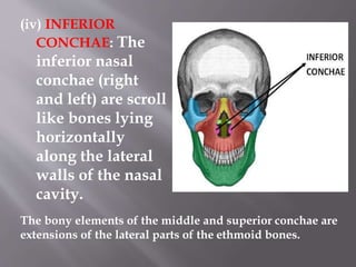
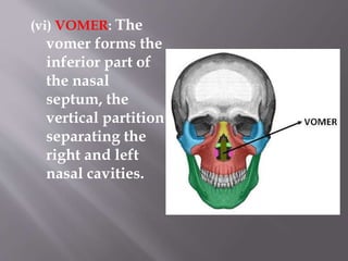
Downloaded 998 times


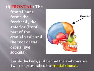
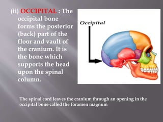
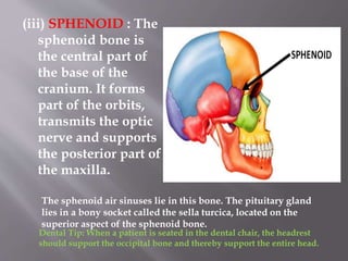


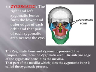


The skull is composed of 22 bones that form the cranium (housing the brain) and face. The 8 bones of the cranium are the frontal, occipital, sphenoid, ethmoid, and two each of the parietal and temporal bones. These bones protect the brain and support structures like the spinal cord. The 14 bones of the face are the mandible, maxillae, zygomatic, lacrimal, nasal, inferior conchae, palatine, and vomer bones, which form structures like the nose and jaws.


