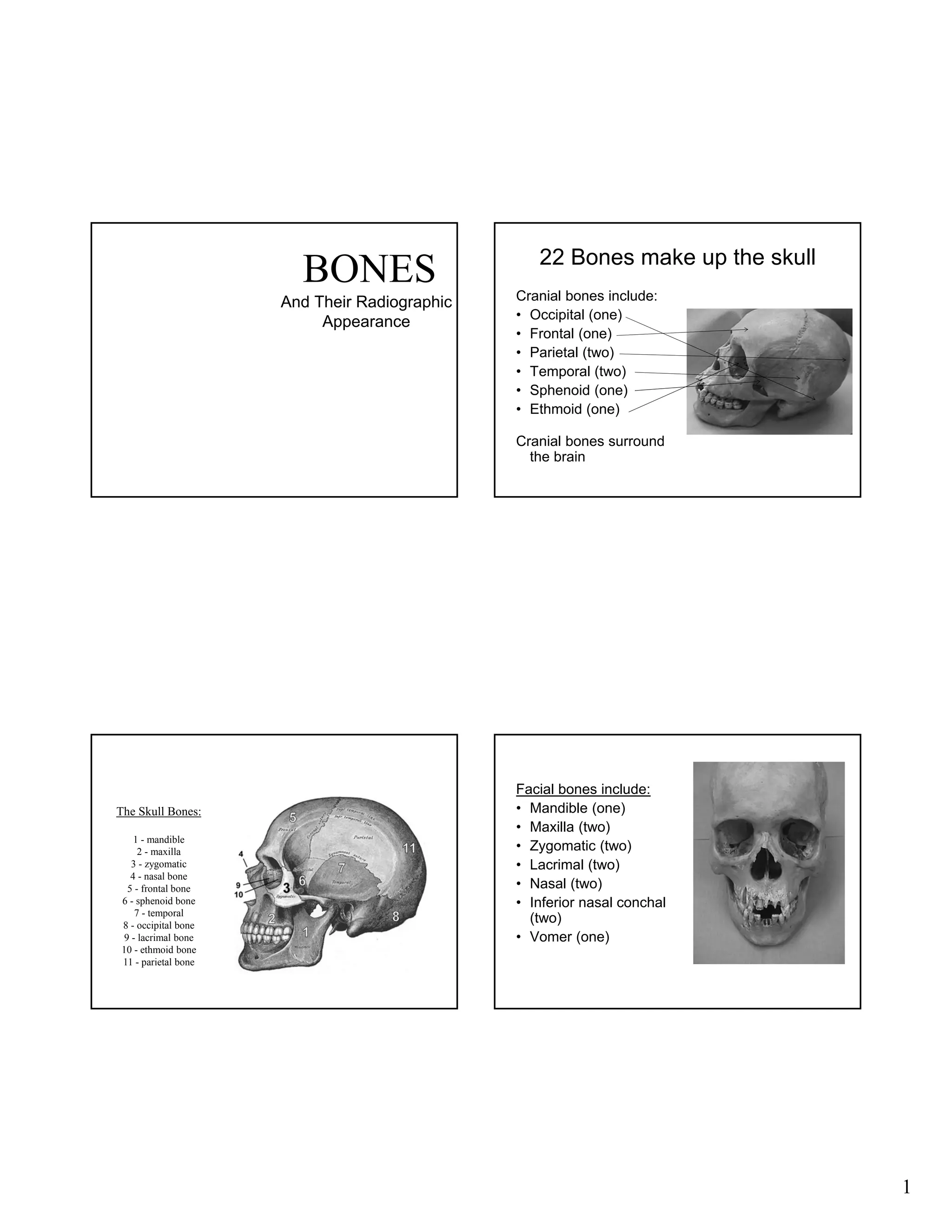
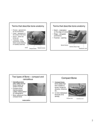
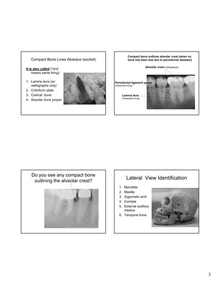
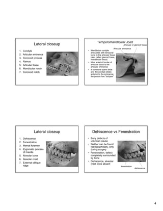
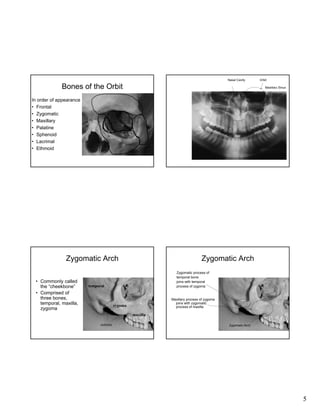
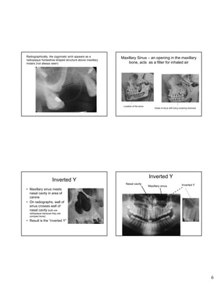
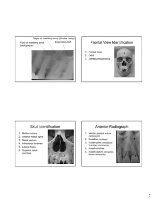
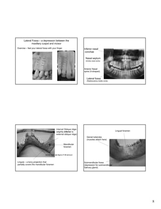
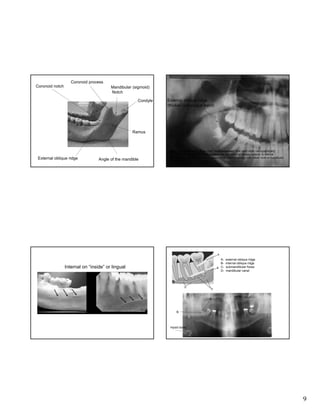
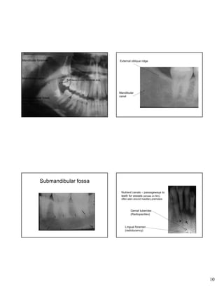
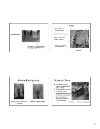
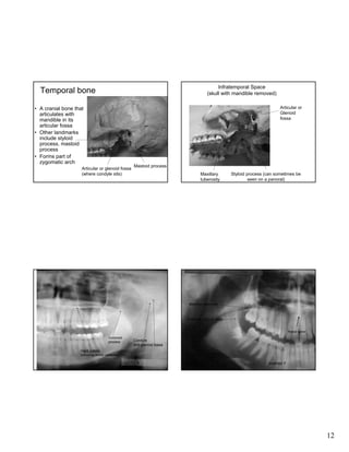
BONES
And Their Radiographic
Appearance

22 Bones make up the skull
Cranial bones include:
• Occipital (one)
• Frontal (one)
• Parietal (two)
• Temporal (two)
• Sphenoid (one)
• Ethmoid (one)
Cranial bones surround
the brain

The Skull Bones:
1 - mandible
2 - maxilla
3 - zygomatic
4 - nasal bone
5 - frontal bone
6 - sphenoid bone
7 - temporal
8 - occipital bone
9 - lacrimal bone
10 - ethmoid bone
11 - parietal bone

Facial bones include:
• Mandible (one)
• Maxilla (two)
• Zygomatic (two)
• Lacrimal (two)
• Nasal (two)
• Inferior nasal conchal
(two)
• Vomer (one)

1
Terms that describe bone anatomy
• Process – general term
for any prominence
• Fossa - depression on
the surface of the bone
• Suture – where two
bones join
• Tuberosity – bony
prominance, usually
where muscle attaches,
ie maxillary tuberosity
suture

Terms that describe bone anatomy
• Notch – indentation
on the edge of a bone
• Ridge – elongated
prominence
• Foramen - opening

Mental foramen
External oblique ridge
Temporal fossa

Mastoid process

Two types of Bone – compact and
cancellous
• Cancellous bone
(also called spongy
bone) makes up
center of bones
• Contains bone
marrow spaces
(called “trabeculation”
on radiographs)
• More radiolucent than
compact bone

Mandibular notch

Compact Bone
1. Compact boneforms plates that
form outside of
bones, linings for
alveolus, foramina,
etc…
2. Is more radiopaque
because of its
density
Compact bone

Cancellous bone

trabeculation

2
Compact bone outlines alveolar crest (when no
bone has been lost due to periodontal disease!)

Compact Bone Lines Alveolus (socket)

Alveolar crest (radiopaque)

It is also called (*and
means same thing):
1. Lamina dura (on
radiographs only)
2. Cribriform plate
3. Cortical bone
4. Alveolar bone proper

Periodontal ligament space
(radiolucent lining)

Lamina dura
(radiopaque lining)

Do you see any compact bone
outlining the alveolar crest?

Lateral View Identification
1.
2.
3.
4.
5.

Mandible
Maxilla
Zygomatic arch
Condyle
External auditory
meatus
6. Temporal bone

3
Temporomandibular Joint

Lateral closeup
1.
2.
3.
4.
5.
6.
7.

Condyle
Articular eminence
Coronoid process
Ramus
Articular fossa
Mandibular notch
Coronoid notch

Articular or gleniod fossa
Articular eminance
• Mandibular condyle
articulates with temporal
bone in the articular fossa
(also called glenoid fossa,
mandibular fossa)
• Most anterior border of
articular fossa is the
articular eminance
• If someone opens wide
and the condyle slides
anterior to the eminance,
the person has “lockjaw”

6

7

Lateral closeup
Dehiscence
Fenestration
Mental foramen
Zygomatic process
of maxilla
5. Alveolar bone
6. Alveolar crest
7. External oblique
ridge

Dehiscence vs Fenestration

1.
2.
3.
4.

7

• Bony defects of
unknown cause
• Neither can be found
radiographically, only
during surgery
• Fenestration, defect
completely surrounded
by bone
• Dehiscence, alveolar
crest bone absent
fenestration

dehiscence

4
Nasal Cavity

Bones of the Orbit

Orbit
Maxillary Sinus

In order of appearance
• Frontal
• Zygomatic
• Maxillary
• Palatine
• Sphenoid
• Lacrimal
• Ethmoid

Zygomatic Arch
• Commonly called
the “cheekbone”
• Comprised of
three bones,
temporal, maxilla,
zygoma

Zygomatic Arch
Zygomatic process of
temporal bone
joins with temporal
process of zygoma

temporal

Maxillary process of zygoma
joins with zygomatic
process of maxilla

zygoma
maxilla
sutures

Zygomatic Arch

5
Radiographically, the zygomatic arch appears as a
radiopaque horseshoe shaped structure above maxillary
molars (not always seen)

Maxillary Sinus – an opening in the maxillary
bone, acts as a filter for inhaled air

Location of the sinus

Inside of sinus with bony covering removed

Inverted Y

Inverted Y
Nasal cavity

Maxillary sinus

Inverted Y

• Maxillary sinus meets
nasal cavity in area of
canine
• On radiographs, wall of
sinus crosses wall of
nasal cavity (both are
radiopaque because they are
compact bone)

• Result is the “inverted Y”

6
Septa of maxillary sinus (divides cavity)
Zygomatic Arch

Floor of maxillary sinus
(radiopaque)

Frontal View Identification
1. Frontal bone
2. Orbit
3. Mental protuberance

Skull Identification
1.
2.
3.
4.
5.
6.

Midline suture
Anterior Nasal spine
Nasal septum
Infraorbital foramen
Lateral fossa
Superior nasal
conchae

Anterior Radiograph
1. Median palatal suture

5

(radiolucent)
6

2. Noseline (cartilage)
3. Nasal spine (radiopaque
V-shaped prominence)

4. Nasal conchae
5. Nasal septum (elongated,
thicker radiopacity)

7
Lateral Fossa – a depression between the
maxillary cuspid and incisor
Exercise – feel your lateral fossa with your finger

Inferior nasal
conchae
Nasal septum
(divides nasal cavity)

Anterior Nasal
spine (V-shaped)

Lateral fossa
(Radiolucency inside circle)

Internal Oblique ridge(slightly inferior to
external oblique ridge)

Lingual foramen
Genial tubercles
(muscles attach here)

Mandibular
foramen

Mental ridge- see figure 27-56 Iannucci

Lingula – a bony projection that
partially covers the mandibular foramen

Submandibular fossa
(depression for submandibular
Salivary gland)

8
Coronoid process
Coronoid notch

Mandibular (sigmoid)
Notch
Condyle

External oblique ridge
(thicker radiopaque band)

Ramus

External oblique ridge

Angle of the mandible

Note: External/internal ridges often “superimposed” over each other radiographically;
therefore difficult to differentiate between the two; external always superior to internal
oblique ridge (mylohyoid muscle attachment); internal usually runs below roots of mandibular
molars (see figure 26-62 Haring)

ABCD-

Internal on “inside” or lingual

external oblique ridge
internal oblique ridge
submandibular fossa
mandibular canal

A

B

Hyoid bone

9
Mandibular foramen

Mandibular canal

External oblique ridge

Soft tissue outline- retromolar area

Submandibular fossa

Mandibular
canal

(large radiolucency
within the circle)

Submandibular fossa
Nutrient canals – passageways to
teeth for vessels (arrows on film),
often seen around maxillary premolars

Genial tubercles
(Radiopacities)

Lingual foramen
(radiolucency)

10
Palate
Nasopalatine or
Incisive foramen
Median palatine suture

Mental Foramen

Anterior or Greater
palatine foramen

Mylohyoid ridge or internal oblique ridge
(see down by roots of teeth)– actually
on lingual of mandible

Posterior or Lessor
palatine foramen
Hamulus

Palatal Radiographs

Nasopalatine or Incisive
foramen

Median Palatal suture

Sphenoid Bone
• A butterfly shaped
cranial bone posterior
to the palate
• It forms part of the orbit
• It’s hamulus can
sometimes be seen on
third molar radiograph
• Some muscles of
masticaton attach to
Pterygoid plate
Hamulus

Lateral pterygoid plate

11
Infratemporal Space
(skull with mandible removed)

Temporal bone

Articular or
Glenoid
fossa

• A cranial bone that
articulates with
mandible in its
articular fossa
• Other landmarks
include styloid
process, mastoid
process
• Forms part of
zygomatic arch
Articular or glenoid fossa
(where condyle sits)

Mastoid process
Maxillary
tuberosity

Styloid process (can sometimes be
seen on a panoral)

Maxillary tuberosity

External oblique ridge
Nasal spine
Coronoid
process

Condyle
and glenoid fossa

Hard palate
(horizontal thicker radiopaque line)

Inverted Y

12

Bones post 11

  • 1.
    BONES And Their Radiographic Appearance 22Bones make up the skull Cranial bones include: • Occipital (one) • Frontal (one) • Parietal (two) • Temporal (two) • Sphenoid (one) • Ethmoid (one) Cranial bones surround the brain The Skull Bones: 1 - mandible 2 - maxilla 3 - zygomatic 4 - nasal bone 5 - frontal bone 6 - sphenoid bone 7 - temporal 8 - occipital bone 9 - lacrimal bone 10 - ethmoid bone 11 - parietal bone Facial bones include: • Mandible (one) • Maxilla (two) • Zygomatic (two) • Lacrimal (two) • Nasal (two) • Inferior nasal conchal (two) • Vomer (one) 1
  • 2.
    Terms that describebone anatomy • Process – general term for any prominence • Fossa - depression on the surface of the bone • Suture – where two bones join • Tuberosity – bony prominance, usually where muscle attaches, ie maxillary tuberosity suture Terms that describe bone anatomy • Notch – indentation on the edge of a bone • Ridge – elongated prominence • Foramen - opening Mental foramen External oblique ridge Temporal fossa Mastoid process Two types of Bone – compact and cancellous • Cancellous bone (also called spongy bone) makes up center of bones • Contains bone marrow spaces (called “trabeculation” on radiographs) • More radiolucent than compact bone Mandibular notch Compact Bone 1. Compact boneforms plates that form outside of bones, linings for alveolus, foramina, etc… 2. Is more radiopaque because of its density Compact bone Cancellous bone trabeculation 2
  • 3.
    Compact bone outlinesalveolar crest (when no bone has been lost due to periodontal disease!) Compact Bone Lines Alveolus (socket) Alveolar crest (radiopaque) It is also called (*and means same thing): 1. Lamina dura (on radiographs only) 2. Cribriform plate 3. Cortical bone 4. Alveolar bone proper Periodontal ligament space (radiolucent lining) Lamina dura (radiopaque lining) Do you see any compact bone outlining the alveolar crest? Lateral View Identification 1. 2. 3. 4. 5. Mandible Maxilla Zygomatic arch Condyle External auditory meatus 6. Temporal bone 3
  • 4.
    Temporomandibular Joint Lateral closeup 1. 2. 3. 4. 5. 6. 7. Condyle Articulareminence Coronoid process Ramus Articular fossa Mandibular notch Coronoid notch Articular or gleniod fossa Articular eminance • Mandibular condyle articulates with temporal bone in the articular fossa (also called glenoid fossa, mandibular fossa) • Most anterior border of articular fossa is the articular eminance • If someone opens wide and the condyle slides anterior to the eminance, the person has “lockjaw” 6 7 Lateral closeup Dehiscence Fenestration Mental foramen Zygomatic process of maxilla 5. Alveolar bone 6. Alveolar crest 7. External oblique ridge Dehiscence vs Fenestration 1. 2. 3. 4. 7 • Bony defects of unknown cause • Neither can be found radiographically, only during surgery • Fenestration, defect completely surrounded by bone • Dehiscence, alveolar crest bone absent fenestration dehiscence 4
  • 5.
    Nasal Cavity Bones ofthe Orbit Orbit Maxillary Sinus In order of appearance • Frontal • Zygomatic • Maxillary • Palatine • Sphenoid • Lacrimal • Ethmoid Zygomatic Arch • Commonly called the “cheekbone” • Comprised of three bones, temporal, maxilla, zygoma Zygomatic Arch Zygomatic process of temporal bone joins with temporal process of zygoma temporal Maxillary process of zygoma joins with zygomatic process of maxilla zygoma maxilla sutures Zygomatic Arch 5
  • 6.
    Radiographically, the zygomaticarch appears as a radiopaque horseshoe shaped structure above maxillary molars (not always seen) Maxillary Sinus – an opening in the maxillary bone, acts as a filter for inhaled air Location of the sinus Inside of sinus with bony covering removed Inverted Y Inverted Y Nasal cavity Maxillary sinus Inverted Y • Maxillary sinus meets nasal cavity in area of canine • On radiographs, wall of sinus crosses wall of nasal cavity (both are radiopaque because they are compact bone) • Result is the “inverted Y” 6
  • 7.
    Septa of maxillarysinus (divides cavity) Zygomatic Arch Floor of maxillary sinus (radiopaque) Frontal View Identification 1. Frontal bone 2. Orbit 3. Mental protuberance Skull Identification 1. 2. 3. 4. 5. 6. Midline suture Anterior Nasal spine Nasal septum Infraorbital foramen Lateral fossa Superior nasal conchae Anterior Radiograph 1. Median palatal suture 5 (radiolucent) 6 2. Noseline (cartilage) 3. Nasal spine (radiopaque V-shaped prominence) 4. Nasal conchae 5. Nasal septum (elongated, thicker radiopacity) 7
  • 8.
    Lateral Fossa –a depression between the maxillary cuspid and incisor Exercise – feel your lateral fossa with your finger Inferior nasal conchae Nasal septum (divides nasal cavity) Anterior Nasal spine (V-shaped) Lateral fossa (Radiolucency inside circle) Internal Oblique ridge(slightly inferior to external oblique ridge) Lingual foramen Genial tubercles (muscles attach here) Mandibular foramen Mental ridge- see figure 27-56 Iannucci Lingula – a bony projection that partially covers the mandibular foramen Submandibular fossa (depression for submandibular Salivary gland) 8
  • 9.
    Coronoid process Coronoid notch Mandibular(sigmoid) Notch Condyle External oblique ridge (thicker radiopaque band) Ramus External oblique ridge Angle of the mandible Note: External/internal ridges often “superimposed” over each other radiographically; therefore difficult to differentiate between the two; external always superior to internal oblique ridge (mylohyoid muscle attachment); internal usually runs below roots of mandibular molars (see figure 26-62 Haring) ABCD- Internal on “inside” or lingual external oblique ridge internal oblique ridge submandibular fossa mandibular canal A B Hyoid bone 9
  • 10.
    Mandibular foramen Mandibular canal Externaloblique ridge Soft tissue outline- retromolar area Submandibular fossa Mandibular canal (large radiolucency within the circle) Submandibular fossa Nutrient canals – passageways to teeth for vessels (arrows on film), often seen around maxillary premolars Genial tubercles (Radiopacities) Lingual foramen (radiolucency) 10
  • 11.
    Palate Nasopalatine or Incisive foramen Medianpalatine suture Mental Foramen Anterior or Greater palatine foramen Mylohyoid ridge or internal oblique ridge (see down by roots of teeth)– actually on lingual of mandible Posterior or Lessor palatine foramen Hamulus Palatal Radiographs Nasopalatine or Incisive foramen Median Palatal suture Sphenoid Bone • A butterfly shaped cranial bone posterior to the palate • It forms part of the orbit • It’s hamulus can sometimes be seen on third molar radiograph • Some muscles of masticaton attach to Pterygoid plate Hamulus Lateral pterygoid plate 11
  • 12.
    Infratemporal Space (skull withmandible removed) Temporal bone Articular or Glenoid fossa • A cranial bone that articulates with mandible in its articular fossa • Other landmarks include styloid process, mastoid process • Forms part of zygomatic arch Articular or glenoid fossa (where condyle sits) Mastoid process Maxillary tuberosity Styloid process (can sometimes be seen on a panoral) Maxillary tuberosity External oblique ridge Nasal spine Coronoid process Condyle and glenoid fossa Hard palate (horizontal thicker radiopaque line) Inverted Y 12