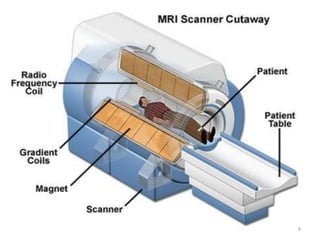
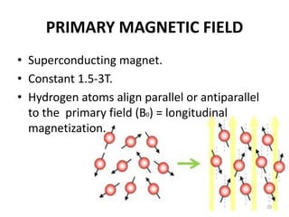
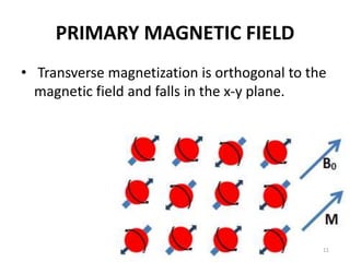
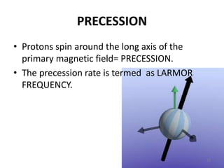
MAGNETIC RESONANCE
IMAGING
QURATULAIN MUGHAL
ISRA UNIVERSITY
BATCH IV
1
CONTENTS
• What is MRI
• MRI equipments
• Indications
• Contraindications
• How it help in diagnosis
• Findings in MRI
• Advantages
• Disadvantages
• Functional MRI
2
DEFINITION
“MRI IS MAGNET RADIO FREQUENCY IMAGING
TECHNIQUE USING A MAGNETIC FIELD AND
RADIOFREQUENCY.”
3
HISTORY OF MRI
• Discovered simultaneously by two physicists:
• Felix Bloch
• Edward Mills Purcell
• First clinical images is obtained by:
• Paul Lauterbur
• Peter Mansfield
• Raymond Damadian
4
MAGNETIC FIELD
• Magnetic flux density is measured in Tesla(T).
• Clinical MRI performed at 1.5-3T.
5
MRI PRINCIPLE
• MRI is based on the principle of nuclear magnetic resonance (NMR)
Two basic principles of NMR
1. Atoms with an odd number of protons or neutrons have spin.
2. A moving electric charge, be it positive or negative, produces a
magnetic field.
• Human body is built of only 26 elements.
• Body has many such atoms that can act as good MR nuclei (1H, 13C,
19F, 23Na)
• Hydrogen nuclei is one of them which is not only positively
charged, but also has magnetic spin.
• MRI utilizes this magnetic spin property of protons of hydrogen to
elicit images.
6
HYDROGEN ATOMS
• Randomly protons are orientated with no applied field.
• So there’s no overall magnetic field.
7
MRI COMPONENTS
• Primary magnet
• Gradient magnets
• Radiofrequency (RF) coils
• Computer system
8
9
PRIMARY MAGNETIC FIELD
• Superconducting magnet.
• Constant 1.5-3T.
• Hydrogen atoms align parallel or antiparallel
to the primary field (B0) = longitudinal
magnetization.
10
PRIMARY MAGNETIC FIELD
• Transverse magnetization is orthogonal to the
magnetic field and falls in the x-y plane.
11
PRECESSION
• Protons spin around the long axis of the
primary magnetic field= PRECESSION.
• The precession rate is termed as LARMOR
FREQUENCY.
12
PRECESSION
• When protons process together, this is known
as in phase.
• When protons process separately, this is
known as out of phase.
13
PRECESSION
• Frequency changes with in proportion to
magnetic field strength.
• At 1.5T=63.9MHz
14
GRADIENT COILS
• It generate secondary magnetic filed over the
primary field.
• They are located within the boa of the primary
magnet.
15
GRADIENT COILS
• They are arrange in opposition to each other
to produce positive and negative pulse.
• The arrangement of these gradient pulse gives
MRI the capacity to image directionally along
the x, y and z axis.
• Alter the primary magnetic field thereby
changing the precessing frequency between
slices.
• They responsible for loud noises of MRI.
16
•Allow spatial encoding for MRI images
in the x, y & z axis i.e. localization.
• Z: axial images
• Y: coronal images
• X: sagittal images
17
18
RADIOFREQUENCY (RF) COILS
• Use to transmit RF pulse (causes the net
magnetic moment of the nuclei) and receiving
signals in MRI.
• They come in many designs alter to best suits
each body part.
19
RELAXATION
• RF coils is use to receives signals to create
images.
• When the protons returns to their prior state
this is known as relaxation.
1. Longitudinal (T1)- parallel to B0 (z axis).
2. Transverse (T2)- perpendicular to B0 (x-y axis)
20
• Different tissues have different relaxation
times.
• These relaxation time differences is used to
generate image contrast.
21
TR AND TE
• TE (echo time) : time interval in which signals
are measured after RF excitation.
• TR (repetition time) : the time between two
excitations is called repetition time.
By varying the TR and TE one can obtain T1WI
and T2WI.
• In general a short TR (<1000ms) and short TE
(<45 ms) scan is T1WI.
• Long TR (>2000ms) and long TE (>45ms) scan is
T2WI.
22
T1-weighted contrast
23
T2-weighted contrast
24
25
COMPUTER SYSTEM
• RF signals covert to analog or digital
conversion mathematical conversions
will form MR images in the screen.
26
KIND OF IMAGES
• T1WI
• T2WI
• DWI
• ADC(Apparent diffusion
coefficient)
• Functional MRI
• MRA(angiography)
• MRV( Magnetic
resonance venography)
• FLAIR
• STIR (Short TI Inversion
Recovery)
27
MAGNETIC RESONANCE
ANGIOGRAPHY
28
FLUID-ATTENUATED INVERSION
RECOVERY
29
DIFFUSION WEIGHTED IMAGING
30
ADC(APPARENT DIFFUSION
COEFFICIENT)
31
MRV
32
STIR (Short Tesla Inversion Recovery)
33
HOW MRI IS DONE?
34
In a Tunnel, of course!
35
CONTRAINDICATIONS
36
37
38
39
MRI INDICATIONS
40
41
42
43
The Uses of the MRI
• Diagnosing: MS; strokes; infections of
the brain/spine/CNS; tendonitis
• Visualising: Injuries; torn ligaments –
especially in areas difficult to see like
the wrist, ankle or knee
• Evaluating: Masses in soft tissue; cysts;
bone tumours or disc problems.
44
FINDINGS IN MRI
When contrast agent is not used an MRI can show:
• The shape, size, appearance, and location of
organs, bones, and joints
• The presence of abnormal growths
• Signs of inflammation or infection
When contrast agent is used MRI can show:
• size and location of benign or malignant growths
• enlarged lymph nodes
• changes in blood flow
• extracellular volume
45
BENEFITS OF MRI
• MRI is non-invasive and does not use radiation
• MRI does not involve radiation
• MRI contrasting agent is less likely to produce an allergic reaction that may
occur when iodine-based substances are used for x-rays and CT scans
• MRI gives extremely clear, detailed images of soft-tissue structures that
other imaging techniques cannot achieve
• MRI can easily create hundreds of images from almost any direction and in
any orientation
• Unlike techniques that examine small parts of the body (i.e. ultrasound or
mammography) MRI exams can cover large portions of the body
• MRI can determine if a cancer has spread, and help determine the best
treatment
46
DISADVANTAGES OF MRI
• MRI is expensive
• MRI will not be able to find all cancers (i.e. breast cancers indicated by
microcalcifications)
• MRI cannot always distinguish between malignant tumors or benign
disease (such as breast fibroadenomas), which could lead to a false
positive results
• MRI is not painful, but the patient must remain still in an enclosed
machine, which may be a problem for claustrophobic patients
• An undetected metal implant in a patient’s body may be affected by the
strong magnet of the MRI unit
• There is a small chance that a patient could develop an allergic reaction to
the contrasting agent, or that a skin infection could develop at the site of
injection
• If a patient chooses to be sedated for the scanning, there is a slight risk
associated with using the sedation medication
47
FUNCTIONAL MAGNETIC RESONANCE
IMAGING
• Functional MRI (fmri) is a functional
neuroimaging procedure using MRI technology
that measures brain activity by detecting changes
associated with blood flow.
• This technique relies on the fact that cerebral
blood flow and neuronal activation are coupled.
• When an area of the brain is in use, blood flow to
that region also increases.
• The primary form of fMRI uses the blood-oxygen-
level dependent (BOLD) contrast.
48
49
50
REFERENCES
• Slideshare.com
• Wikipedia
51

Radiology ppt

  • 1.
  • 2.
    CONTENTS • What isMRI • MRI equipments • Indications • Contraindications • How it help in diagnosis • Findings in MRI • Advantages • Disadvantages • Functional MRI 2
  • 3.
    DEFINITION “MRI IS MAGNETRADIO FREQUENCY IMAGING TECHNIQUE USING A MAGNETIC FIELD AND RADIOFREQUENCY.” 3
  • 4.
    HISTORY OF MRI •Discovered simultaneously by two physicists: • Felix Bloch • Edward Mills Purcell • First clinical images is obtained by: • Paul Lauterbur • Peter Mansfield • Raymond Damadian 4
  • 5.
    MAGNETIC FIELD • Magneticflux density is measured in Tesla(T). • Clinical MRI performed at 1.5-3T. 5
  • 6.
    MRI PRINCIPLE • MRIis based on the principle of nuclear magnetic resonance (NMR) Two basic principles of NMR 1. Atoms with an odd number of protons or neutrons have spin. 2. A moving electric charge, be it positive or negative, produces a magnetic field. • Human body is built of only 26 elements. • Body has many such atoms that can act as good MR nuclei (1H, 13C, 19F, 23Na) • Hydrogen nuclei is one of them which is not only positively charged, but also has magnetic spin. • MRI utilizes this magnetic spin property of protons of hydrogen to elicit images. 6
  • 7.
    HYDROGEN ATOMS • Randomlyprotons are orientated with no applied field. • So there’s no overall magnetic field. 7
  • 8.
    MRI COMPONENTS • Primarymagnet • Gradient magnets • Radiofrequency (RF) coils • Computer system 8
  • 9.
  • 10.
    PRIMARY MAGNETIC FIELD •Superconducting magnet. • Constant 1.5-3T. • Hydrogen atoms align parallel or antiparallel to the primary field (B0) = longitudinal magnetization. 10
  • 11.
    PRIMARY MAGNETIC FIELD •Transverse magnetization is orthogonal to the magnetic field and falls in the x-y plane. 11
  • 12.
    PRECESSION • Protons spinaround the long axis of the primary magnetic field= PRECESSION. • The precession rate is termed as LARMOR FREQUENCY. 12
  • 13.
    PRECESSION • When protonsprocess together, this is known as in phase. • When protons process separately, this is known as out of phase. 13
  • 14.
    PRECESSION • Frequency changeswith in proportion to magnetic field strength. • At 1.5T=63.9MHz 14
  • 15.
    GRADIENT COILS • Itgenerate secondary magnetic filed over the primary field. • They are located within the boa of the primary magnet. 15
  • 16.
    GRADIENT COILS • Theyare arrange in opposition to each other to produce positive and negative pulse. • The arrangement of these gradient pulse gives MRI the capacity to image directionally along the x, y and z axis. • Alter the primary magnetic field thereby changing the precessing frequency between slices. • They responsible for loud noises of MRI. 16
  • 17.
    •Allow spatial encodingfor MRI images in the x, y & z axis i.e. localization. • Z: axial images • Y: coronal images • X: sagittal images 17
  • 18.
  • 19.
    RADIOFREQUENCY (RF) COILS •Use to transmit RF pulse (causes the net magnetic moment of the nuclei) and receiving signals in MRI. • They come in many designs alter to best suits each body part. 19
  • 20.
    RELAXATION • RF coilsis use to receives signals to create images. • When the protons returns to their prior state this is known as relaxation. 1. Longitudinal (T1)- parallel to B0 (z axis). 2. Transverse (T2)- perpendicular to B0 (x-y axis) 20
  • 21.
    • Different tissueshave different relaxation times. • These relaxation time differences is used to generate image contrast. 21
  • 22.
    TR AND TE •TE (echo time) : time interval in which signals are measured after RF excitation. • TR (repetition time) : the time between two excitations is called repetition time. By varying the TR and TE one can obtain T1WI and T2WI. • In general a short TR (<1000ms) and short TE (<45 ms) scan is T1WI. • Long TR (>2000ms) and long TE (>45ms) scan is T2WI. 22
  • 23.
  • 24.
  • 25.
  • 26.
    COMPUTER SYSTEM • RFsignals covert to analog or digital conversion mathematical conversions will form MR images in the screen. 26
  • 27.
    KIND OF IMAGES •T1WI • T2WI • DWI • ADC(Apparent diffusion coefficient) • Functional MRI • MRA(angiography) • MRV( Magnetic resonance venography) • FLAIR • STIR (Short TI Inversion Recovery) 27
  • 28.
  • 29.
  • 30.
  • 31.
  • 32.
  • 33.
    STIR (Short TeslaInversion Recovery) 33
  • 34.
    HOW MRI ISDONE? 34
  • 35.
    In a Tunnel,of course! 35
  • 36.
  • 37.
  • 38.
  • 39.
  • 40.
  • 41.
  • 42.
  • 43.
  • 44.
    The Uses ofthe MRI • Diagnosing: MS; strokes; infections of the brain/spine/CNS; tendonitis • Visualising: Injuries; torn ligaments – especially in areas difficult to see like the wrist, ankle or knee • Evaluating: Masses in soft tissue; cysts; bone tumours or disc problems. 44
  • 45.
    FINDINGS IN MRI Whencontrast agent is not used an MRI can show: • The shape, size, appearance, and location of organs, bones, and joints • The presence of abnormal growths • Signs of inflammation or infection When contrast agent is used MRI can show: • size and location of benign or malignant growths • enlarged lymph nodes • changes in blood flow • extracellular volume 45
  • 46.
    BENEFITS OF MRI •MRI is non-invasive and does not use radiation • MRI does not involve radiation • MRI contrasting agent is less likely to produce an allergic reaction that may occur when iodine-based substances are used for x-rays and CT scans • MRI gives extremely clear, detailed images of soft-tissue structures that other imaging techniques cannot achieve • MRI can easily create hundreds of images from almost any direction and in any orientation • Unlike techniques that examine small parts of the body (i.e. ultrasound or mammography) MRI exams can cover large portions of the body • MRI can determine if a cancer has spread, and help determine the best treatment 46
  • 47.
    DISADVANTAGES OF MRI •MRI is expensive • MRI will not be able to find all cancers (i.e. breast cancers indicated by microcalcifications) • MRI cannot always distinguish between malignant tumors or benign disease (such as breast fibroadenomas), which could lead to a false positive results • MRI is not painful, but the patient must remain still in an enclosed machine, which may be a problem for claustrophobic patients • An undetected metal implant in a patient’s body may be affected by the strong magnet of the MRI unit • There is a small chance that a patient could develop an allergic reaction to the contrasting agent, or that a skin infection could develop at the site of injection • If a patient chooses to be sedated for the scanning, there is a slight risk associated with using the sedation medication 47
  • 48.
    FUNCTIONAL MAGNETIC RESONANCE IMAGING •Functional MRI (fmri) is a functional neuroimaging procedure using MRI technology that measures brain activity by detecting changes associated with blood flow. • This technique relies on the fact that cerebral blood flow and neuronal activation are coupled. • When an area of the brain is in use, blood flow to that region also increases. • The primary form of fMRI uses the blood-oxygen- level dependent (BOLD) contrast. 48
  • 49.
  • 50.
  • 51.