Structure of Prokaryotic Cell
•Download as PPTX, PDF•
5 likes•2,754 views

Functional anatomy prokaryotic cell structure

Recommended
More Related Content
What's hot
What's hot (20)
Similar to Structure of Prokaryotic Cell
Similar to Structure of Prokaryotic Cell (20)
More from Pulipati Sowjanya
More from Pulipati Sowjanya (20)
Recently uploaded
Recently uploaded (20)
Structure of Prokaryotic Cell
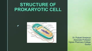
- 1. z STRUCTURE OF PROKARYOTIC CELL Dr. Pulipati Sowjanya Associate Professor Vignan Pharmacy College Guntur
- 2. z Contents Appendages- flagella, pili, fimbrae Cell envelope- glycocalyx, cell wall, cell membrane Cytoplasm- ribosomes, granules, nucleoid/chromosome.
- 5. Shapes and Arrangement of Bacteria
- 6. zChemical Composition of Bacteria • • Water - 70% Dry weight - 30% composed of: – – – • • • • – – – DNA - 5% MW 2,000,000,000 RNA - 12% protein- 70% found in: Ribosomes(10,000) – RNA Protein particles - MW 3,000,000 Enzymes Surface structures polysaccharides - 5% lipids - 6% phospholipids - 4%
- 7. z Prokaryote Cell Structure 1. Appendages- flagella, pili/fimbrae 2. Cell envelope- glycocalyx, cell wall , cell membrane 3. Cytoplasm- ribosomes, granules, nucleoid/chromosome.
- 8. z Bacterial Appendages • Pili (pl), pilus (s) – – only found in gram negative bacteria tubulare, hairlike structures of protein larger and more rare than fimbriae. • - - 2 types of pili attachment pilus - allow bacteria to attach to other cells/ solid surfaces sex pilus, - transfer from one bacterial cell to another- conjugation.
- 9. z Fimbriae • fimbriae (pl) fimbria (s) – Adhesion to cells and surfaces – Responsible for biofilms. Escherichia coli
- 10. Comparison of Pili & Fimbriae
- 11. z Flagella • Flagella (pl), flagellum(s) – long appendages which rotate by means of a "motor" located just under the cytoplasmic membrane. – bacteria may have one, a few, or many flagella in different positions on the cell. • Advantages - chemotaxis - positive and negative. - motility • All spirilla, half of bacilli, rare cocci.
- 12. z Flagella
- 13. z Flagella Three morphological regions • – – Helical filament long outermost region; composes up to 90% of its length contains the globular (roughly spherical) protein flagellin arranged in several chains and form a helix around a hollow core – • Hooked or curved area filament is attached; consists of a different protein • – – – Basal body terminal portion of the flagellum fix the flagellum to the cell wall and plasma membrane composed of a central rod inserted into a series of rings Gram negative - 2 pairs of rings • • Outer pair - fixed to the outer membrane and peptidoglycan layer Inner pair - fixed to the plasma membrane (SM ring) Gram positive - only inner pair is present
- 14. z Types of Flagellar Arrangements
- 15. z Motility • – – – Types of bacterial motility run or swim - when a bacterium moves in one direction for a length of time tumbles - periodic, abrupt random changes in direction swarming - rapid wavelike growth across a solid culture medium • Mechanism of flagellar movement - relative rotation of the rings in the basal body of the flagellum Antigenicity – flagellar or H antigen - useful in the serological identification of serotypes of Salmonella organisms
- 16. z Flagellar Movement Doetsch & Sjoblad described flagellar movement that flagella function as a propeller of a boat. If the rotation of flagella is in anti clock-wise direction, the bacterial cell moves in clock – wise direction.
- 17. z Spirochetal Movement Tuft of axial fibrils/ endoflagella that arise at the ends of the cell under the outer membrane and spiral around the cell Found in Spirochetes and are similar to flagella, but are located between the cell wall and an outer membrane, and are attached to one end of the organism. Gliding Movement Some bacteria like cyanobacteria & mycoplasmas show gliding movement when come in contact with solid surface
- 18. z Motility of Bacteria Two ways by which motility can be demonstrated: • direct or microscopic – hanging drop preparation or wet mount preparation by dark field mycroscope – Distinguishes: • Brownian movement - when the bacteria show molecular movement • true motility - if a bacterium describes a rotatory, undulatory or sinuous movement • indirect or macroscopic – Stab inoculation of the semisolid media • nonmotile - growth is limited at the point of inoculation • motile - growth is diffuse or moves away from the line of inoculation; turbidity of the medium
- 19. z Detection of Motility • Direct • Indirect Presence mobile bacteria
- 20. z Taxis Chemotaxis: Movement of bacteria towards chemical attractant and away from repellant Aerotaxis: Movement of bacteria based on requirement of aeration. Aerobic Anaerobic Microaerophilic
- 21. z Taxis Phototaxis: Phototaxis is the movement of an organism in response to light: that is, the response to variation in light intensity and direction. Negative phototaxis, or movement away from a light source. Positive phototaxis, or movement towards a light source, is advantageous for phototrophic organisms as they can orient themselves most efficiently to receive light for photosynthesis. Many phytoflagellates, e.g. Euglena, and the chloroplasts of higher plants positively phototactic, moving towards a light source.
- 22. z Taxis Magnetotaxis: Magnetotaxis is the movement of an organism towards or away from magnetic field. Magnetotactic bacteria possess a chain of magnetite (Fe2O4) particles known as magnetosomes. Figure: Aquaspirillum magnetotacticum
- 23. z 2. Bacterial Surface Structure - cell envelope A. Glycocalyx - some extracellular material secreted by many bacterial cells in the form of: a. capsule - attached tightly to the bacterium and has definite boundaries. b. slime layer - loosely associated with the bacterium and can be easily washed off Compositions: - - layer of polysaccharide proteins - sometimes
- 24. Differences between Capsule and Slime Layer
- 25. z Functions of the Capsule • Protection • Identification • Vaccine preparation • Tissue attachment
- 26. z Medical Importance rapid serological identification of: • Several groups of streptococci • Meningococcus • Hemophilus influenzae • Klebsiella pneumoniae • Yersinia and Bacillus species
- 27. z Functions of the Capsule • Protection • Identification • Vaccine preparation • Tissue attachment • Antibiotic barrier
- 28. z Cell wall Peptidoglycan (polysaccharides + protein), • Support and shape of a bacterial cell. The three primary shapes in bacteria are: • coccus (spherical), • • • bacillus (rod-shaped) spirillum (spiral). Mycoplasma are bacteria that have no cell wall and therefore have no definite shape.
- 29. z Cell wall peptidoglycan (polysaccharides + protein) Components of the peptidoglycan layer: Repeating glycan chains - N acetyl glucosamine (NAG) and N acetyl muramic acid (NAM) joined with 1,4 – glycosidic bond. A set of identical tetrapeptide side chains attached to N- acetylmuramic acid A set of identical peptide cross bridges
- 30. z Peptidoglycan
- 31. z Differences in Cell Wall Structure • Basis of Gram Stain Reaction – Hans Christian Gram- 1884 • Differential Stain – Gram Positive vs Gram Negative Cells • Gram Positive Cells- – Thick peptidoglycan layer with embedded teichoic acids • Gram Negative Cells- – Thin peptidoglycan layer, outer membrane of lipopolysaccharide.
- 32. z Gram Positive Cell Wall • Multilayered peptidoglycan covered by surface layer (S-layer). • Teichoic acid – ribitol phosphate or glycerol phosphate. • Teichuronic acid – long chains of alternating glucuronic acid and N- acetylgalactosamine linked with 1-3 glycosidic bond. • Lipoteichoic acid – teichoic acid originated from cell membrane.
- 33. Gram Positive Cell Wall • The peptidoglycan chains contains tetrapeptides consisting L-lysine-D- alanine-L-lysine-D-alanine residues. • In some members like Staphylococcus aureus the tetrapeptide chain is connected through pentaglycine chains. Fig.: Organisation of peptidoglycan layer of Staphylococcus aureus. 1. L-lysine, 2. D-alanine, 3. L-lysine, 4. D-alanine, G-glycine
- 34. Acid fast bacteria • M. tuberculae & M. leprae show the property of acid fastness. • When these bacteria are stained with carbol-fuchsin and then washed with dilute acid, the bacteria retain the stain. • Non- acid fast organisms destained by this treatment. • The acid fastness property was found to be due to the presence of mycolic acid lipid in cell walls.
- 35. z Gram Negative Cell Wall
- 36. Gram Negative Cell Wall • The distinctive feature of Gram-ve cell wall is the presence of an outer membrane. • Presence of few layers of peptidoglycan layer. • Peptidoglycan is present in periplasmic space and covalently linked with lipoproteins in the outer membrane. • Teichoic acid or teichuronic acid chains are absent. • Outer membrane is a bilayered structure composed of lipopolysaccharides, lipoproteins & phospholipids.
- 37. Gram Negative Cell Wall • The lipopolysaccharides has 3 components – Lipid A embedded in the outer membrane, core polysaccharide lying on the membrane surface and polysaccharide side chains (O- antigen) projecting outside the membrane. • Outer membrane possess pores contains a special protein porin. • These pores allow the entry of molecules into the cytoplasm • Outer membrane – several proteins • Receptor protein – entire outer membrane • Braun’s protein – restricted to the inner layer.
- 38. Gram Negative Cell Wall • The peptidoglycan layer of Gram –ve bacterial cell wall contains tetrapeptide chain contains meso-diaminopimelic acid (m-DAP) in place of L-lysine and pentaglycine chain is absent. • The tetrapeptide chains are directly cross linked between m- DAP & D-alanine of adjacent peptidoglycan chains. • The outer membrane protects the bacteria from the action of lysozyme. • Lysozyme present in saliva, tears and other body fluids. It breaks the glycosidic bond between NAG & NAM.
- 39. Differential Response to Gram Stain • The crystal-violet iodine complex is deposited on the cytoplasmic membrane. • Washing with alcohol removes the stain in Gram-ve bacteria, because of thin layer of peptidoglycan. • Gram+ve bacteria contains thick layer of peptidoglycan, hence it retains crystal violet stain.
- 40. Functions of Cell Wall Maintenance of the shape (due to rigidity of peptidoglycan). Protects the cytoplasmic membrane cell contents Rigidity Cell wall is osmotically insensitive Hypotonic solution – cell burst. Hypertonic solution – cell shrank. Isotonic solution – bacteria is life. It acts as barrier for diffusion to certain molecules. The O-antigen determines the antigen specificity of Gram negative bacteria.
- 41. z Cytoplasmic Membrane Phospholipid bilayer “Fluid mosaic” model Embedded proteins for active transport Enzymes for energy generation Photosynthetic pigments
- 42. z Cell membrane General structure is phospholipid bilayer Contain both hydrophobic and hydrophilic components. Fatty acids point inward to form hydrophobic environment; hydrophilic portions remain exposed to external environment or the cytoplasm
- 45. z Cell membrane Two types of proteins Peripheral- are loosely associated to the membrane and can be easily separated. Generally they make up between 20 and 30% of the total membrane proteins Integral proteins- are amphipathic like the lipids, much more strongly associated to the membrane, and make up about 70 to 80% of total proteins
- 46. z Cell membrane Figure: Presence of steroids in plasma membrane (A) and chemical structure of a steroid (B) In some microorganisms such as mycoplasmas & fungi, sterols are found to be associated within the plasma membrane. The sterols are structurally different from the lipids.
- 47. z Selective permeability to different molecules. Active transport aided by permease. Play a role in DNA replication. Cell wall biosynthesis. Mesosomes ----- cell division. Function of Cytoplasmic Membrane
- 48. z Mesosomes Figure: The bacterial mesosome Mesosomes are invaginated structures formed by infoldings of inner membrane of plasma membrane. Salton & Owen suggested that the mesosomes are formed due to vesicularization of outer half of the lipid bilayer. Functions: Their exact function is unknown but they are supposed to take part in respiration. They play an important role in cell division. Mesosomes begin the formation of septum and attach the bacterial DNA to the cell membrane.
- 49. z CYTOPLASM Ribosomes: Thousands of ribosomes are present that gives granular appearance of the cytoplasm. During protein synthesis ribosomes form a chain connected by the m-RNA, such strings of ribosomes are known as polysomes. Figure: The prokaryotic ribosome. (a) A small 30S subunit and (b) a large 50S subunit © the complete 70S prokaryotic ribosome
- 50. z CYTOPLASM Bacterial Nucleus (Nucleoid): It consists of single, long supercoiled, circular, dsDNA molecule which is associated with RNA and some proteins. The supercoiling is induced by topoisomerase enzyme Figure: The process of folding and super coiling of bacterial chromosome
- 51. z CYTOPLASM Cytoplasmic inclusions: Volutin granules: insoluble polyphosphates – source of reserve phosphate. Polymer of β-hydroxybutyric acid (PHB): a fat substance – source of carbon & energy. Sulphur granules: found in photosynthetic bacteria Parasporal body: possess insecticidal property Ex: Bacillus thuringiensis Figure: Sulphur granules found in Chromatium vinosum
- 52. z CYTOPLASM Magnetosomes: Magnetotaxis is the movement of an organism towards or away from magnetic field. Magnetotactic bacteria possess a chain of magnetite (Fe2O4) particles known as magnetosomes. Figure: Aquaspirillum magnetotacticum
- 53. z Endospores The spore is externally covered by a loose structure called exosporium which is formed by the remnants of mother cell protoplast. Inside the exosporium several layers of spore coats are present. Next to the coat layers a thick zone called cortex is present. Below the cortex lies the primordial cell wall enclosing the inner membrane. The inner membrane surrounds the core protoplast of the spore
- 55. z Endospore Formation Endospore is formed by accumulation of Ca++ in the mother cell. In vegetative cells, Ca++ level is low but during sporulation, Ca++ is actively transported from the medium. From the mother cell cytoplasm, Ca++ is transported by facilitated diffusion into the forespore. The forespore contains dipicolinic acid and it forms calcium dipicolinate complex. The concentration of calcium dipicolinate is high and it results in heat resistance of the bacterial endospores.
- 56. z Exospore Formation Cells of methane-oxidizing genus Methylosinus forms exospores i.e., spores external to the vegetative cells by budding at one end of the cell. These are desiccation and heat resistant but they do not contain dipicolinic acid. Examples of exospores include Conidiospores, streptomyces, actinobacteria and diverse groups of fungi, algae and Cyanobacteria.
- 57. Streptomyces MicrobisporaMicromonospora Conidia are formed externally and found in most species of Actinomycetes. In Streptomyces, Micromonospora, Microbispora conidia are formed in the aerial hyphae. The conidia are long, straight, spiral or coiled chains of conidia formed from the tip of conidiophores. Exospore Formation
- 58. z REFERENCES 1. Prescott, Harley and Klein's, Microbiology. 5th edition. 42-72. 2. Gerard J. Tortora, Berdell R. Funke, Christine L. Case. Microbiology – An Introduction. 10th edition. Pearson. 76-97. 3. Michael J. Pelczar, JR., E.C.S. Chan, Noel R. Krieg. Microbiology. 5th edition. 73-97.
- 59. z It is easy to solve a problem that everyone sees, but it’s hard to solve a problem that no one sees
Editor's Notes
- 3