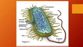
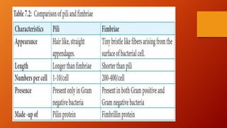
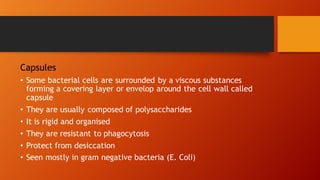
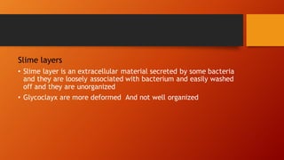
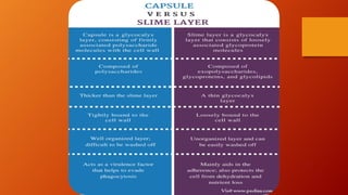
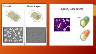
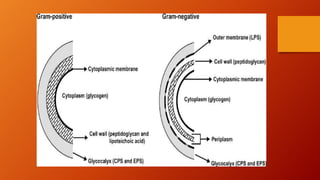
The document provides an overview of bacteria, distinguishing between gram-positive and gram-negative types based on their staining properties and structural characteristics. It describes various bacterial structures including flagella, pili, capsules, and cell walls, as well as the formation and significance of endospores in adverse conditions. Key components such as ribosomes and the unique structures of gram-negative bacteria are also discussed, highlighting their adaptations and functions.


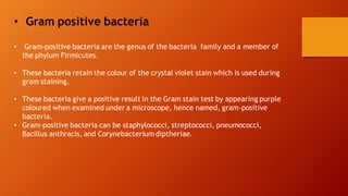


![sturcture of bacteria lecture 3[1].pptx](https://cdn.slidesharecdn.com/ss_thumbnails/sturctureofbacterialecture31-240128072427-20b3d95c-thumbnail.jpg?width=640&height=640&fit=bounds)


![Polymer [ बहुलक ] Chemistry Notes PDF - Irfanullah Mehar - JJ Sir Chemistry.pdf](https://cdn.slidesharecdn.com/ss_thumbnails/polymerchemistrynotespdf-irfanullahmehar-jjsirchemistry-260210172118-3f9b37f7-thumbnail.jpg?width=640&height=640&fit=bounds)


