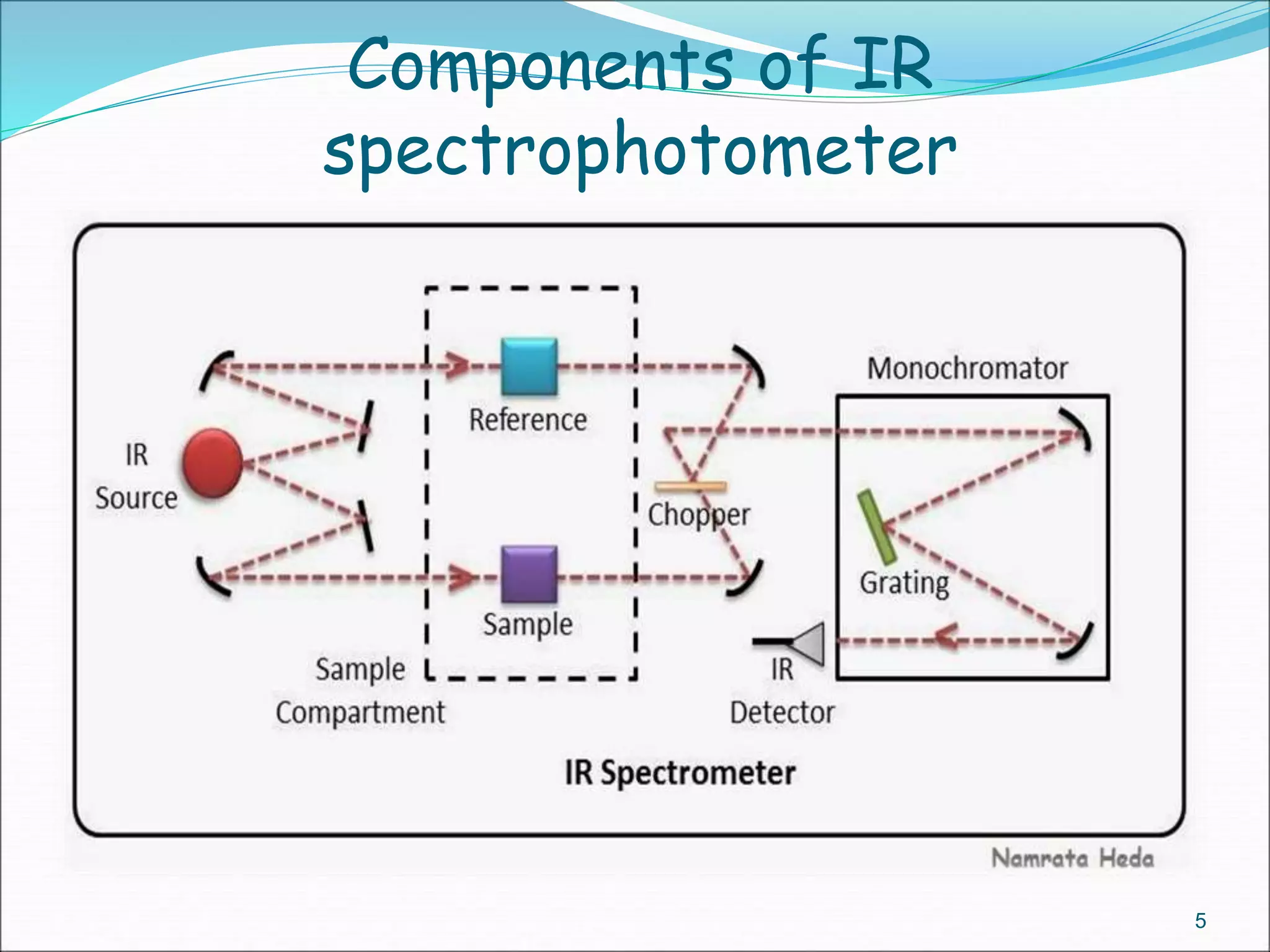
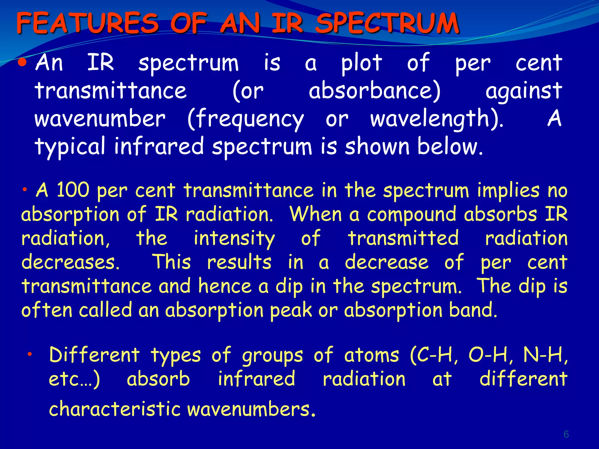
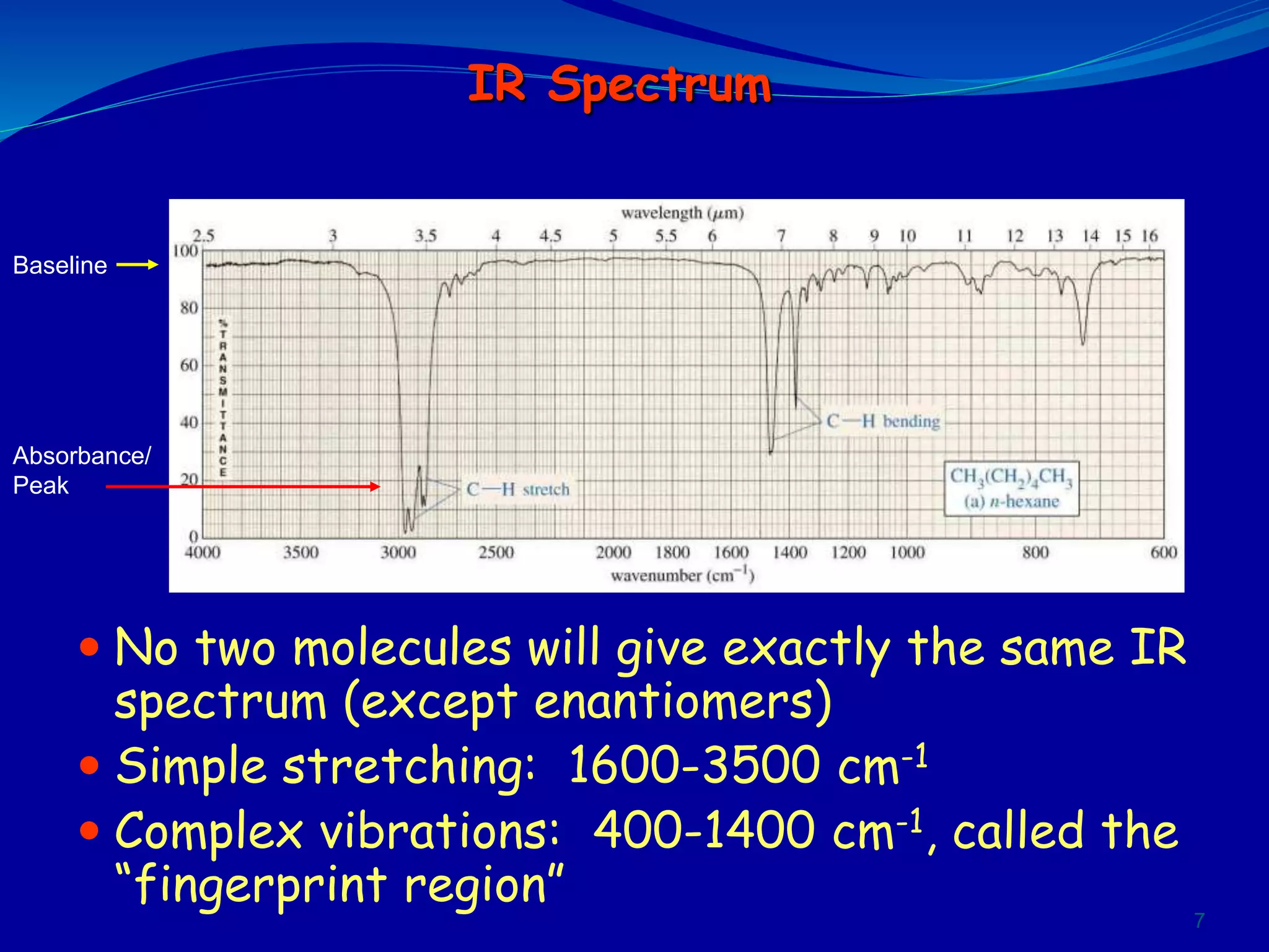
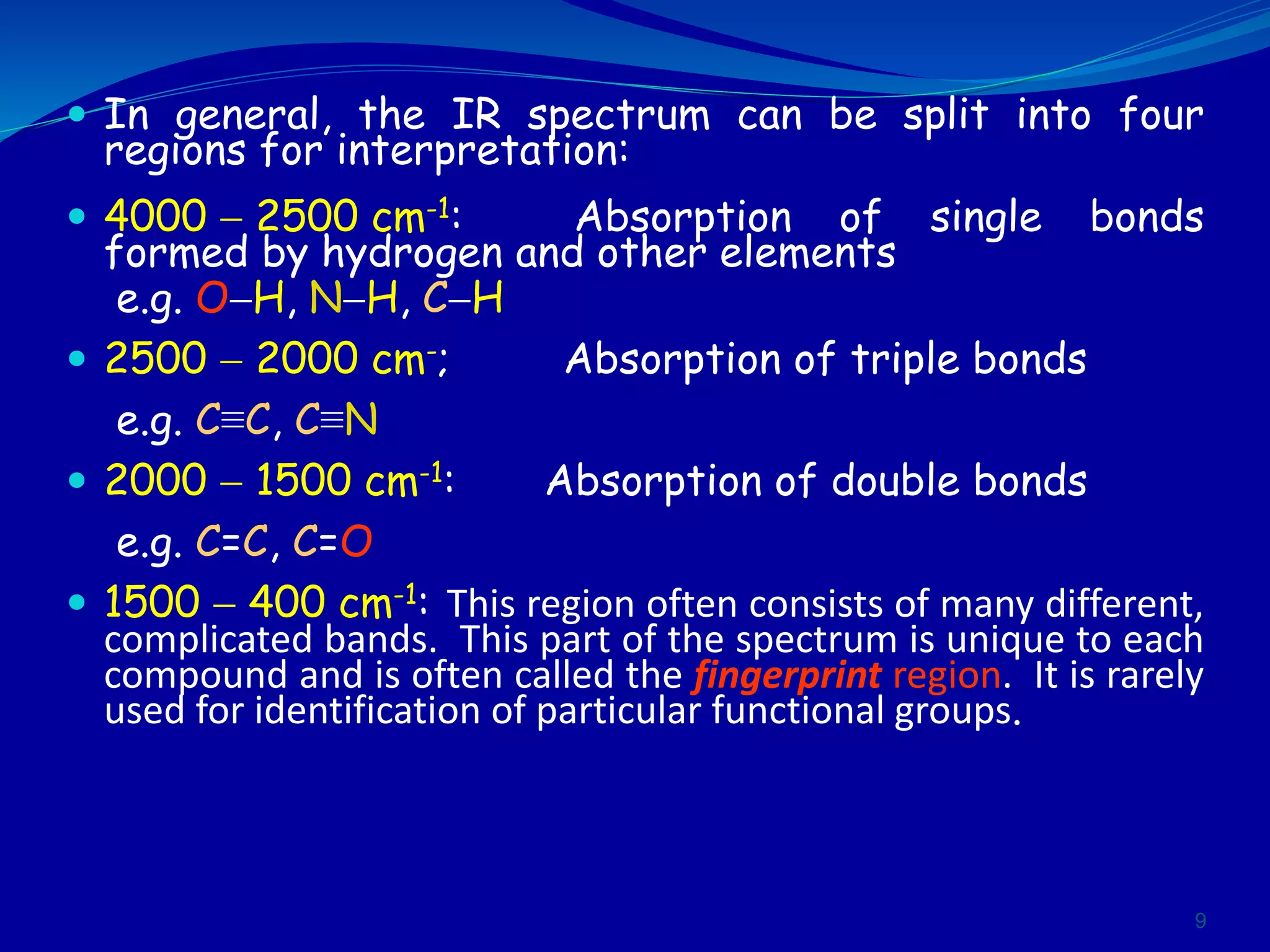
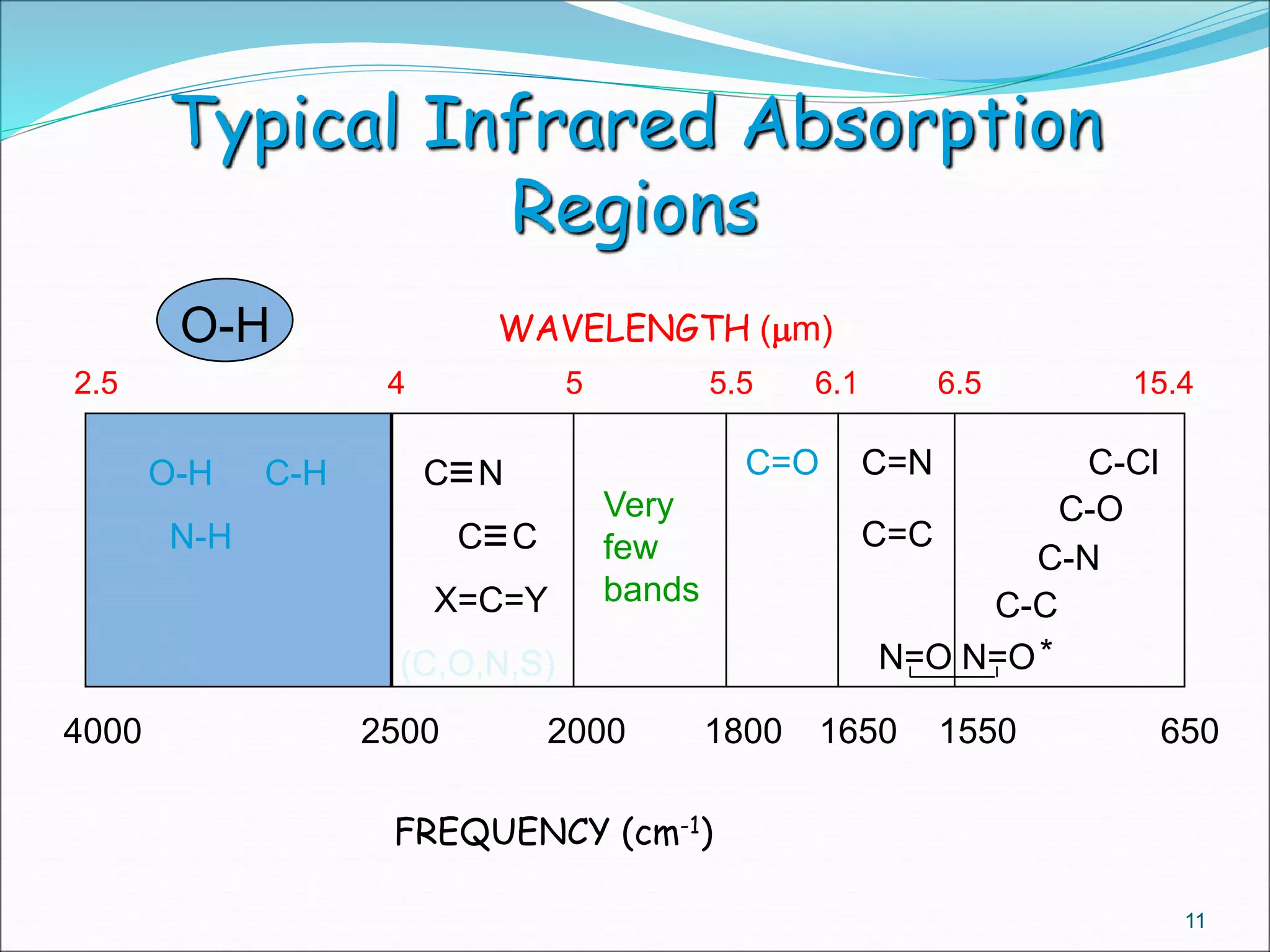
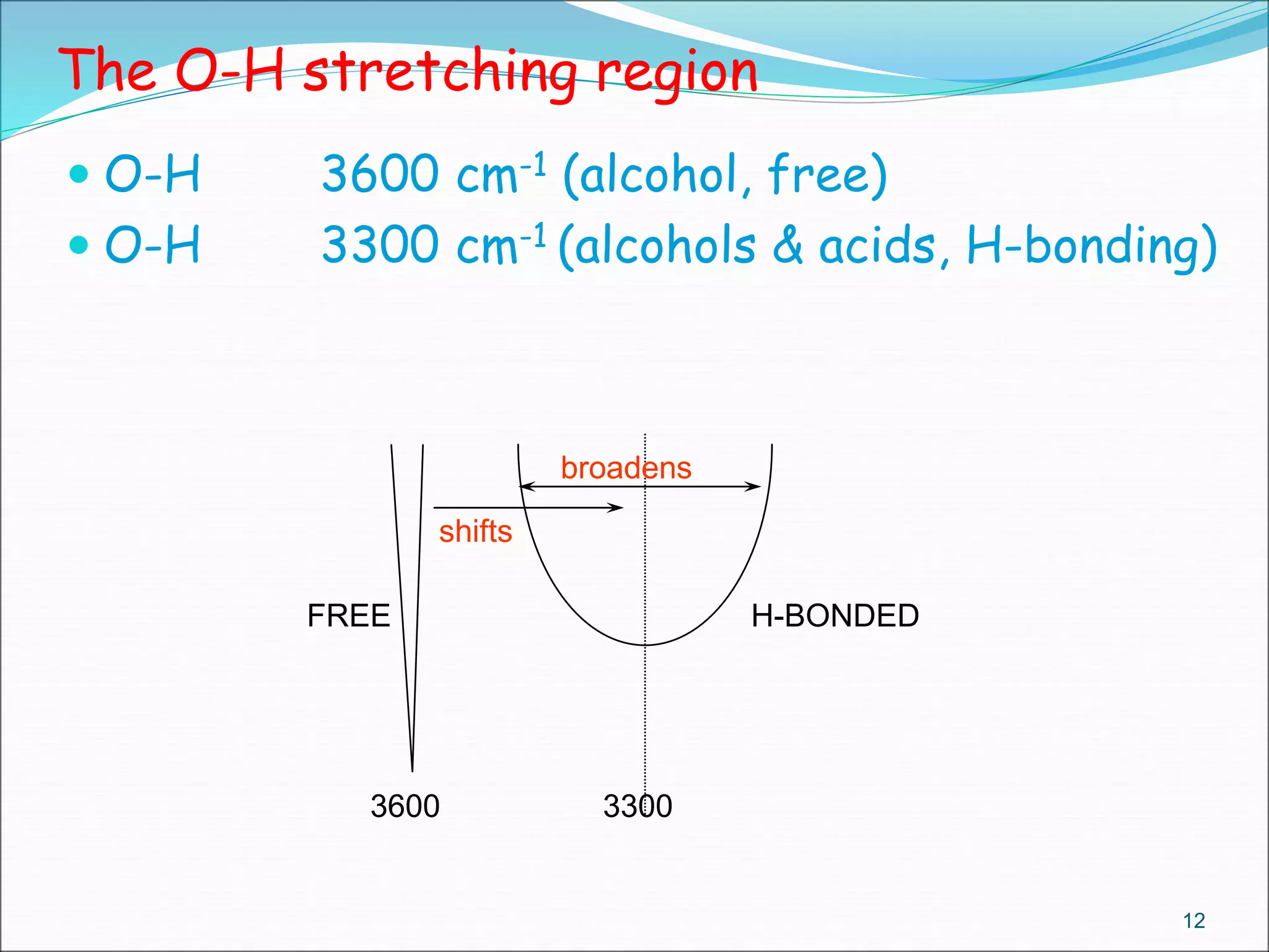
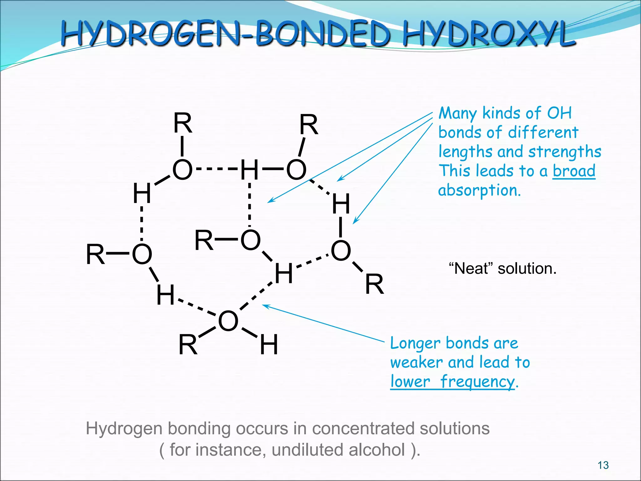
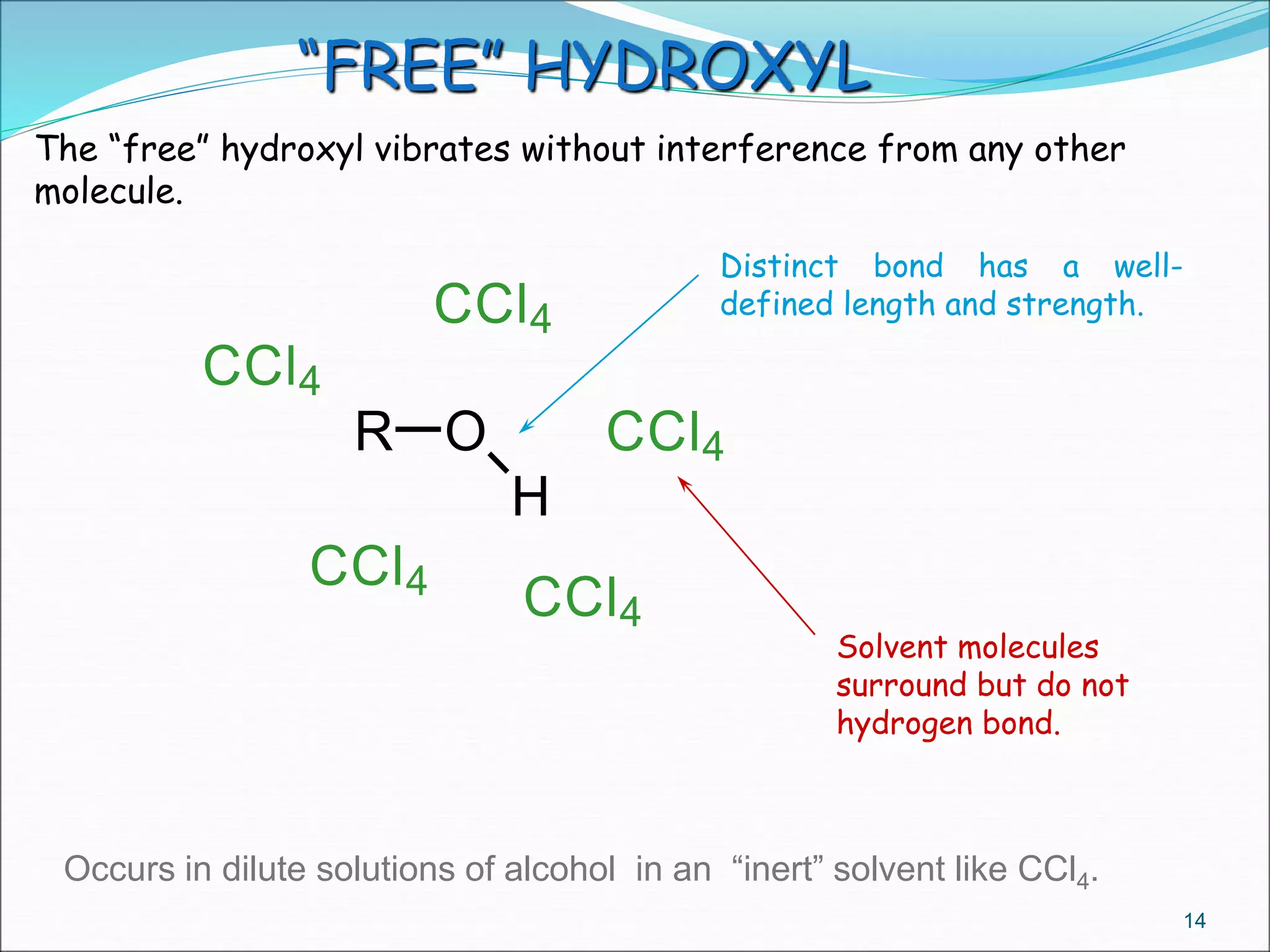
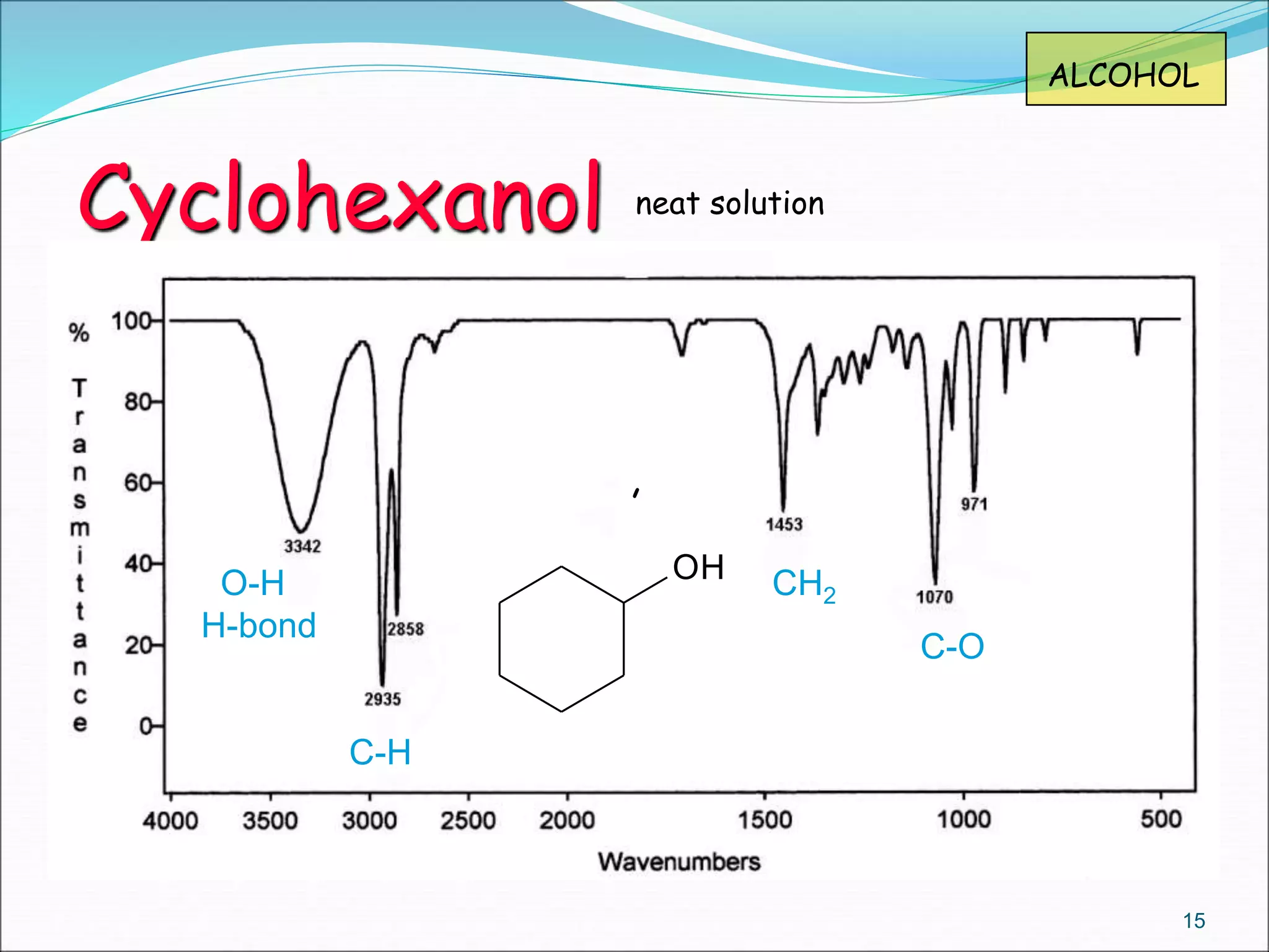
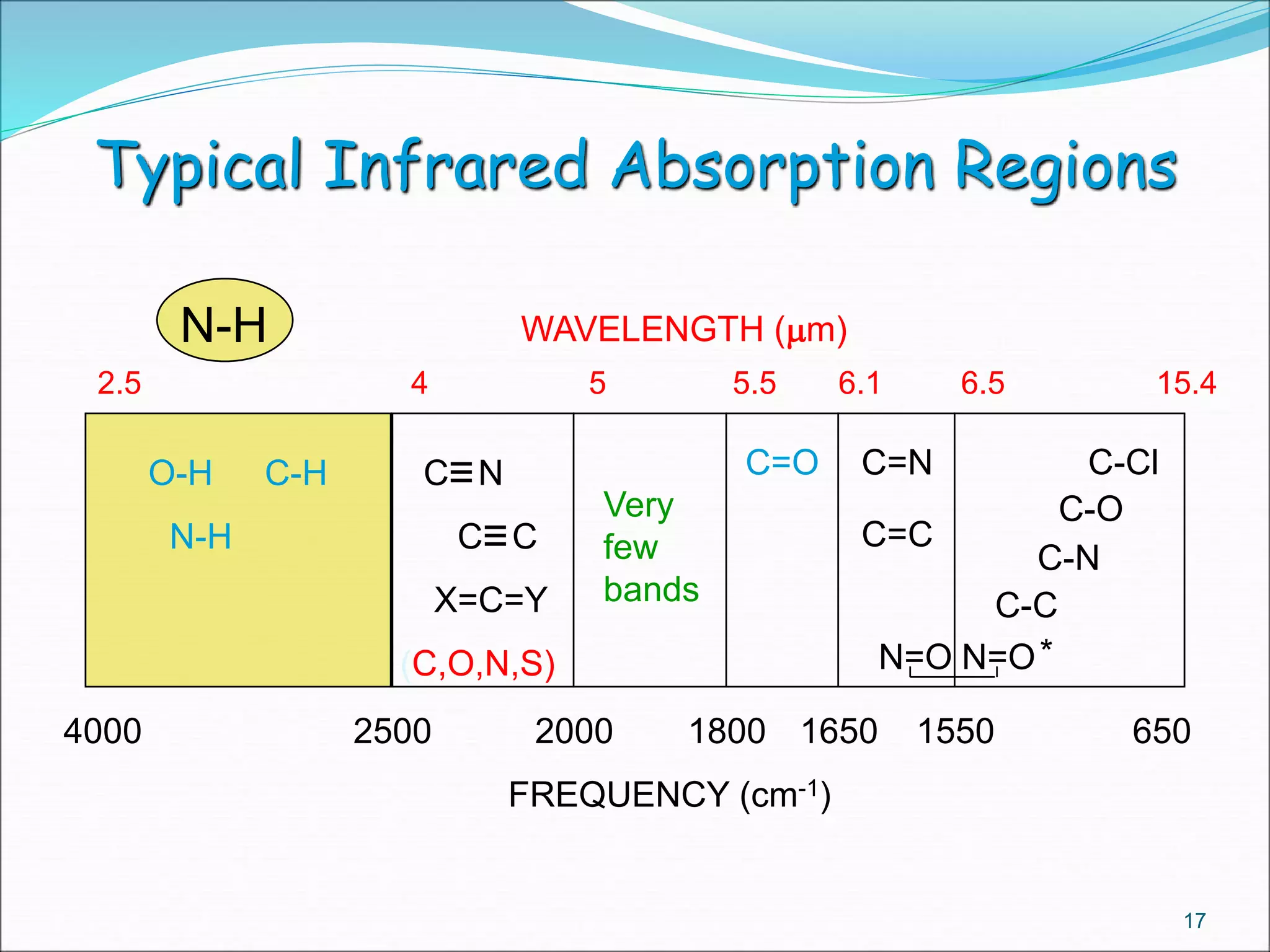
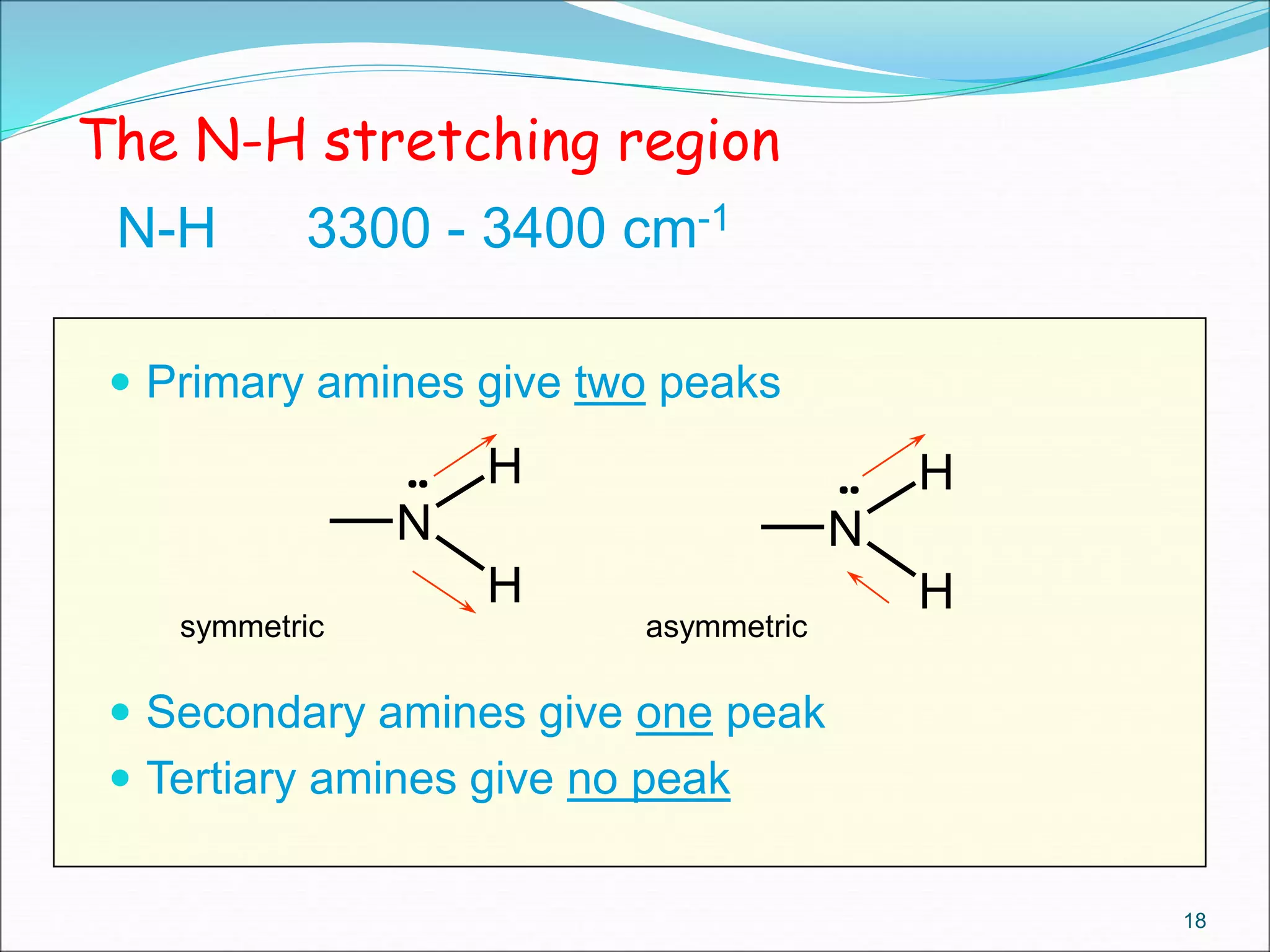
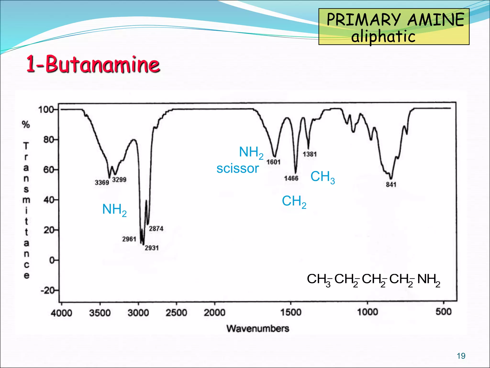
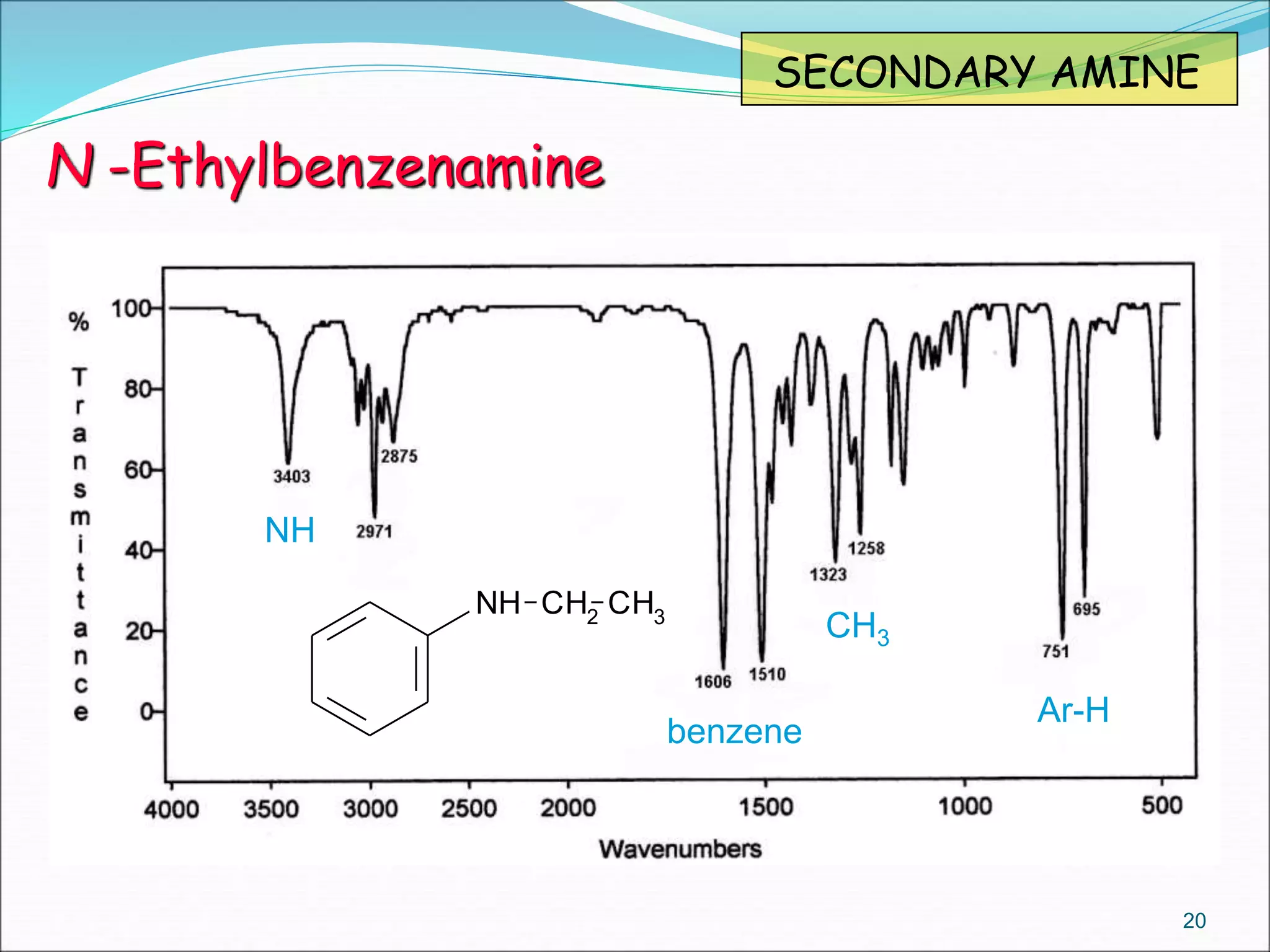
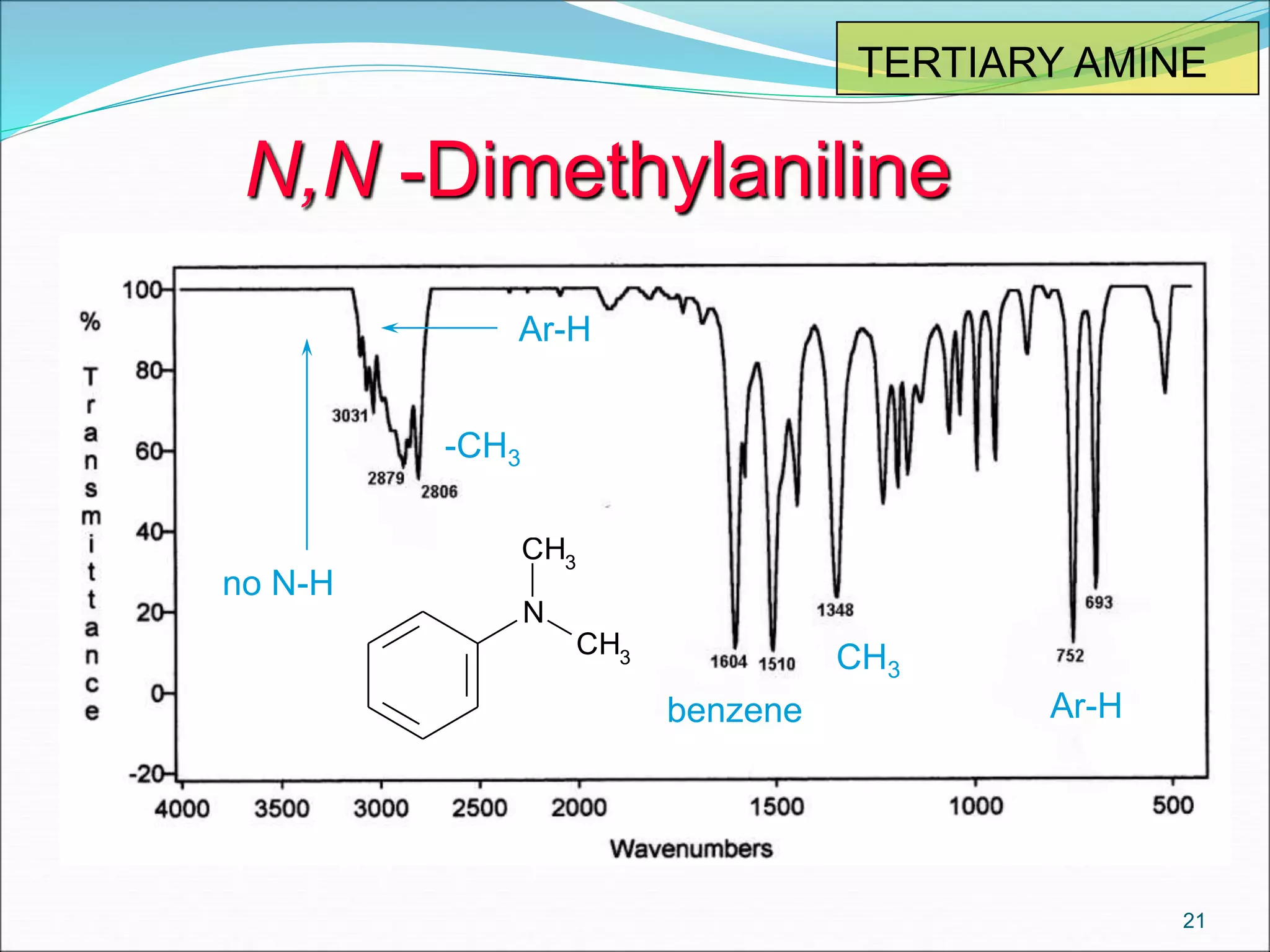
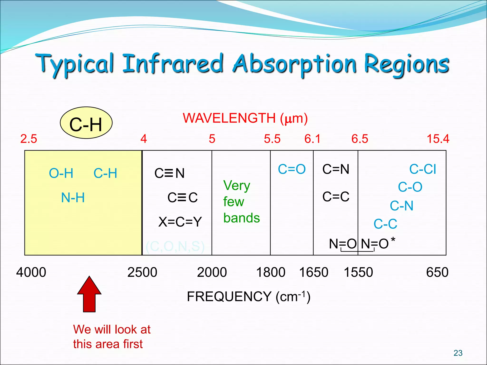
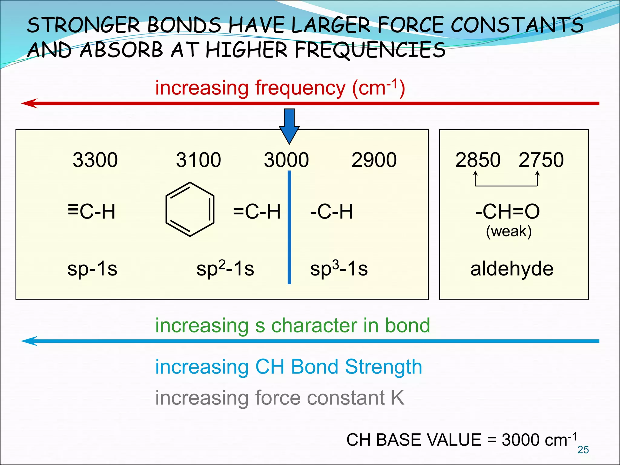
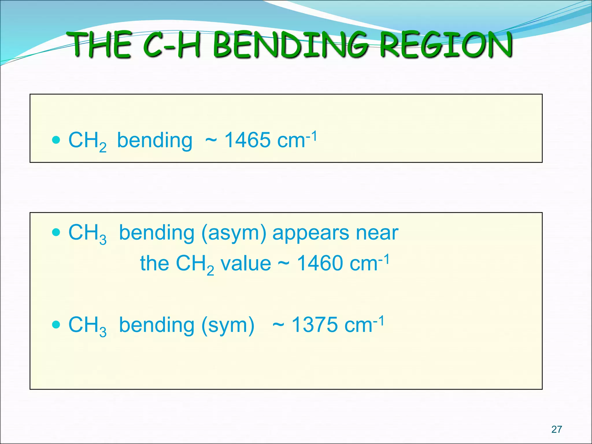
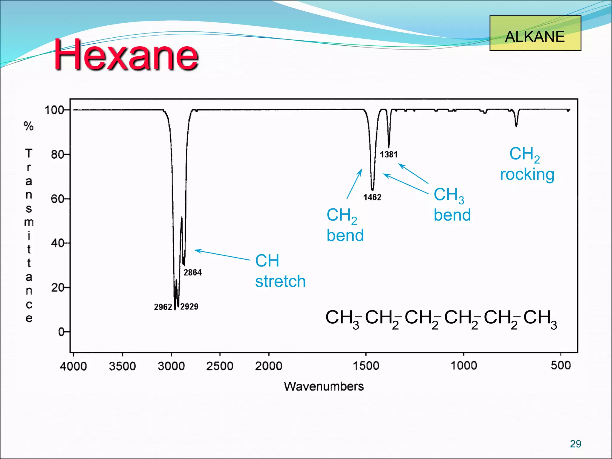
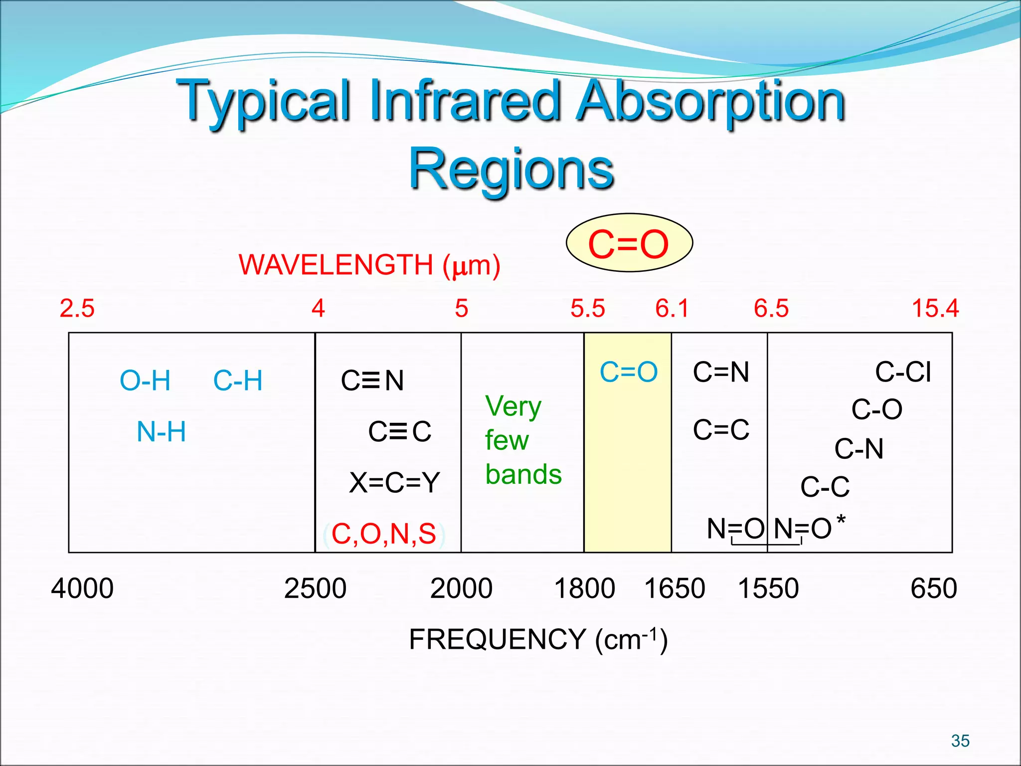
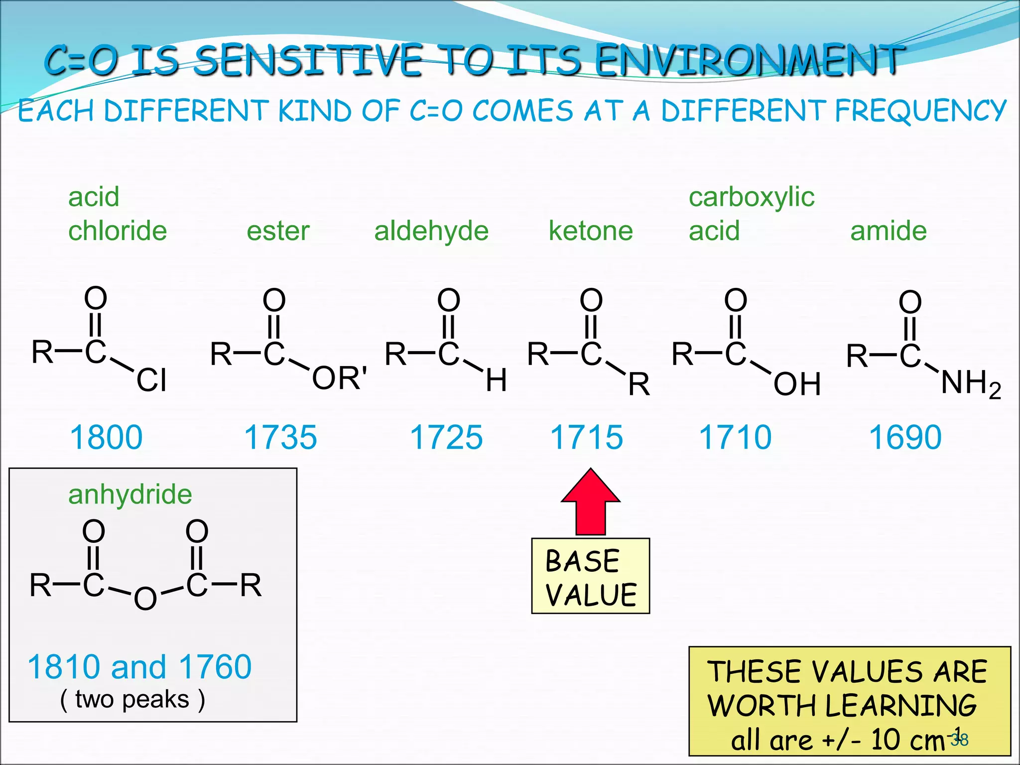
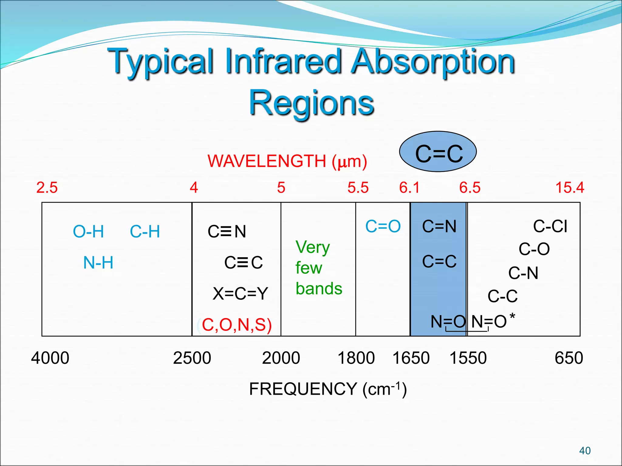
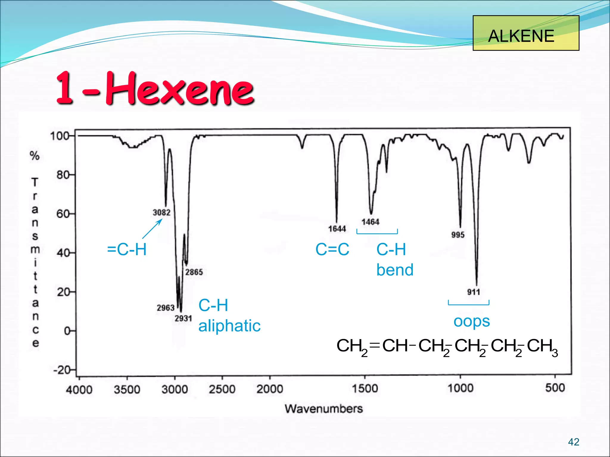
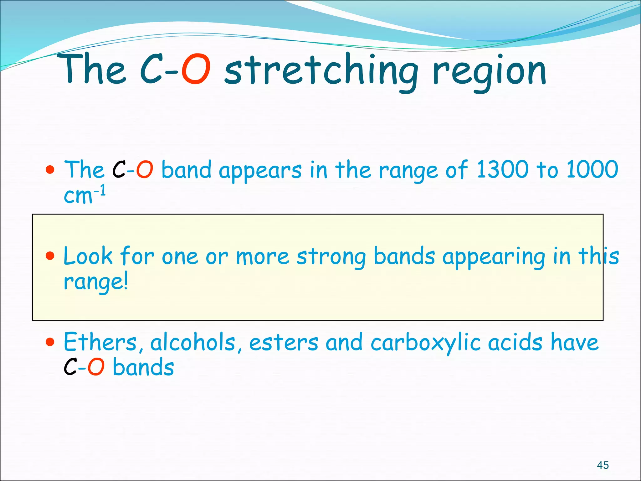
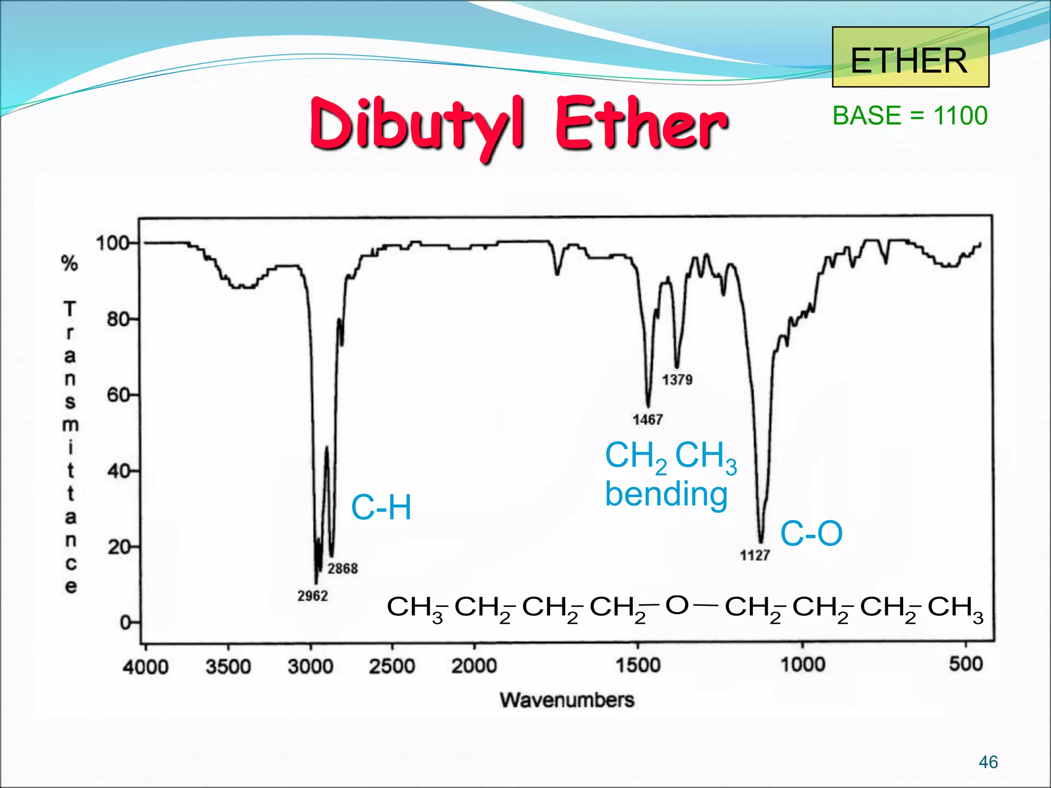
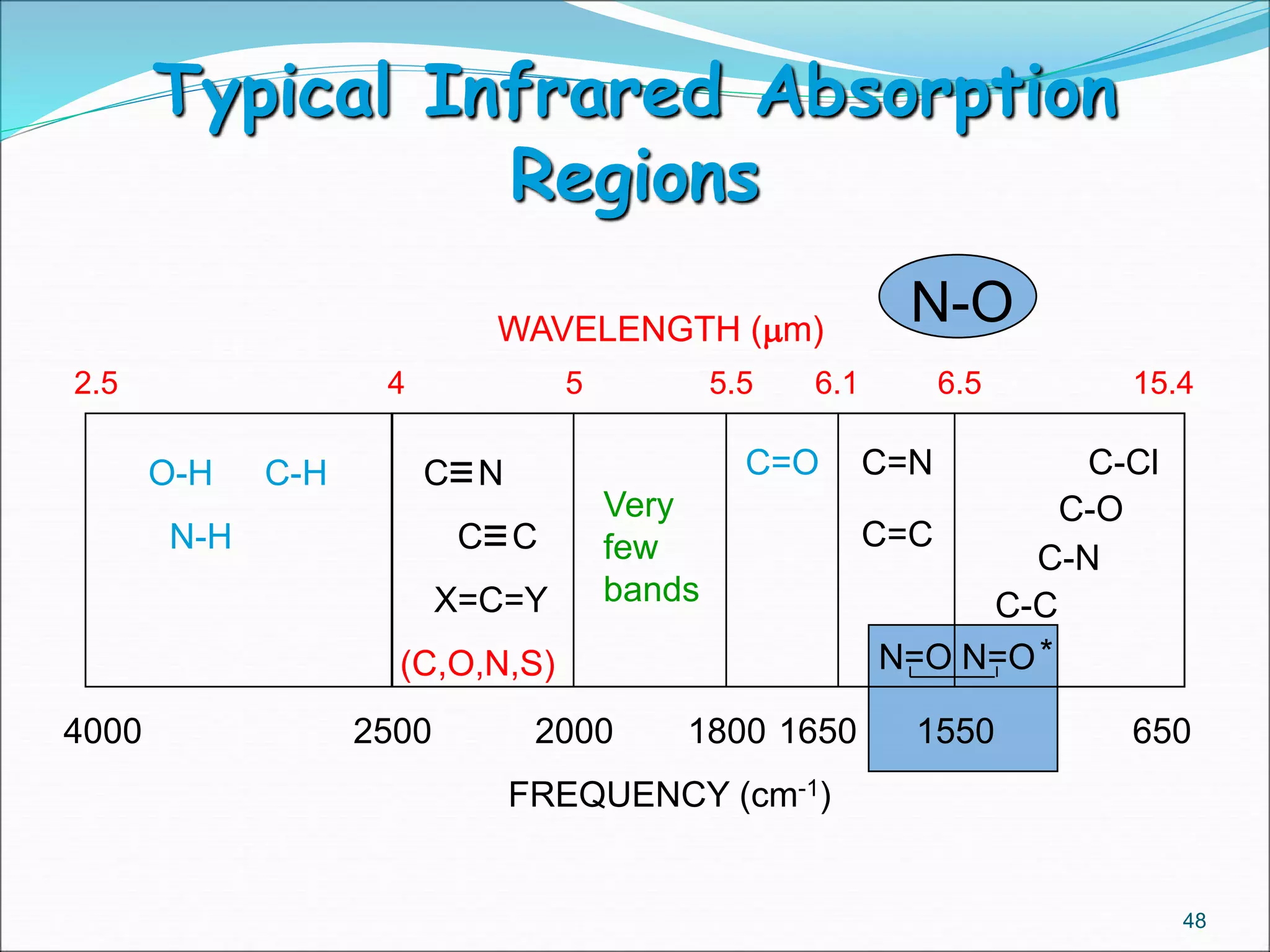
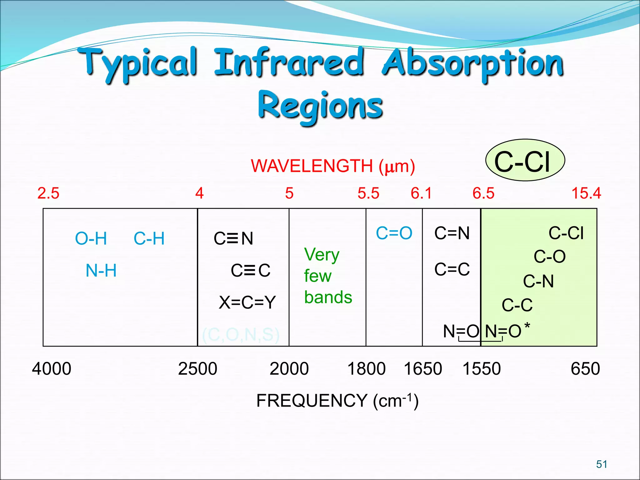
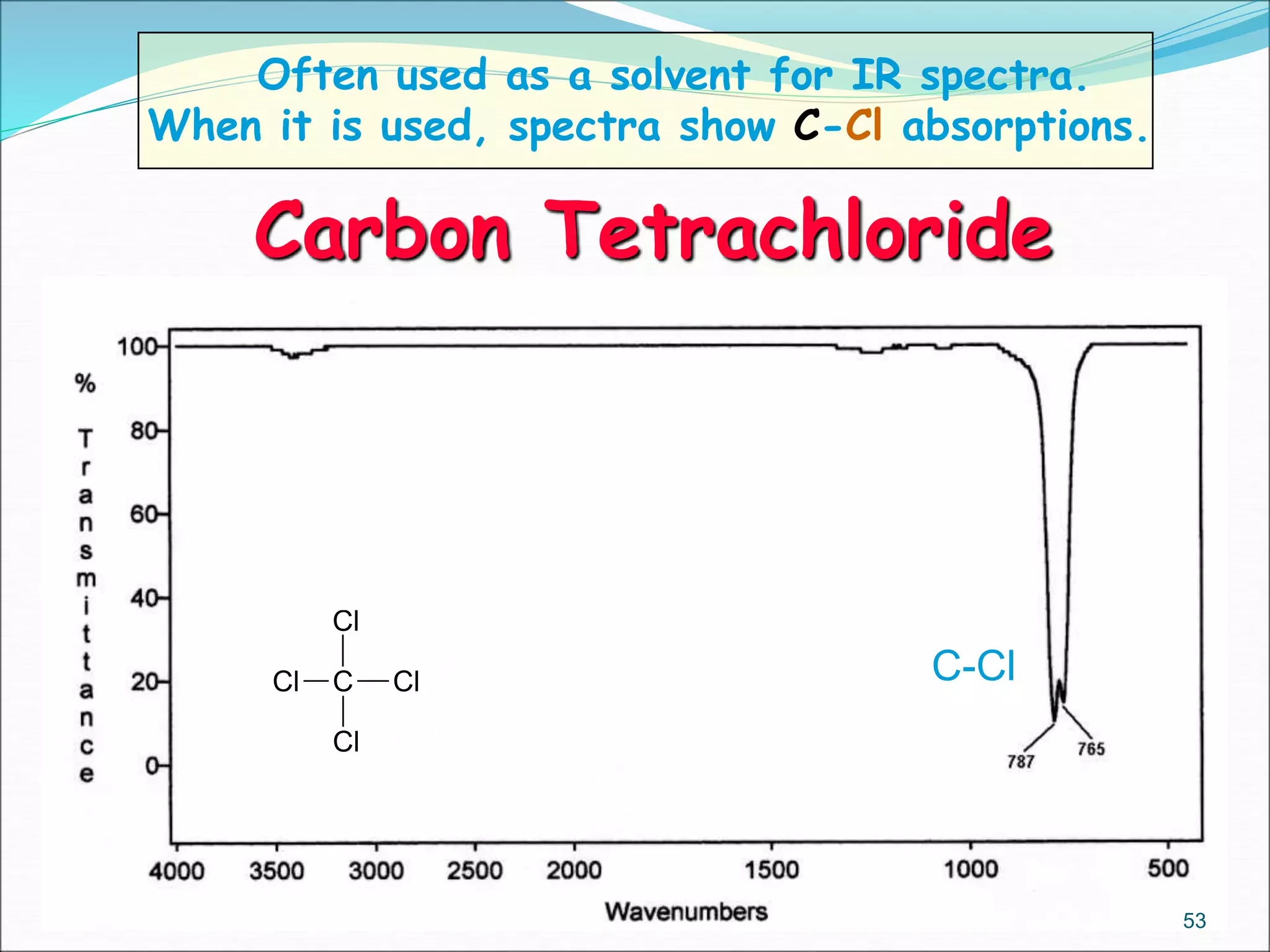
ACHARYA NAGARJUNA UNIVERSITY COLLEGE OF
PHARMACEUTICAL SCIENCES
SEMINAR PRESENTED BY
O.SASIVARDHAN
Roll.no:Y15MPH326
IN PHARMACEUTICAL ANALYSIS
A SEMINAR ON INTERPRETATION OF IR
SPECTROSCOPY
CONTENTS
 PRINCIPLE
 MODES OF VIBRATIONS
 COMPONENTS OF IR INSTRUMENT
 FEATURES OF IR SPECTRA
 USE OF IR SPECTRA
 INTERPRITATION OF VARIOUS FUNCTIONAL
GROUPS
 CONCLUSION
 REFERENCES
2
PRINCIPLE
 Infrared spectroscopy is based on a simple fact
that a chemical substance shows marked selective
absorption in IR region.
 After absorption of IR radiation the molecules of
chemical substance vibrate at many rates of
vibration giving rise to closely packed absorption
spectrum called as IR absorption spectrum which
may extend over a wide wavelength range.
 REQUIREMENTS
3
Modes of vibration
It can be divided into two principal groups.
 1.Stretching vibrations: changes in bond length.
a) Symmetrical stretching:
b) Asymmetrical stretching
 2.Bending vibration; changes in bond angle.
In plane bending:
 i) Scissoring
 ii) Rocking
Out of plane bending:
 i ) Wagging
 ii ) Twisting
4
Components of IR
spectrophotometer
5
 An IR spectrum is a plot of per cent
transmittance (or absorbance) against
wavenumber (frequency or wavelength). A
typical infrared spectrum is shown below.
6
• A 100 per cent transmittance in the spectrum implies no
absorption of IR radiation. When a compound absorbs IR
radiation, the intensity of transmitted radiation
decreases. This results in a decrease of per cent
transmittance and hence a dip in the spectrum. The dip is
often called an absorption peak or absorption band.
FEATURES OF AN IR SPECTRUM
• Different types of groups of atoms (C-H, O-H, N-H,
etc…) absorb infrared radiation at different
characteristic wavenumbers.
 No two molecules will give exactly the same IR
spectrum (except enantiomers)
 Simple stretching: 1600-3500 cm-1
 Complex vibrations: 400-1400 cm-1, called the
“fingerprint region”
7
Baseline
Absorbance/
Peak
IR Spectrum
 Identification of functional groups on a
molecule – this is a very important tool in
organic chemistry
 Spectral matching can be done by computer
software and library spectra
 Since absorbance follows Beer’s Law, can do
quantitative analysis
8
Use of IR spectra
 In general, the IR spectrum can be split into four
regions for interpretation:
 4000  2500 cm-1: Absorption of single bonds
formed by hydrogen and other elements
e.g. OH, NH, CH
 2500  2000 cm-; Absorption of triple bonds
e.g. C≡C, C≡N
 2000  1500 cm-1: Absorption of double bonds
e.g. C=C, C=O
 1500  400 cm-1: This region often consists of many different,
complicated bands. This part of the spectrum is unique to each
compound and is often called the fingerprint region. It is rarely
used for identification of particular functional groups.
9
O-H STRETCH
10
INTERPRETATION OF
VARIOUS FUNCTIONAL
GROUPS
Typical Infrared Absorption
Regions
O-H
2.5 4 5 5.5 6.1 6.5 15.4
4000 2500 2000 1800 1650 1550 650
FREQUENCY (cm-1)
WAVELENGTH (mm)
O-H C-H
N-H
C=O C=N
Very
few
bands
C=C
C-Cl
C-O
C-N
C-CX=C=Y
(C,O,N,S)
C N
C C
N=O N=O*
11
 O-H 3600 cm-1 (alcohol, free)
 O-H 3300 cm-1 (alcohols & acids, H-bonding)
12
3600 3300
H-BONDEDFREE
broadens
shifts
The O-H stretching region
O
H
R
O
H
R
R O
H
R
OH
H
R O
R H
O
HYDROGEN-BONDED HYDROXYL
Many kinds of OH
bonds of different
lengths and strengths
This leads to a broad
absorption.
Longer bonds are
weaker and lead to
lower frequency.
Hydrogen bonding occurs in concentrated solutions
( for instance, undiluted alcohol ).
“Neat” solution.
13
“FREE” HYDROXYL
R O
H
CCl4
CCl4
CCl4
CCl4
CCl4
Distinct bond has a well-
defined length and strength.
Occurs in dilute solutions of alcohol in an “inert” solvent like CCl4.
Solvent molecules
surround but do not
hydrogen bond.
The “free” hydroxyl vibrates without interference from any other
molecule.
14
Cyclohexanol
15
OHO-H
H-bond
C-H
C-O
CH2
ALCOHOL
neat solution
,
N-H STRETCH
16
Typical Infrared Absorption Regions
N-H
2.5 4 5 5.5 6.1 6.5 15.4
4000 2500 2000 1800 1650 1550 650
FREQUENCY (cm-1)
WAVELENGTH (mm)
O-H C-H
N-H
C=O C=N
Very
few
bands
C=C
C-Cl
C-O
C-N
C-CX=C=Y
(C,O,N,S)
C N
C C
N=O N=O*
17
The N-H stretching region
 Primary amines give two peaks
 Secondary amines give one peak
 Tertiary amines give no peak
18
N
H
H
N
H
H
symmetric asymmetric
N-H 3300 - 3400 cm-1
CH3
CH2
CH2
CH2
NH2
NH2
NH2
scissor
CH2
CH3
PRIMARY AMINE
aliphatic
1-Butanamine
19
NH CH2 CH3
NH
benzene
Ar-H
CH3
SECONDARY AMINE
N -Ethylbenzenamine
20
N
CH3
CH3
no N-H
benzene
CH3
Ar-H
Ar-H
-CH3
TERTIARY AMINE
N,N -Dimethylaniline
21
C-H STRETCH
22
Typical Infrared Absorption Regions
C-H
2.5 4 5 5.5 6.1 6.5 15.4
4000 2500 2000 1800 1650 1550 650
FREQUENCY (cm-1)
WAVELENGTH (mm)
O-H C-H
N-H
C=O C=N
Very
few
bands
C=C
C-Cl
C-O
C-N
C-CX=C=Y
(C,O,N,S)
C N
C C
N=O N=O*
We will look at
this area first
23
•C-H aldehyde, two peaks (both weak)
~ 2850 and 2750 cm-1
3000 divides
UNSATURATED
SATURATED
•C-H sp stretch ~ 3300 cm-1
•C-H sp2 stretch > 3000 cm-1
•C-H sp3 stretch < 3000 cm-1
The C-H stretching region
BASE VALUE = 3000 cm-1
24
3000
-C-H=C-H
31003300
=C-H=
2900 2850 2750
-CH=O
(weak)
increasing CH Bond Strength
sp3-1ssp2-1ssp-1s
increasing frequency (cm-1)
aldehyde
increasing s character in bond
increasing force constant K
STRONGER BONDS HAVE LARGER FORCE CONSTANTS
AND ABSORB AT HIGHER FREQUENCIES
CH BASE VALUE = 3000 cm-1
25
C-H BENDING
26
 CH2 bending ~ 1465 cm-1
 CH3 bending (asym) appears near
the CH2 value ~ 1460 cm-1
 CH3 bending (sym) ~ 1375 cm-1
27
THE C-H BENDING REGION
C
H
H
C
H
H
C
H
H
C
H
H
C
HH
C
HH
Scissoring Wagging
Rocking Twisting
Bending
Vibrations
~1465 cm-1
~720 cm-1
~1250 cm-1
~1250 cm-1
in-plane out-of-plane
METHYLENE GROUP BENDING VIBRATIONS
28
Hexane
29
CH3
CH2
CH2
CH2
CH2
CH3
CH
stretch
CH2
bend
CH3
bend
CH2
rocking
ALKANE
C N AND C C STRETCH
30
Typical Infrared Absorption
Regions
C=N
C=C
=
=2.5 4 5 5.5 6.1 6.5 15.4
4000 2500 2000 1800 1650 1550 650
FREQUENCY (cm-1)
WAVELENGTH (mm)
O-H C-H
N-H
C=O C=N
Very
few
bands
C=C
C-Cl
C-O
C-N
C-CX=C=Y
(C,O,N,S)
C N
C C
N=O N=O*
31
The triple bond stretching region
 C N 2250 cm-1
 C C 2150 cm-1
32
=
=
The cyano group often gives a strong, sharp peak
due to its large dipole moment.
The carbon-carbon triple bond gives a sharp peak,
but it is often weak due to a lack of a dipole. This is
especially true if it is at the center of a symmetric
molecule.
R C C R
Propanenitrile
33
CH3
CH2
C N
C=N=
NITRILE
BASE = 2250
C=O STRETCHING
34
Typical Infrared Absorption
Regions
C=O
2.5 4 5 5.5 6.1 6.5 15.4
4000 2500 2000 1800 1650 1550 650
FREQUENCY (cm-1)
WAVELENGTH (mm)
O-H C-H
N-H
C=O C=N
Very
few
bands
C=C
C-Cl
C-O
C-N
C-CX=C=Y
(C,O,N,S)
C N
C C
N=O N=O*
35
 This region stretches from about 1800 to 1650
cm-1 - RIGHT IN THE MIDDLE OF THE SPECTRUM
 The base value is 1715 cm-1 (ketone)
 The bands are very strong !!! due to the large
C=O dipole moment.
36
 C=O is often one of the strongest peaks in
the spectrum
THE CARBONYL STRETCHING REGION
2-Butanone
CH3 C CH2 CH3
O
KETONE
C=O
C-H
overtone
2x C=O
CH bend
BASE = 1715
1715
C=O
37
CR
O
H
CR
O
O C R
O
CR
O
Cl
CR
O
OR'
CR
O
R
CR
O
NH2
CR
O
OH
169017101715172517351800
1810 and 1760
BASE
VALUE
acid
chloride ester aldehyde
carboxylic
acid amideketone
anhydride
( two peaks )
EACH DIFFERENT KIND OF C=O COMES AT A DIFFERENT FREQUENCY
C=O IS SENSITIVE TO ITS ENVIRONMENT
THESE VALUES ARE
WORTH LEARNING
all are +/- 10 cm-138
C=C STRETCHING
39
Typical Infrared Absorption
Regions
C=C
2.5 4 5 5.5 6.1 6.5 15.4
4000 2500 2000 1800 1650 1550 650
FREQUENCY (cm-1)
WAVELENGTH (mm)
O-H C-H
N-H
C=O C=N
Very
few
bands
C=C
C-Cl
C-O
C-N
C-CX=C=Y
(C,O,N,S)
C N
C C
N=O N=O*
40
The C=C stretching region
41
 C=C double bond at 1650 cm-1 is often weak or not
even seen.
 C=C benzene ring shows peak(s) near 1600 and 1400
cm-1 , one or two at each value - CONJUGATION LOWERS
THE VALUE.
 When C=C is conjugated with C=O it is stronger and
comes at a lower frequency.
1-Hexene
42
CH2
CH CH2
CH2
CH2
CH3
ALKENE
oops
C=C=C-H
C-H
aliphatic
C-H
bend
Typical Infrared Absorption
Regions
C-O
2.5 4 5 5.5 6.1 6.5 15.4
4000 2500 2000 1800 1650 1550 650
FREQUENCY (cm-1)
WAVELENGTH (mm)
O-H C-H
N-H
C=O C=N
Very
few
bands
C=C
C-Cl
C-O
C-N
C-CX=C=Y
(C,O,N,S)
C N
C C
N=O N=O*
43
C-O STRETCHING
44
The C-O stretching region
 The C-O band appears in the range of 1300 to 1000
cm-1
 Look for one or more strong bands appearing in this
range!
 Ethers, alcohols, esters and carboxylic acids have
C-O bands
45
CH3 CH2 CH2 CH2
O CH2 CH2 CH2 CH3
ETHER
C-O
BASE = 1100
CH2 CH3
bendingC-H
Dibutyl Ether
46
N=O STRETCHING
47
Typical Infrared Absorption
Regions
N-O
2.5 4 5 5.5 6.1 6.5 15.4
4000 2500 2000 1800 1650 1550 650
FREQUENCY (cm-1)
WAVELENGTH (mm)
O-H C-H
N-H
C=O C=N
Very
few
bands
C=C
C-Cl
C-O
C-N
C-CX=C=Y
(C,O,N,S)
C N
C C
N=O N=O*
48
The N=O stretching region
 N=O stretching -- 1550 and 1350 cm-1 asymmetric
and symmetric stretchings
 Often the 1550 cm-1 peak is stronger than the other
one
49
CH3
CH
CH3
NO2
NITROALKANE
N=O
N=O
C-H
gem-dimethyl
2-Nitropropane
50
Typical Infrared Absorption
Regions
C-Cl
2.5 4 5 5.5 6.1 6.5 15.4
4000 2500 2000 1800 1650 1550 650
FREQUENCY (cm-1)
WAVELENGTH (mm)
O-H C-H
N-H
C=O C=N
Very
few
bands
C=C
C-Cl
C-O
C-N
C-CX=C=Y
(C,O,N,S)
C N
C C
N=O N=O*
51
The C-X stretching region
 C-Cl 785 to 540 cm-1,
often hard to find amongst the fingerprint bands!!
 C-Br and C-I
appear outside the useful range of infrared
spectroscopy.
 C-F bonds can be found easily, but are not that
common.
52
CCl Cl
Cl
Cl
Often used as a solvent for IR spectra.
When it is used, spectra show C-Cl absorptions.
C-Cl
Carbon Tetrachloride
53
Conclusion
 The IR interpretation is the qualitative tool
widely useful in pharmaceutical ,chemical and
fertilize industry’s to identify the functional
groups.
54
REFERENCES
 Instrumental methods of analysis,7th edition,
Willard Merritt Dean Settle, Pg. no. 305-310
 Instrumental methods of chemical analysis, 26th
edition, B.K.Sharma, Pg.No.262-264
 Instrumental methods of chemical analysis, 5th
edition, Gurdeep.R.Chatwal & Sham.K.Anand,
Pg.No.2.29,2.30,2.43-2.45
 Organic spectroscopy, third edition, William
Kemp, pg. no. 51
 www.google.com
 www.pubmed.com
55
56

INTERPRETATION OF IR SPECTROSCOPY

  • 1.
    ACHARYA NAGARJUNA UNIVERSITYCOLLEGE OF PHARMACEUTICAL SCIENCES SEMINAR PRESENTED BY O.SASIVARDHAN Roll.no:Y15MPH326 IN PHARMACEUTICAL ANALYSIS A SEMINAR ON INTERPRETATION OF IR SPECTROSCOPY
  • 2.
    CONTENTS  PRINCIPLE  MODESOF VIBRATIONS  COMPONENTS OF IR INSTRUMENT  FEATURES OF IR SPECTRA  USE OF IR SPECTRA  INTERPRITATION OF VARIOUS FUNCTIONAL GROUPS  CONCLUSION  REFERENCES 2
  • 3.
    PRINCIPLE  Infrared spectroscopyis based on a simple fact that a chemical substance shows marked selective absorption in IR region.  After absorption of IR radiation the molecules of chemical substance vibrate at many rates of vibration giving rise to closely packed absorption spectrum called as IR absorption spectrum which may extend over a wide wavelength range.  REQUIREMENTS 3
  • 4.
    Modes of vibration Itcan be divided into two principal groups.  1.Stretching vibrations: changes in bond length. a) Symmetrical stretching: b) Asymmetrical stretching  2.Bending vibration; changes in bond angle. In plane bending:  i) Scissoring  ii) Rocking Out of plane bending:  i ) Wagging  ii ) Twisting 4
  • 5.
  • 6.
     An IRspectrum is a plot of per cent transmittance (or absorbance) against wavenumber (frequency or wavelength). A typical infrared spectrum is shown below. 6 • A 100 per cent transmittance in the spectrum implies no absorption of IR radiation. When a compound absorbs IR radiation, the intensity of transmitted radiation decreases. This results in a decrease of per cent transmittance and hence a dip in the spectrum. The dip is often called an absorption peak or absorption band. FEATURES OF AN IR SPECTRUM • Different types of groups of atoms (C-H, O-H, N-H, etc…) absorb infrared radiation at different characteristic wavenumbers.
  • 7.
     No twomolecules will give exactly the same IR spectrum (except enantiomers)  Simple stretching: 1600-3500 cm-1  Complex vibrations: 400-1400 cm-1, called the “fingerprint region” 7 Baseline Absorbance/ Peak IR Spectrum
  • 8.
     Identification offunctional groups on a molecule – this is a very important tool in organic chemistry  Spectral matching can be done by computer software and library spectra  Since absorbance follows Beer’s Law, can do quantitative analysis 8 Use of IR spectra
  • 9.
     In general,the IR spectrum can be split into four regions for interpretation:  4000  2500 cm-1: Absorption of single bonds formed by hydrogen and other elements e.g. OH, NH, CH  2500  2000 cm-; Absorption of triple bonds e.g. C≡C, C≡N  2000  1500 cm-1: Absorption of double bonds e.g. C=C, C=O  1500  400 cm-1: This region often consists of many different, complicated bands. This part of the spectrum is unique to each compound and is often called the fingerprint region. It is rarely used for identification of particular functional groups. 9
  • 10.
  • 11.
    Typical Infrared Absorption Regions O-H 2.54 5 5.5 6.1 6.5 15.4 4000 2500 2000 1800 1650 1550 650 FREQUENCY (cm-1) WAVELENGTH (mm) O-H C-H N-H C=O C=N Very few bands C=C C-Cl C-O C-N C-CX=C=Y (C,O,N,S) C N C C N=O N=O* 11
  • 12.
     O-H 3600cm-1 (alcohol, free)  O-H 3300 cm-1 (alcohols & acids, H-bonding) 12 3600 3300 H-BONDEDFREE broadens shifts The O-H stretching region
  • 13.
    O H R O H R R O H R OH H R O RH O HYDROGEN-BONDED HYDROXYL Many kinds of OH bonds of different lengths and strengths This leads to a broad absorption. Longer bonds are weaker and lead to lower frequency. Hydrogen bonding occurs in concentrated solutions ( for instance, undiluted alcohol ). “Neat” solution. 13
  • 14.
    “FREE” HYDROXYL R O H CCl4 CCl4 CCl4 CCl4 CCl4 Distinctbond has a well- defined length and strength. Occurs in dilute solutions of alcohol in an “inert” solvent like CCl4. Solvent molecules surround but do not hydrogen bond. The “free” hydroxyl vibrates without interference from any other molecule. 14
  • 15.
  • 16.
  • 17.
    Typical Infrared AbsorptionRegions N-H 2.5 4 5 5.5 6.1 6.5 15.4 4000 2500 2000 1800 1650 1550 650 FREQUENCY (cm-1) WAVELENGTH (mm) O-H C-H N-H C=O C=N Very few bands C=C C-Cl C-O C-N C-CX=C=Y (C,O,N,S) C N C C N=O N=O* 17
  • 18.
    The N-H stretchingregion  Primary amines give two peaks  Secondary amines give one peak  Tertiary amines give no peak 18 N H H N H H symmetric asymmetric N-H 3300 - 3400 cm-1
  • 19.
  • 20.
    NH CH2 CH3 NH benzene Ar-H CH3 SECONDARYAMINE N -Ethylbenzenamine 20
  • 21.
  • 22.
  • 23.
    Typical Infrared AbsorptionRegions C-H 2.5 4 5 5.5 6.1 6.5 15.4 4000 2500 2000 1800 1650 1550 650 FREQUENCY (cm-1) WAVELENGTH (mm) O-H C-H N-H C=O C=N Very few bands C=C C-Cl C-O C-N C-CX=C=Y (C,O,N,S) C N C C N=O N=O* We will look at this area first 23
  • 24.
    •C-H aldehyde, twopeaks (both weak) ~ 2850 and 2750 cm-1 3000 divides UNSATURATED SATURATED •C-H sp stretch ~ 3300 cm-1 •C-H sp2 stretch > 3000 cm-1 •C-H sp3 stretch < 3000 cm-1 The C-H stretching region BASE VALUE = 3000 cm-1 24
  • 25.
    3000 -C-H=C-H 31003300 =C-H= 2900 2850 2750 -CH=O (weak) increasingCH Bond Strength sp3-1ssp2-1ssp-1s increasing frequency (cm-1) aldehyde increasing s character in bond increasing force constant K STRONGER BONDS HAVE LARGER FORCE CONSTANTS AND ABSORB AT HIGHER FREQUENCIES CH BASE VALUE = 3000 cm-1 25
  • 26.
  • 27.
     CH2 bending~ 1465 cm-1  CH3 bending (asym) appears near the CH2 value ~ 1460 cm-1  CH3 bending (sym) ~ 1375 cm-1 27 THE C-H BENDING REGION
  • 28.
    C H H C H H C H H C H H C HH C HH Scissoring Wagging Rocking Twisting Bending Vibrations ~1465cm-1 ~720 cm-1 ~1250 cm-1 ~1250 cm-1 in-plane out-of-plane METHYLENE GROUP BENDING VIBRATIONS 28
  • 29.
  • 30.
    C N ANDC C STRETCH 30
  • 31.
    Typical Infrared Absorption Regions C=N C=C = =2.54 5 5.5 6.1 6.5 15.4 4000 2500 2000 1800 1650 1550 650 FREQUENCY (cm-1) WAVELENGTH (mm) O-H C-H N-H C=O C=N Very few bands C=C C-Cl C-O C-N C-CX=C=Y (C,O,N,S) C N C C N=O N=O* 31
  • 32.
    The triple bondstretching region  C N 2250 cm-1  C C 2150 cm-1 32 = = The cyano group often gives a strong, sharp peak due to its large dipole moment. The carbon-carbon triple bond gives a sharp peak, but it is often weak due to a lack of a dipole. This is especially true if it is at the center of a symmetric molecule. R C C R
  • 33.
  • 34.
  • 35.
    Typical Infrared Absorption Regions C=O 2.54 5 5.5 6.1 6.5 15.4 4000 2500 2000 1800 1650 1550 650 FREQUENCY (cm-1) WAVELENGTH (mm) O-H C-H N-H C=O C=N Very few bands C=C C-Cl C-O C-N C-CX=C=Y (C,O,N,S) C N C C N=O N=O* 35
  • 36.
     This regionstretches from about 1800 to 1650 cm-1 - RIGHT IN THE MIDDLE OF THE SPECTRUM  The base value is 1715 cm-1 (ketone)  The bands are very strong !!! due to the large C=O dipole moment. 36  C=O is often one of the strongest peaks in the spectrum THE CARBONYL STRETCHING REGION
  • 37.
    2-Butanone CH3 C CH2CH3 O KETONE C=O C-H overtone 2x C=O CH bend BASE = 1715 1715 C=O 37
  • 38.
    CR O H CR O O C R O CR O Cl CR O OR' CR O R CR O NH2 CR O OH 169017101715172517351800 1810and 1760 BASE VALUE acid chloride ester aldehyde carboxylic acid amideketone anhydride ( two peaks ) EACH DIFFERENT KIND OF C=O COMES AT A DIFFERENT FREQUENCY C=O IS SENSITIVE TO ITS ENVIRONMENT THESE VALUES ARE WORTH LEARNING all are +/- 10 cm-138
  • 39.
  • 40.
    Typical Infrared Absorption Regions C=C 2.54 5 5.5 6.1 6.5 15.4 4000 2500 2000 1800 1650 1550 650 FREQUENCY (cm-1) WAVELENGTH (mm) O-H C-H N-H C=O C=N Very few bands C=C C-Cl C-O C-N C-CX=C=Y (C,O,N,S) C N C C N=O N=O* 40
  • 41.
    The C=C stretchingregion 41  C=C double bond at 1650 cm-1 is often weak or not even seen.  C=C benzene ring shows peak(s) near 1600 and 1400 cm-1 , one or two at each value - CONJUGATION LOWERS THE VALUE.  When C=C is conjugated with C=O it is stronger and comes at a lower frequency.
  • 42.
  • 43.
    Typical Infrared Absorption Regions C-O 2.54 5 5.5 6.1 6.5 15.4 4000 2500 2000 1800 1650 1550 650 FREQUENCY (cm-1) WAVELENGTH (mm) O-H C-H N-H C=O C=N Very few bands C=C C-Cl C-O C-N C-CX=C=Y (C,O,N,S) C N C C N=O N=O* 43
  • 44.
  • 45.
    The C-O stretchingregion  The C-O band appears in the range of 1300 to 1000 cm-1  Look for one or more strong bands appearing in this range!  Ethers, alcohols, esters and carboxylic acids have C-O bands 45
  • 46.
    CH3 CH2 CH2CH2 O CH2 CH2 CH2 CH3 ETHER C-O BASE = 1100 CH2 CH3 bendingC-H Dibutyl Ether 46
  • 47.
  • 48.
    Typical Infrared Absorption Regions N-O 2.54 5 5.5 6.1 6.5 15.4 4000 2500 2000 1800 1650 1550 650 FREQUENCY (cm-1) WAVELENGTH (mm) O-H C-H N-H C=O C=N Very few bands C=C C-Cl C-O C-N C-CX=C=Y (C,O,N,S) C N C C N=O N=O* 48
  • 49.
    The N=O stretchingregion  N=O stretching -- 1550 and 1350 cm-1 asymmetric and symmetric stretchings  Often the 1550 cm-1 peak is stronger than the other one 49
  • 50.
  • 51.
    Typical Infrared Absorption Regions C-Cl 2.54 5 5.5 6.1 6.5 15.4 4000 2500 2000 1800 1650 1550 650 FREQUENCY (cm-1) WAVELENGTH (mm) O-H C-H N-H C=O C=N Very few bands C=C C-Cl C-O C-N C-CX=C=Y (C,O,N,S) C N C C N=O N=O* 51
  • 52.
    The C-X stretchingregion  C-Cl 785 to 540 cm-1, often hard to find amongst the fingerprint bands!!  C-Br and C-I appear outside the useful range of infrared spectroscopy.  C-F bonds can be found easily, but are not that common. 52
  • 53.
    CCl Cl Cl Cl Often usedas a solvent for IR spectra. When it is used, spectra show C-Cl absorptions. C-Cl Carbon Tetrachloride 53
  • 54.
    Conclusion  The IRinterpretation is the qualitative tool widely useful in pharmaceutical ,chemical and fertilize industry’s to identify the functional groups. 54
  • 55.
    REFERENCES  Instrumental methodsof analysis,7th edition, Willard Merritt Dean Settle, Pg. no. 305-310  Instrumental methods of chemical analysis, 26th edition, B.K.Sharma, Pg.No.262-264  Instrumental methods of chemical analysis, 5th edition, Gurdeep.R.Chatwal & Sham.K.Anand, Pg.No.2.29,2.30,2.43-2.45  Organic spectroscopy, third edition, William Kemp, pg. no. 51  www.google.com  www.pubmed.com 55
  • 56.