Downloaded 1,106 times

![TYPES OF HUS
Classified into 2 main categories, depending on whether it is associated with Shiga-like toxin or not.
1. TYPICAL HUS:
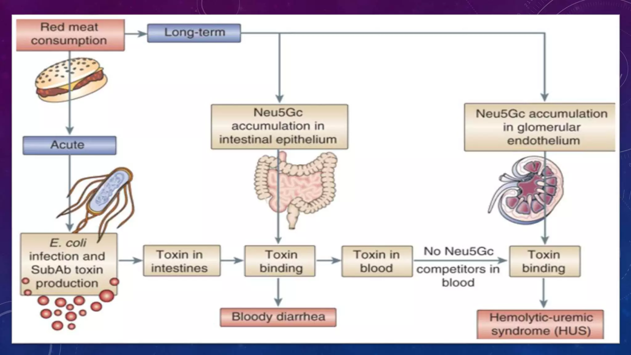
• Typical HUS follows a diarrheal infection often caused by E. coli OH157:H7. Infection related Shiga toxin
producing E.coli/Shigella Pneumococcal infection HIV Typical Other viral or bacterial infections. Only the
diarrheal form of HUS is considered to be typical HUS and is usually a disease of infants and children
younger than 3 years of age
2. ATYPICAL HUS
• caused by exposure to certain medications (eg ciclosporin, tacrolimus), genetic mutations in the
complement pathway[4] and systemic conditions, including lupus, cancer and pregnancy.](https://image.slidesharecdn.com/hemolyticuremicsyndrome-141212190757-conversion-gate01/75/Hemolytic-uremic-syndrome-4-2048.jpg)


Hemolytic uremic syndrome (HUS) is a disease characterized by microangiopathic hemolytic anemia, thrombocytopenia, and acute kidney injury. It is most commonly caused by infections from Shiga toxin-producing bacteria like E. coli O157:H7. The Shiga toxin damages endothelial cells and causes blood clots to form in the kidneys. Treatment involves fluid replacement, dialysis, and plasma exchange to support kidney function and replace lost blood cells. While HUS prognosis is generally good, some children may have long term kidney damage or rarely die from severe complications.


