Hemolytic uremic syndrome
•Download as PPTX, PDF•
5 likes•1,460 views

Hemolytic uremic syndrome

Recommended
More Related Content
What's hot
What's hot (20)
Similar to Hemolytic uremic syndrome
Similar to Hemolytic uremic syndrome (20)
More from Rivindu Wickramanayake
More from Rivindu Wickramanayake (20)
Recently uploaded
Recently uploaded (20)
Hemolytic uremic syndrome
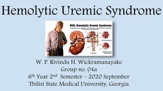
- 1. W. P. Rivindu H. Wickramanayake Group no. 04a 6th Year 2nd Semester – 2020 September Tbilisi State Medical University, Georgia Hemolytic Uremic Syndrome
- 2. ● HUS is thrombotic microangiopathy characterized by the presence of the triad of symptoms: thrombocytopenia, acute renal impairment, and microangiopathic hemolytic anemia. ● Thrombotic microangiopathy is the formation of platelet microthrombi in walls of small blood vessels (arterioles and capillaries), causing platelet consumption leading to thrombocytopenia, nonimmune hemolytic anemia (Coombs negative), and acute renal injury. ● Although traditionally thought as a triad, HUS presentation may vary based on different etiology. ● HUS can be a life-threatening condition requiring prompt diagnosis and treatment. ● One of the leading causes of acute renal injury in the pediatric population. Introduction
- 3. ● HUS has two variants termed typical and atypical HUS. ● The typical variant is caused by Shiga-like toxin (verotoxin) produced by E. coli (O157: H7) and Shiga toxin by Shigella dysenteriae. ● The atypical form is linked to bacteria, medication, or immune processes capable of endothelial damage. ● Streptococcus pneumoniae is a common bacterial infection associated with atypical HUS, while other causes include Mycoplasma pneumoniae, Clostridium difficile, HIV, histoplasmosis, and coxsackievirus and H1N1 Influenza A. ● Common drugs associated with HUS include mitomycin C, cyclosporine, cisplatin, cocaine, quinine, and rarely FK506 (tacrolimus) and interferon-alpha. Etiology
- 4. ● Most often associated with children under ten years old, with the majority occurring in those less than 5 years old. ● More than 90% of the typical HUS is caused by Shiga toxin- producing Escherichia coli (STEC). ● The yearly incidence of typical HUS is 3 cases per 100,000. ● Atypical HUS occurs more sporadically with an estimated incidence of 10 cases per 1,000,000. ● About 40% of the atypical HUS is associated with S. pneumoniae. ● The estimated fatality rate for both atypical and typical HUS is less than 5%. Epidemiology
- 5. ● Typically associated with bacterial infection resulting from the consumption of undercooked beef or unpasteurized milk. ● The Shiga toxin formed by the E. coli in typical HUS is then absorbed by the villi in the intestines, allowing the toxin to enter the bloodstream. ● The toxin then binds to glycosphingolipid found on multiple different cells throughout the body. The damage that ensues leads to an increase in thrombin and fibrin levels resulting in microthrombi being laid down. ● These microthrombi lead to platelet consumption, causing thrombocytopenia. ● Microthrombi present in the blood vessel also leads to the mechanical breakdown of red blood cells, causing hemolytic anemia. ● As the red blood cells are hemolyzed, there is a further increase in platelet use, causing thrombocytopenia. ● The Shiga toxin has a high affinity for globotriaosylceramide (Gb3) membrane receptor present in glomerular endothelium & tubular cells, causing widespread damage resulting in glomerular necrosis, cellular apoptosis & microangiopathic thrombosis leading to acute renal injury. Pathophysiology
- 6. Continued; ● Atypical HUS is a broad term used for those patients who develop HUS not associated with a Shiga-toxin illness. ● A common etiology is associated with S. pneumoniae that produces a neuraminidase enzyme. ● This enzyme targets N-acetylneuraminic acid found on the surface of red blood cells. ● When the enzyme cleaves N-acetylneuraminic acid, a T antigen is exposed, initiating an immune response. ● Another proposed mechanism includes increased expression and binding of pneumococcal surface proteins (Tuf and PspC) with human plasminogen leading to bacterial surface plasmin activation and widespread endothelial damage exposing subendothelial matrix and subsequent thrombotic microangiopathy. ● HIV, Mycoplasma pneumoniae, histoplasmosis, and coxsackievirus have also been linked to atypical HUS.
- 7. Continued; ● Another form of atypical HUS is called atypical familial HUS. ● In short, this disease results from a change in the genetic makeup for part of the complement pathway resulting in unregulated endothelial damage. ● As research continues, multiple different genetic mutations have been linked to atypical HUS. ● These patients are at higher risk for recurrent episodes of atypical HUS. ● A rare form of HUS associated with pregnancy and postpartum is linked to widespread complement activation triggered by pregnancy, and thus patients benefit from eculizumab (anti-C5 humanized monoclonal antibody).
- 8. ● Of the kidney shows fibrin deposition in the capillary lumen with extensive necrosis of the renal cortex and widespread renal arterial thrombosis. ● Glomerular mesangial expansion and endothelial proliferation are part of early histological changes while proliferation and aneurysmal dilatation of hilar arterioles with the double contour of the basement of the glomeruli are part of the late changes. ● Immunofluorescence demonstrates glomerular IgM, C3, and C1q deposition. ● Electron microscopy shows swollen mesangium with intraluminal platelet microthrombi and subendothelial space widening filled with fibrinogen. ● Obliterating endarteritis may be seen later. Histopathology
- 9. ● Is most often associated with a gastrointestinal illness; thus, patients presenting early in the course of the disease will complain of fever, abdominal pain, nausea, vomiting, and diarrhea. ● Diarrhea is often bloody and is within 3 days of diarrhea onset. Bloody diarrhea that ensues can be secondary to colitis from the invasion of the gastrointestinal cells or ischemia related to a vascular lesion. ● After a week of such gastrointestinal symptoms, patients may start to develop symptoms more closely related to the triad that defines HUS: specifically, symptoms related to anemia (syncope, shortness of breath, and jaundice) and kidney impairment (oliguria/anuria, hematuria). ● The skin exam may show small ecchymosis and mucosal bleeding. History and Physical
- 10. ● Late in the course of the infection, patients may develop seizures and encephalopathy related to ongoing uremia and other electrolyte derangements. ● In rare cases, the colitis can be severe enough to cause intestinal necrosis and perforation. ● In atypical HUS, a gastrointestinal illness is not usually the initial insult, and a thorough history and physical is required to find the offending agent. ● If S. pneumoniae is the source, the patient may site a recent respiratory illness. ● A review of any new medications or chronic autoimmune diseases is necessary. ● These patients typically have decreased urine output, pallor, and scattered ecchymosis. Continued;
- 11. ● The diagnosis requires a high index of suspicion based on symptoms, travel history, and dietary history. ● A complete blood count (CBC), comprehensive metabolic panel (CMP), and a urinalysis may aid in the diagnosis. ● The CBC shows both anemia and thrombocytopenia. ● Hemoglobin is often less than 10 g/dl. ● A peripheral smear reveals characteristic schistocytes. ● The Coombs test is negative, consistent with mechanical hemolysis. ● The CMP reveals elevated creatinine consistent with acute kidney injury. ● It also shows signs of acute hemolysis with elevated indirect bilirubin and elevated lactate dehydrogenase. ● As the disease progresses, hyponatremia and hyperkalemia may develop as the patient enters into renal failure. ● Urinalysis reveals hematuria and proteinuria. Evaluation
- 12. ● Begins with good supportive care. The patients are often fluid depleted and thus benefit from several fluid boluses. ● A careful assessment of the patient’s overall kidney function so as to avoid giving too much fluid in the setting of kidney failure. A subset of patients will go on to develop oliguric renal failure (<0.5 ml/kg/hr x72 hr) and require dialysis. ● Most experts do not recommend antibiotics and antiperistaltic agents to treat diarrheal illness. They believe that this will increase the complications associated with the E. coli infection. ● Patients should have their anemia corrected with packed RBCs when hemoglobin reaches 7 to 8 g/dl or hematocrit less than 18%. ● The treatment of thrombocytopenia is often unnecessary, given the continued consumption during the disease. ● Furthermore, there is concern that platelet transfusion leads to worsening thrombosis. Exceptions to this are those with active bleeding or before a medical procedure. ● Patients diagnosed with HUS should be admitted to the hospital for further monitoring. ● Those with encephalopathy or need for dialysis should be admitted to an intensive care unit. ● Familial atypical HUS requires consultation with hematology for possible plasmapheresis. Treatment / Management
- 13. ● Conditions that may present with hemolytic anemia, thrombocytopenia, & acute kidney injury 1) Thrombotic thrombocytopenic purpura (TTP): - TTP is thrombotic microangiopathy characterized by a pentad of hemolytic anemia, thrombocytopenia, renal dysfunction, fever, and neurological dysfunction. - It is due to deficiency or mutation in "a disintegrin and metalloproteinase with a thrombospondin type 1 motif member 13" (ADAMTS13) and usually has the adult-onset of symptoms. Differential Diagnosis
- 14. 2) Disseminated intravascular coagulation (DIC): - DIC is the systemic activation of the coagulation cascade and is characterized by abnormal coagulation studies, including prolonged prothrombin time and activated partial thromboplastin time and elevated D dimer and fibrin degradation products which are usually normal in HUS. - Patient with DIC usually has a very serious underlying illness like septic shock, trauma, malignancy, etc. Continued;
- 15. 3) HELLP syndrome: - It is characterized by hemolysis of red blood cells, elevated liver enzymes, and low platelet count occurring in pregnancy and severe preeclampsia. 4) Systemic vasculitis: - These patients typically present with inflammatory signs like fever, rash, and arthralgia and lack prodromal diarrhea. Continued;
- 16. ● The long-term complication rate from typical HUS is low, and most patients make a full recovery. Yearly evaluation for proteinuria, raised blood pressure, and renal insufficiency should be done for at least 5 years. ● Those patients who require dialysis have the highest risk of long-term chronic kidney disease. ● Patients who develop significant colitis, as evidenced by fever, leukocytosis, and severe abdominal pain, are at a higher risk of developing abdominal strictures. ● Those diagnosed with familial atypical HUS are at the highest risk of developing end-stage renal disease (ESRD) requiring routine dialysis. ● Familial atypical HUS patients also require several rounds of plasma exchange, placing the patient at risk for line infections. ● Other rare complications include transient or permanent diabetes mellitus, seizures, coma, stroke, abnormal liver functions, and hepatomegaly. Complications
- 17. ● Typically depends on the prompt initiation of treatment. ● Acute complications like acute renal injury, coma, and death, as well as progression to chronic renal failure, can be prevented with timely intervention. ● Overall mortality from HUS is less than 5%, while long term renal complications occur in 5% to 25% of the children with HUS. Prognosis 1. https://www.ncbi.nlm.nih.gov/books/NBK556038/ 2. Noris M, Remuzzi G. Hemolytic uremic syndrome. J. Am. Soc. Nephrol. 2005 Apr;16(4):1035-50. [PubMed] 3. Golubovic E, Miljkovic P, Zivic S, Jovancic D, Kostic G. Hemolytic uremic syndrome associated with novel influenza A H1N1 infection. Pediatr. Nephrol. 2011 Jan;26(1):149-50. [PubMed] 4. Fitzpatrick MM, Shah V, Trompeter RS, Dillon MJ, Barratt TM. Long term renal outcome of childhood haemolytic uraemic syndrome. BMJ. 1991 Aug 31;303(6801):489-92. [PMC free article] [PubMed] References