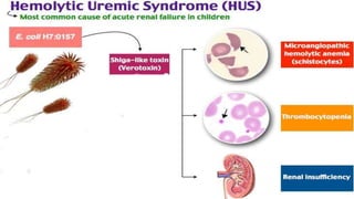
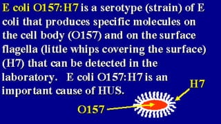
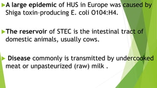
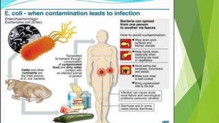
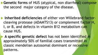
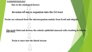
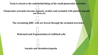
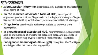
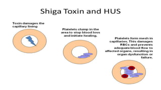
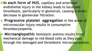
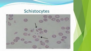
Hemolytic uremic
syndrome
By
Dr.
Ahmed zuhry
assiut university
Is acommon cause of community acquired acute
kidney injury in young children.
It characterized by the triad of:
 microangiopathic hemolytic anemia.
 thrombocytopenia.
renal in-sufficiency.
ETIOLOGY
 The most common form of HUS is caused by toxin-producing
Escherichia coli that cause prodromal acute enteritis and is commonly
termed diarrhea-associated HUS.
 In Asia and in southern Africa, the toxin of Shigella
dysenteriae type 1 is causative,
 In Western countries, verotoxin or Shiga-like toxin producing
E. coli (STEC) is the usual cause.
 Several serotypes of E. coli can produce the toxin; O157:H7 is
most common Europe and the Americas.
A large epidemic of HUS in Europe was caused by
Shiga toxin–producing E. coli O104:H4.
The reservoir of STEC is the intestinal tract of
domestic animals, usually cows.
 Disease commonly is transmitted by undercooked
meat or unpasteurized (raw) milk .
Genetic forms of HUS (atypical, non diarrheal) compose
the second major category of the disease.
 Inherited deficiencies of either von Willebrand factor–
cleaving protease (ADAMTS13) or complement factor H,
I, or B, and defects in vitamin B12 metabolism can
cause HUS.
 A specific genetic defect has not been identified in
approximately 50% of familial cases transmitted in
classic mendelian autosomal dominant or recessive
patterns.
 A major feature characteristic of genetic forms of HUS
is the absence of a preceding diarrhea prodrome.
HUS can be superimposed on any disease associated
with microvascular injury, including malignant
hypertension, systemic lupus erythematosus, and anti
phospholipid syndrome .
 It can also occur following bone marrow or solid organ
transplantation.
PATHOLOGY
 Early glomerular changes include:
 Thickening of the capillary walls caused by swelling of
endothelial cells and accumulation of fibrillar material between
endothelial cells and the underlying basement membrane, causing
narrowing of the capillary lumens.
 Platelet–fibrin thrombi are often seen in glomerular capillaries.
 Thrombi are also seen in afferent arterioles and small
arteries with fibrinoid necrosis of the arterial wall,
leading to renal cortical necrosis from vascular
occlusion.
Safouh, M.D.
CPNT
Late findings include:
 glomerular sclerosis secondary to either severe
direct glomerular involvement or glomerular
ischemia from arteriolar involvement.
PATHOPHYSIOLOGY
Due to the etiological factors
Invasion of micro organism into the GI tract
Toxins are released from the microorganism mainly from Ecoli and shigella
The toxin bind and destroy the colonic epithelial mucosal cells resulting in bloody
diarrhea.
Toxin is enter into the blood stream
Toxin is attach to the endothelial lining of the small glomerular arterioles
Glomerular arterioles become injured, swollen and occluded with platelet deposits
and fibrin clot
The circulating RBC cells are forced through the occluded arterioles
Deformed and fragmentation of redblood cells
Anemia and thrombocytopenia
PATHOGENESIS
 Microvascular injury with endothelial cell damage is characteristic
of all forms of HUS.
 In the diarrhea-associated form of HUS, enteropathic
organisms produce either Shiga toxin or the highly homologous Shiga-
like verotoxin both of which directly cause endothelial cell damage.
 Shiga toxin can directly activate platelets to promote their
aggregation.
 In pneumococcal-associated HUS, neuraminidase cleaves sialic
acid on membranes of endothelial cells, red cells, and platelets to
reveal the underlying cryptic Thomsen-Friedenreich (T) antigen.
 Endogenous immunoglobulin M (IgM) recognizes the T antigen
and triggers the microvascular angiopathy.
In each form of HUS, capillary and arteriolar
endothelial injury in the kidney leads to localized
thrombosis, particularly in glomeruli, causing a direct
decrease in glomerular filtration.
 Progressive platelet aggregation in the areas of
microvascular injury results in consumptive
thrombocytopenia.
 Microangiopathic hemolytic anemia results from
mechanical damage to red blood cells as they pass
through the damaged and thrombotic microvasculature.
CLINICAL MANIFESTATIONS
 Onset of HUS occurs a few days after onset of gastroenteritis
with ;
 fever, vomiting, abdominal pain, and diarrhea.
 The diarrhea is often bloody.
 Oliguria can be present in early stages but may be masked by
ongoing diarrhea.
 Proteinuria.
 Hypertension.
 Dehydration or Volume overload, depending on whether the
enteritis or renal insufficiency from HUS predominates, and the
amount of fluid that has been administered.
 CNS involvement occurs in ≤20% of cases.
Lethargy
Irritability
Seizures
Paresis
Coma
Cerebral edema
resulting from focal ischemia secondary to
microvascular CNS thrombosis.
DIAGNOSIS
 The diagnosis is made by the combination of:
 microangiopathic hemolytic anemia with schistocytes,
thrombocytopenia, and some degree of kidney involvement.
 The anemia can be mild at presentation, but rapidly
progresses.
 Thrombocytopenia is an invariable finding in the acute
phase, with platelet counts usually 20,000-100,000/mm3.
 Partial thromboplastin and prothrombin times are usually
normal.
 The Coombs test is negative, with the exception of
pneumococci-induced HUS, where the Coombs test is usually
positive.
 Leukocytosis is often present and significant.
 Urinalysis typically shows microscopic hematuria and low-
grade proteinuria.
 The renal insufficiency can vary from mild elevations in serum
blood urea nitrogen and creatinine to acute, anuric kidney
failure.
PROGNOSIS
 The mortality for diarrhea-associated HUS is <5% in most major
medical centers.
 Up to half of patients may require dialysis support during the acute
phase of the disease.
 Most recover renal function completely, but of surviving patients,
 5% remain dependent on dialysis,
 up to 30% are left with some degree of chronic renal insufficiency.
 Pneumococci associated HUS causes increased patient morbidity,
with mortality reported as 20%.
The familial, genetic forms of HUS can be insidiously
progressive or relapsing diseases and have a poor prognosis .
TREATMENT
There is no evidence that any therapy
directed at arresting the disease
process of the most common,
diarrhea-associated form of HUS
provides benefit.
Supportive care:
 careful management of fluid and electrolytes.
prompt correction of volume deficit.
 control of hypertension.
 Early dialysis if the patient becomes significantly
oliguric or anuric, particularly with hyperkalemia.
Early intravenous volume expansion before the onset of
oligoanuria may be nephroprotective in diarrhea-
associated HUS.
Red cell transfusions.
 Antibiotic therapy to clear enteric toxigenic organisms
(STEC) can result in increased toxin release, potentially
exacerbating the disease, and therefore is not recommended.
Eculizumab is an anti-C5 antibody that inhibits
complement activation, a pathway that contributes to active
disease in some forms of atypical familial HUS; this pathway
may contribute to the process in STEC-HUS.
Eculizumab is FDA approved for the treatment of atypical
HUS.
Cost:-
Eculizumab: 27 000 US$ for the first 4
weeks only.
‫هللا‬ ‫بحمد‬ ‫تم‬
Hemolytic uremic syndrome

Hemolytic uremic syndrome

  • 2.
  • 3.
    Is acommon causeof community acquired acute kidney injury in young children. It characterized by the triad of:  microangiopathic hemolytic anemia.  thrombocytopenia. renal in-sufficiency.
  • 5.
    ETIOLOGY  The mostcommon form of HUS is caused by toxin-producing Escherichia coli that cause prodromal acute enteritis and is commonly termed diarrhea-associated HUS.  In Asia and in southern Africa, the toxin of Shigella dysenteriae type 1 is causative,  In Western countries, verotoxin or Shiga-like toxin producing E. coli (STEC) is the usual cause.  Several serotypes of E. coli can produce the toxin; O157:H7 is most common Europe and the Americas.
  • 8.
    A large epidemicof HUS in Europe was caused by Shiga toxin–producing E. coli O104:H4. The reservoir of STEC is the intestinal tract of domestic animals, usually cows.  Disease commonly is transmitted by undercooked meat or unpasteurized (raw) milk .
  • 10.
    Genetic forms ofHUS (atypical, non diarrheal) compose the second major category of the disease.  Inherited deficiencies of either von Willebrand factor– cleaving protease (ADAMTS13) or complement factor H, I, or B, and defects in vitamin B12 metabolism can cause HUS.  A specific genetic defect has not been identified in approximately 50% of familial cases transmitted in classic mendelian autosomal dominant or recessive patterns.
  • 11.
     A majorfeature characteristic of genetic forms of HUS is the absence of a preceding diarrhea prodrome. HUS can be superimposed on any disease associated with microvascular injury, including malignant hypertension, systemic lupus erythematosus, and anti phospholipid syndrome .  It can also occur following bone marrow or solid organ transplantation.
  • 12.
    PATHOLOGY  Early glomerularchanges include:  Thickening of the capillary walls caused by swelling of endothelial cells and accumulation of fibrillar material between endothelial cells and the underlying basement membrane, causing narrowing of the capillary lumens.  Platelet–fibrin thrombi are often seen in glomerular capillaries.  Thrombi are also seen in afferent arterioles and small arteries with fibrinoid necrosis of the arterial wall, leading to renal cortical necrosis from vascular occlusion.
  • 13.
  • 14.
    Late findings include: glomerular sclerosis secondary to either severe direct glomerular involvement or glomerular ischemia from arteriolar involvement.
  • 15.
    PATHOPHYSIOLOGY Due to theetiological factors Invasion of micro organism into the GI tract Toxins are released from the microorganism mainly from Ecoli and shigella The toxin bind and destroy the colonic epithelial mucosal cells resulting in bloody diarrhea. Toxin is enter into the blood stream
  • 16.
    Toxin is attachto the endothelial lining of the small glomerular arterioles Glomerular arterioles become injured, swollen and occluded with platelet deposits and fibrin clot The circulating RBC cells are forced through the occluded arterioles Deformed and fragmentation of redblood cells Anemia and thrombocytopenia
  • 17.
    PATHOGENESIS  Microvascular injurywith endothelial cell damage is characteristic of all forms of HUS.  In the diarrhea-associated form of HUS, enteropathic organisms produce either Shiga toxin or the highly homologous Shiga- like verotoxin both of which directly cause endothelial cell damage.  Shiga toxin can directly activate platelets to promote their aggregation.  In pneumococcal-associated HUS, neuraminidase cleaves sialic acid on membranes of endothelial cells, red cells, and platelets to reveal the underlying cryptic Thomsen-Friedenreich (T) antigen.  Endogenous immunoglobulin M (IgM) recognizes the T antigen and triggers the microvascular angiopathy.
  • 19.
    In each formof HUS, capillary and arteriolar endothelial injury in the kidney leads to localized thrombosis, particularly in glomeruli, causing a direct decrease in glomerular filtration.  Progressive platelet aggregation in the areas of microvascular injury results in consumptive thrombocytopenia.  Microangiopathic hemolytic anemia results from mechanical damage to red blood cells as they pass through the damaged and thrombotic microvasculature.
  • 20.
    CLINICAL MANIFESTATIONS  Onsetof HUS occurs a few days after onset of gastroenteritis with ;  fever, vomiting, abdominal pain, and diarrhea.  The diarrhea is often bloody.  Oliguria can be present in early stages but may be masked by ongoing diarrhea.  Proteinuria.  Hypertension.
  • 21.
     Dehydration orVolume overload, depending on whether the enteritis or renal insufficiency from HUS predominates, and the amount of fluid that has been administered.
  • 22.
     CNS involvementoccurs in ≤20% of cases. Lethargy Irritability Seizures Paresis Coma Cerebral edema resulting from focal ischemia secondary to microvascular CNS thrombosis.
  • 23.
    DIAGNOSIS  The diagnosisis made by the combination of:  microangiopathic hemolytic anemia with schistocytes, thrombocytopenia, and some degree of kidney involvement.  The anemia can be mild at presentation, but rapidly progresses.  Thrombocytopenia is an invariable finding in the acute phase, with platelet counts usually 20,000-100,000/mm3.
  • 26.
     Partial thromboplastinand prothrombin times are usually normal.  The Coombs test is negative, with the exception of pneumococci-induced HUS, where the Coombs test is usually positive.  Leukocytosis is often present and significant.  Urinalysis typically shows microscopic hematuria and low- grade proteinuria.  The renal insufficiency can vary from mild elevations in serum blood urea nitrogen and creatinine to acute, anuric kidney failure.
  • 27.
    PROGNOSIS  The mortalityfor diarrhea-associated HUS is <5% in most major medical centers.  Up to half of patients may require dialysis support during the acute phase of the disease.  Most recover renal function completely, but of surviving patients,  5% remain dependent on dialysis,  up to 30% are left with some degree of chronic renal insufficiency.  Pneumococci associated HUS causes increased patient morbidity, with mortality reported as 20%. The familial, genetic forms of HUS can be insidiously progressive or relapsing diseases and have a poor prognosis .
  • 28.
    TREATMENT There is noevidence that any therapy directed at arresting the disease process of the most common, diarrhea-associated form of HUS provides benefit.
  • 29.
    Supportive care:  carefulmanagement of fluid and electrolytes. prompt correction of volume deficit.  control of hypertension.  Early dialysis if the patient becomes significantly oliguric or anuric, particularly with hyperkalemia. Early intravenous volume expansion before the onset of oligoanuria may be nephroprotective in diarrhea- associated HUS. Red cell transfusions.
  • 30.
     Antibiotic therapyto clear enteric toxigenic organisms (STEC) can result in increased toxin release, potentially exacerbating the disease, and therefore is not recommended. Eculizumab is an anti-C5 antibody that inhibits complement activation, a pathway that contributes to active disease in some forms of atypical familial HUS; this pathway may contribute to the process in STEC-HUS. Eculizumab is FDA approved for the treatment of atypical HUS. Cost:- Eculizumab: 27 000 US$ for the first 4 weeks only.
  • 31.