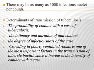
MYCOBACTERIUM TUBERCULOSIS
 Tuberculosis, one of the oldest diseases known to affect
humans, is caused by bacteria belonging to the
Mycobacterium tuberculosis complex.
 The disease usually affects the lungs, although in up to
one-third of cases other organs are involved
 If untreated, the disease may be fatal within 5 years in
more than half of cases
 Transmission usually takes place through the airborne
spread of droplet nuclei produced by patients with
infectious pulmonary tuberculosis
 M. tuberculosis is a rod-shaped, non-spore-forming, thin
aerobic bacterium measuring 0.5 m by 3 m.
 Once stained, the bacilli cannot be decolorized by acid
alcohol, a characteristic justifying their classification as
acid-fast bacilli.
 Other organisms showing acid fastness include ;
Nocardia and Rhodococcus, Legionella micdadei, and
the protozoa Isospora and Cryptosporidium.
 Pathogenesis of tuberculosis
 From exposure to infection;
 M. tuberculosis is most commonly transmitted from a
patient with infectious pulmonary tuberculosis to other
persons by droplet nuclei, which are aerosolized by
coughing, sneezing, or speaking.
 The tiny droplets dry rapidly; the smallest (10 m in
diameter) may remain suspended in the air for several
hours and may gain direct access to the terminal air
passages when inhaled.
 There may be as many as 3000 infectious nuclei
per cough.
 Determinants of transmission of tuberculosis;
1. The probability of contact with a case of
tuberculosis,
2. the intimacy and duration of that contact,
3. the degree of infectiousness of the case
4. Crowding in poorly ventilated rooms is one of
the most important factors in the transmission of
tubercle bacilli, since it increases the intensity of
contact with a case
 Patients who have cavitary pulmonary disease or
tuberculosis of the respiratory tract (endobronchial or
laryngeal tuberculosis produce sputa containing as many
as 105 AFB/mL.
 Patients with sputum smear–negative/culture-positive
tuberculosis are less infectious,
and those with culture-negative pulmonary disease and
extrapulmonary tuberculosis are essentially noninfectious.
 From infection to disease;
 the risk of developing disease after being infected
depends largely on endogenous factors, such as
1. the individual’s innate susceptibility to disease
2. and level of function of cell-mediated immunity.
 Clinical illness directly following infection is classified as
primary tuberculosis and is common among children up
to 4 years of age.
 Dormant bacilli, however, may persist for years before
reactivating to produce secondary (or postprimary)
tuberculosis, which is often infectious.
 Risk factor for active tuberculosis;
Recent infection (1 year) 12.9
Fibrotic lesions (spontaneously healed) 12-20
Comorbidity
HIV infection
Silicosis
Chronic renal failure / hemodialysis
Diabetes
Intravenous drug abuse
Immunosuppresant treatment
Gastrectomy
Jejunoileal bypass
Post transplantation period
100
30
20-25
2-4
10-30
10
2-5
30-60
20-70
Malnutrition and severe underweight 2
 PATHOGENESIS AND IMMUNITY
 the majority of inhaled bacilli are trapped in the upper
airways and expelled by ciliated mucosal cells, a fraction
(usually 10%) reach the alveoli.
 There, nonspecifically activated alveolar macrophages
ingest the bacilli.
 The balance between the bactericidal activity of the
macrophage and the number and virulence of the bacilli
determines the events following phagocytosis.
 In the initial stage of host-bacterium interaction, either the
host’s macrophages contain bacillary multiplication by
producing proteolytic enzymes and cytokines or the
bacilli begin to multiply.
 About 2 to 4 weeks after infection, two additional host
responses to M. tuberculosis develop:
1. a tissue-damaging response and
2. a macrophage- activating response.
 The tissue-damaging response is the result of a
delayed-type hypersensitivity (DTH) reaction to
various bacillary antigens; it destroys nonactivated
macrophages that contain multiplying bacilli.
 The macrophage-activating response is a cell-
mediated phenomenon resulting in the activation
of macrophages that are capable of killing and
digesting tubercle bacilli. Although both of these
responses can inhibit mycobacterial growth, it is
the balance between the two that determines the
form of tuberculosis that will develop
subsequently.
 With the development of specific immunity and
the accumulation of large numbers of activated
macrophages at the site of the primary lesion,
granulomatous lesions (tubercles) are formed.
 These lesions consist of lymphocytes and
activated macrophages, such as epithelioid cells
and giant cells.
 Cell-mediated immunity is critical at this early stage. In
the majority of infected individuals, local macrophages
are activated when bacillary antigens processed by
macrophages stimulate T lymphocytes to release a variety
of lymphokines.
 These activated cells aggregate around the lesion’s center
and effectively neutralize tubercle bacilli without causing
further tissue destruction.
 In the central part of the lesion, the necrotic material
resembles soft cheese (caseous necrosis)— a phenomenon
that may also be observed in other conditions, such as
neoplasms.
 Even when healing takes place, viable bacilli may remain
dormant within macrophages or in the necrotic material
for years or even throughout the patient’s lifetime.
 These “healed” lesions in the lung parenchyma and hilar
lymph nodes may later undergo calcification.
 In a minority of cases, the macrophage-activating
response is weak, and mycobacterial growth can be
inhibited only by intensified DTH reactions, which lead
to tissue destruction.
 The lesion tends to enlarge further, and the surrounding
tissue is progressively damaged. At the center of the
lesion, the caseous material liquefies. Bronchial walls as
well as blood vessels are invaded and destroyed, and
cavities are formed.
 The liquefied caseous material, containing large numbers
of bacilli, is drained through bronchi. Within the cavity,
tubercle bacilli multiply well and spread into the airways
and the environment through expectorated sputum.
 In the early stages of infection, bacilli are usually
transported by macrophages to regional lymph nodes,
from which they disseminate widely to many organs and
tissues
 Cell-mediated immunity confers partial protection
against M. tuberculosis,while humoral immunity
has no defined role in protection.
 Clinical manifestations
 Tuberculosis is classified as pulmonary or
extrapulmonary.
 Before the recognition of HIV infection, 80% of
all cases of tuberculosis were limited to the lungs.
PULMONARY TUBERCULOSIS
 Pulmonary tuberculosis can be categorized as primary or
postprimary (secondary).
 PRIMARY DISEASE
 Primary pulmonary tuberculosis results from an initial
infection with tubercle bacilli.
 The lesion forming after infection is usually peripheral
and accompanied by hilar or paratracheal
lymphadenopathy, which may not be detectable on chest
radiography. In the majority of cases, the lesion heals
spontaneously and may later be evident as a small
calcified nodule (Ghon lesion).
 In children and in persons with impaired immunity (e.g.,
those with malnutrition or HIV infection), primary
pulmonary tuberculosis may progress rapidly to clinical
illness.
 The initial lesion increases in size and can evolve in
different ways.
 Pleural effusion, a frequent finding, results from the
penetration of bacilli into the pleural space from an
adjacent subpleural focus.
 In severe cases, the primary site rapidly enlarges, its
central portion undergoes necrosis, and acute cavitation
develops (progressive primary tuberculosis).
 Tuberculosis in young children is almost invariably
accompanied by hilar or mediastinal
lymphadenopathy due to the spread of bacilli from
the lung parenchyma through lymphatic vessels.
 Hematogenous dissemination, which is common
and is often asymptomatic, may result in the most
severe manifestations of primary M. tuberculosis
infection.
POST PRIMARY DISEASE
 Also called adult-type, reactivation, or secondary
tuberculosis, postprimary disease results from
endogenous reactivation of latent infection and is usually
localized to the apical and posterior segments of the upper
lobes, where the high oxygen concentration favors
mycobacterial growth.
 The extent of lung parenchymal involvement varies
greatly, from small infiltrates to extensive cavitary
disease.
 With cavity formation, liquefied necrotic contents are
ultimately discharged into the airways, resulting in
satellite lesions within the lungs that may in turn
undergo cavitation.
 Massive involvement of pulmonary segments or
lobes, with coalescence of lesions, produces
tuberculous pneumonia.
 While up to one-third of untreated patients reportedly
succumb to severe pulmonary tuberculosis within a
few weeks or months after onset, others undergo a
process of spontaneous remission or proceed along a
chronic, progressively debilitating course
(“consumption”).
Mycobacterium tuberculosis

Mycobacterium tuberculosis

  • 1.
  • 2.
     Tuberculosis, oneof the oldest diseases known to affect humans, is caused by bacteria belonging to the Mycobacterium tuberculosis complex.  The disease usually affects the lungs, although in up to one-third of cases other organs are involved  If untreated, the disease may be fatal within 5 years in more than half of cases  Transmission usually takes place through the airborne spread of droplet nuclei produced by patients with infectious pulmonary tuberculosis
  • 3.
     M. tuberculosisis a rod-shaped, non-spore-forming, thin aerobic bacterium measuring 0.5 m by 3 m.  Once stained, the bacilli cannot be decolorized by acid alcohol, a characteristic justifying their classification as acid-fast bacilli.  Other organisms showing acid fastness include ; Nocardia and Rhodococcus, Legionella micdadei, and the protozoa Isospora and Cryptosporidium.
  • 5.
     Pathogenesis oftuberculosis  From exposure to infection;  M. tuberculosis is most commonly transmitted from a patient with infectious pulmonary tuberculosis to other persons by droplet nuclei, which are aerosolized by coughing, sneezing, or speaking.  The tiny droplets dry rapidly; the smallest (10 m in diameter) may remain suspended in the air for several hours and may gain direct access to the terminal air passages when inhaled.
  • 6.
     There maybe as many as 3000 infectious nuclei per cough.  Determinants of transmission of tuberculosis; 1. The probability of contact with a case of tuberculosis, 2. the intimacy and duration of that contact, 3. the degree of infectiousness of the case 4. Crowding in poorly ventilated rooms is one of the most important factors in the transmission of tubercle bacilli, since it increases the intensity of contact with a case
  • 7.
     Patients whohave cavitary pulmonary disease or tuberculosis of the respiratory tract (endobronchial or laryngeal tuberculosis produce sputa containing as many as 105 AFB/mL.  Patients with sputum smear–negative/culture-positive tuberculosis are less infectious, and those with culture-negative pulmonary disease and extrapulmonary tuberculosis are essentially noninfectious.
  • 8.
     From infectionto disease;  the risk of developing disease after being infected depends largely on endogenous factors, such as 1. the individual’s innate susceptibility to disease 2. and level of function of cell-mediated immunity.  Clinical illness directly following infection is classified as primary tuberculosis and is common among children up to 4 years of age.  Dormant bacilli, however, may persist for years before reactivating to produce secondary (or postprimary) tuberculosis, which is often infectious.
  • 9.
     Risk factorfor active tuberculosis; Recent infection (1 year) 12.9 Fibrotic lesions (spontaneously healed) 12-20 Comorbidity HIV infection Silicosis Chronic renal failure / hemodialysis Diabetes Intravenous drug abuse Immunosuppresant treatment Gastrectomy Jejunoileal bypass Post transplantation period 100 30 20-25 2-4 10-30 10 2-5 30-60 20-70 Malnutrition and severe underweight 2
  • 10.
     PATHOGENESIS ANDIMMUNITY  the majority of inhaled bacilli are trapped in the upper airways and expelled by ciliated mucosal cells, a fraction (usually 10%) reach the alveoli.  There, nonspecifically activated alveolar macrophages ingest the bacilli.  The balance between the bactericidal activity of the macrophage and the number and virulence of the bacilli determines the events following phagocytosis.
  • 11.
     In theinitial stage of host-bacterium interaction, either the host’s macrophages contain bacillary multiplication by producing proteolytic enzymes and cytokines or the bacilli begin to multiply.  About 2 to 4 weeks after infection, two additional host responses to M. tuberculosis develop: 1. a tissue-damaging response and 2. a macrophage- activating response.
  • 12.
     The tissue-damagingresponse is the result of a delayed-type hypersensitivity (DTH) reaction to various bacillary antigens; it destroys nonactivated macrophages that contain multiplying bacilli.  The macrophage-activating response is a cell- mediated phenomenon resulting in the activation of macrophages that are capable of killing and digesting tubercle bacilli. Although both of these responses can inhibit mycobacterial growth, it is the balance between the two that determines the form of tuberculosis that will develop subsequently.
  • 13.
     With thedevelopment of specific immunity and the accumulation of large numbers of activated macrophages at the site of the primary lesion, granulomatous lesions (tubercles) are formed.  These lesions consist of lymphocytes and activated macrophages, such as epithelioid cells and giant cells.
  • 14.
     Cell-mediated immunityis critical at this early stage. In the majority of infected individuals, local macrophages are activated when bacillary antigens processed by macrophages stimulate T lymphocytes to release a variety of lymphokines.  These activated cells aggregate around the lesion’s center and effectively neutralize tubercle bacilli without causing further tissue destruction.  In the central part of the lesion, the necrotic material resembles soft cheese (caseous necrosis)— a phenomenon that may also be observed in other conditions, such as neoplasms.
  • 15.
     Even whenhealing takes place, viable bacilli may remain dormant within macrophages or in the necrotic material for years or even throughout the patient’s lifetime.  These “healed” lesions in the lung parenchyma and hilar lymph nodes may later undergo calcification.  In a minority of cases, the macrophage-activating response is weak, and mycobacterial growth can be inhibited only by intensified DTH reactions, which lead to tissue destruction.
  • 16.
     The lesiontends to enlarge further, and the surrounding tissue is progressively damaged. At the center of the lesion, the caseous material liquefies. Bronchial walls as well as blood vessels are invaded and destroyed, and cavities are formed.  The liquefied caseous material, containing large numbers of bacilli, is drained through bronchi. Within the cavity, tubercle bacilli multiply well and spread into the airways and the environment through expectorated sputum.  In the early stages of infection, bacilli are usually transported by macrophages to regional lymph nodes, from which they disseminate widely to many organs and tissues
  • 17.
     Cell-mediated immunityconfers partial protection against M. tuberculosis,while humoral immunity has no defined role in protection.  Clinical manifestations  Tuberculosis is classified as pulmonary or extrapulmonary.  Before the recognition of HIV infection, 80% of all cases of tuberculosis were limited to the lungs.
  • 18.
    PULMONARY TUBERCULOSIS  Pulmonarytuberculosis can be categorized as primary or postprimary (secondary).  PRIMARY DISEASE  Primary pulmonary tuberculosis results from an initial infection with tubercle bacilli.  The lesion forming after infection is usually peripheral and accompanied by hilar or paratracheal lymphadenopathy, which may not be detectable on chest radiography. In the majority of cases, the lesion heals spontaneously and may later be evident as a small calcified nodule (Ghon lesion).
  • 19.
     In childrenand in persons with impaired immunity (e.g., those with malnutrition or HIV infection), primary pulmonary tuberculosis may progress rapidly to clinical illness.  The initial lesion increases in size and can evolve in different ways.  Pleural effusion, a frequent finding, results from the penetration of bacilli into the pleural space from an adjacent subpleural focus.  In severe cases, the primary site rapidly enlarges, its central portion undergoes necrosis, and acute cavitation develops (progressive primary tuberculosis).
  • 20.
     Tuberculosis inyoung children is almost invariably accompanied by hilar or mediastinal lymphadenopathy due to the spread of bacilli from the lung parenchyma through lymphatic vessels.  Hematogenous dissemination, which is common and is often asymptomatic, may result in the most severe manifestations of primary M. tuberculosis infection.
  • 21.
    POST PRIMARY DISEASE Also called adult-type, reactivation, or secondary tuberculosis, postprimary disease results from endogenous reactivation of latent infection and is usually localized to the apical and posterior segments of the upper lobes, where the high oxygen concentration favors mycobacterial growth.  The extent of lung parenchymal involvement varies greatly, from small infiltrates to extensive cavitary disease.
  • 22.
     With cavityformation, liquefied necrotic contents are ultimately discharged into the airways, resulting in satellite lesions within the lungs that may in turn undergo cavitation.  Massive involvement of pulmonary segments or lobes, with coalescence of lesions, produces tuberculous pneumonia.  While up to one-third of untreated patients reportedly succumb to severe pulmonary tuberculosis within a few weeks or months after onset, others undergo a process of spontaneous remission or proceed along a chronic, progressively debilitating course (“consumption”).

Editor's Notes