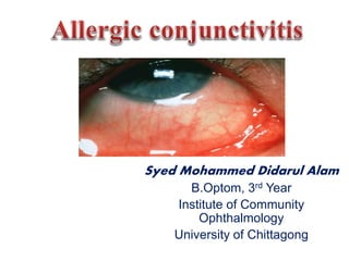
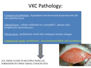
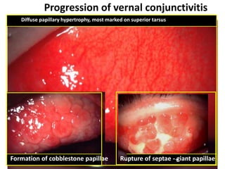
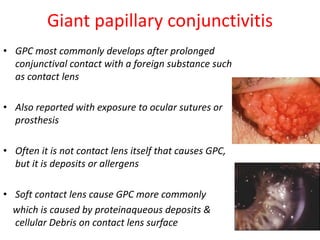
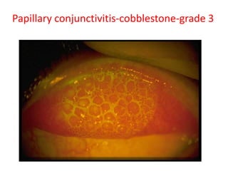
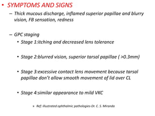
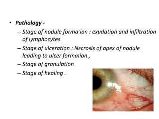
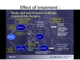
Syed Mohammed Didarul Alam
B.Optom, 3rd Year
Institute of Community
Ophthalmology
University of Chittagong
Allergic conjunctivitis:
• Inflammation of conjunctiva due to allergic or
hypersensitive reaction which may be
immediate (humoral ) or delayed (cellular) to
specific antigens
Allergic Reaction
Allergic conjunctivitis:
1. Simple allergic conjunctivitis
-hay fever conjunctivitis
-seasonal allergic conjunctivitis (SAC)
-perennial allergic conjunctivitis(PAC)
2. Vernal keratoconjunctivitis (VKC)
3. Atopic keratoconjunctivitis (AKC)
4. Giant papillary conjunctivitis (GPC)
5. Phlyctenular keratoconjunctivitis (PKC)
6. Contact dermoconjunctivitis (CDC)
• Mild ,non specific IgE mediated Type I
hypersensitivity reaction
• Etiology :
• Hay fever conjunctivitis : associated with
allergic rhinitis
• Allergens : pollens , grass , animal dandruffs
• SAC: common , d/t: grass pollens
• PAC: not common , d/t: house dust and mites
Pathogenesis
Allergen enters tear film
Comes in contact with conjunctival mast cells that bear
lgE antibodies.
Degranulation of mast cells releases histamine
Histamine promotes vasodilatation & edema
Symptoms: - itching
-Redness
-burning sensation
- watery discharge and
-mild photophobia
Signs : -hyperemia and chemosis
-mild papillary reaction
-oedema of eyelids
• Treatment (severity dependent)
– Elimination of allergens if possible
– cold compresses
– antihistamines oral/ topical (epinistine,fexofenadrine)
– mast cell stabilizers (sodium cromoglycate,lodaximide)
– Combination( ketotifen,patalon,azelastine)
– topical corticosteroids
– Immunosuppressant's (cyclosporin) for steroid
resistant cases
Vernal keratoconjunctivitis or spring catarrh
– Recurrent, Bilateral , self limiting allergic inflammation
of the conjunctiva affecting children and young adults
– more common in males
– allergic disorder in which IgE and cell mediated immune
mechanism play an important role
• Clinical features :
– 98% bilateral, can be asymmetric
– Intense ocular itching, Lacrimation, Photophobia,
blepharospasm, blurred vision, FB sensation , burning
and difficulty opening eyes in the morning.
– Thick mucous ropy discharge , Pseudoptosis due to large
papillae.
– Giant papillae on the superior Palpebral conjunctiva are
the clinical hallmark.
ropy discharge
VKC Pathology:
• Conjunctival epithelium : hyperplasia and downward projections into the
sub epithelial tissue
• Adenoid layer : cellular infiltration by eosinophil's , plasma cells ,
lymphocytes and histiocytes .
• Fibrous layer : proliferation which later undergoes hyaline changes
• Conjunctival vessels: proliferation , increased permeability and vasodilation
ALL THESE LEADS TO MULTIPLE PAPILLAE
FORMATION IN UPPER TARSAL CONJUNCTIVA
• palpebral
• bulbar
• mixed form
Palpebral form
• Diffuse papillary
hypertrophy, > on
superior tarsus
• Papillae have a flat-
topped polygonal
appearance resembling
COBBLESTONES
• Severe cases- Giant
papillae, which may be
coated with mucus
Progression of vernal conjunctivitis
Diffuse papillary hypertrophy, most marked on superior tarsus
Formation of cobblestone papillae Rupture of septae - giant papillae
Limbal / Bulbar form
• May start as a thickening &
opacification of limbus
• Limbal nodules - Mucoid
nodules, which are
gelatinous, elevated
• Horner-Trantas dots –
composed mainly of
eosinophils and epithelial
debris (limbal apices)
Limbal vernal
Tranta's dotsMucoid nodule
Vernal Keratopathy /
Corneal involvement
• Punctate epithelial
erosions to
macroerosions
• Shield ulcers – Oval
ulceration with
thickened, opaque
edges
Progression of vernal Keratopathy
Punctate epithelopathy Epithelial macroerosions
Plaque formation (shield ulcer) Sub epithelial scarring
Treatment
• Topical antihistamine
• Mast cell stabilizers : sodium chromoglycate 2 % drops
4-5 times/day
• Topical steroid : Every 4 hrs. for 2 days followed by 3-4
times a day for 2 weeks .
MONITOR IOP TO PREVENT STEROID INDUCED
GLAUCOMA
• Acetyl cysteine (0.5%)
• Topical cyclosporine (1%): severe unresponsive case
Systemic :
I. Oral antihistamine : for itching
II. Oral steroid : short course for very severe
non responsive case
• Treatment of large papilla supratarsal
injection of long acting steroid or surgical
removal
• General measures: dark goggles , cold
compress , change of place from hot to cold
Atopic keratoconjunctivitis
• AKC is rare bilateral that more common adult(30-
35Years)
• Long history of eczema
• May be associated with atopic dermatitis
• Asthma is also common with AKC
Symptoms : itching , soreness , dry
sensation , mucoid discharge, Hardening
Eyelid, phtophobia or blurred vision
Signs :
lid margins: inflamed with round
posterior borders
conjunctiva : inferiorly involve,watery
discharge,milky appearance , very fine
papilla , hyperaemia scarring with
shrinkage
limbal: limbal involvement similar to
limbal VKC
cornea-punctate epithelial keratitis in
lower half, vascularization , plaque
A :-periocular eczema and corneal haze
C:-progression of the disease; dense pannus entering visual axis
E: symblepharon. F: posterior subcapsular cataract, which can be
associated with atopic keratoconjunctivitis.
Treatment
Local:-
 sodium chromoglycate
 Antihistamine
 Combination( antihistamine & mast cell strabilizer)
 topical steroids ( fluromethalone 0.1%, loteprednol 0.2%)
 Supratarsal steriod injection in severe
others:-
treat facial eczema and lid margin disease
Immunosuppressive agents( cyclosporine,tacrolimus)
if untreated AKC can progress to ulceration, scarring,
cataract, keratoconus, and corneal vascularization.
Giant papillary conjunctivitis
• GPC most commonly develops after prolonged
conjunctival contact with a foreign substance such
as contact lens
• Also reported with exposure to ocular sutures or
prosthesis
• Often it is not contact lens itself that causes GPC,
but it is deposits or allergens
• Soft contact lens cause GPC more commonly
which is caused by proteinaqueous deposits &
cellular Debris on contact lens surface
Papillary conjunctivitis-cobblestone-grade-2(flour)
Papillary conjunctivitis-cobblestone-grade 3
Papillary conjunctivitis-cobblestone-grade-4(flour)
• SYMPTOMS AND SIGNS
– Thick mucous discharge, inflamed superior papillae and blurry
vision, FB sensation, redness
– GPC staging
• Stage 1:itching and decreased lens tolerance
• Stage 2:blurred vision, superior tarsal papillae ( >0.3mm)
• Stage 3:excessive contact lens movement because tarsal
papillae don’t allow smooth movement of lid over CL
• Stage 4:similar appearance to mild VKC
» Ref: illustrated ophthalmic pathologies-Dr. C. S. Miranda
Treatment :
• Removal of cause
• Discontinue contact lens wear &
strong counseling
• Antihistamin
• Mast cell stabilizer
• Disodium chromoglycate
• Steroids can be use for Acute phase
Phlyctenular conjunctivitis :
• Nodular affection occurring as an allergic response by
conjunctiva and corneal epithelium to some
endogenous allergens .
• Etiology -
– Delayed hypersensitivity ( type I ) response to
endogenous microbial proteins : Tuberculous protein
Staphylococcal protein , parasitic protein .
• Pathology -
– Stage of nodule formation : exudation and infiltration
of lymphocytes
– Stage of ulceration : Necrosis of apex of nodule
leading to ulcer formation ,
– Stage of granulation
– Stage of healing .
• Treatment -
– steroid eye drops ,
– Antibiotic drops ( secondary infection )
– specific therapy
• Tuberculosis
• septic focus should be treated
• parasitic infestation - stool examination .
– General measures - improve health of child .
• Contact dermatoconjunctivitis is an
allergic reaction in the conjunctiva and
eyelid skin to medications (or other toxic
products like cosmetics) applied there
• Etiology: delayed type hyper
sensitivity response to prolong contact
with chemicals and ophthalmic
medicines( atropine, neomycin,
soframycin)
Clinical Features:
• Cutaneous involvement: weeping eczema
around the area involved with medication
• Conjunctival response: lower fornix and lower
palpebral conjunctiva
• Treatment:
• Discontinuing of causative chemical or
medications
• Antihistamine
• NSAID
• Topical steroid eye drops
• Steroid ointment in involved surrounding area
Effect of treatment :
REFERENCES
• Comorehensive Ophthalmology-A.K.Khurana
• Clinical ophthalmology-Jack.J.Kanski
• Lippincott’s microbiology
• Internet
Allergic conjunctivitis

Allergic conjunctivitis

  • 1.
    Syed Mohammed DidarulAlam B.Optom, 3rd Year Institute of Community Ophthalmology University of Chittagong
  • 2.
    Allergic conjunctivitis: • Inflammationof conjunctiva due to allergic or hypersensitive reaction which may be immediate (humoral ) or delayed (cellular) to specific antigens
  • 3.
  • 4.
    Allergic conjunctivitis: 1. Simpleallergic conjunctivitis -hay fever conjunctivitis -seasonal allergic conjunctivitis (SAC) -perennial allergic conjunctivitis(PAC) 2. Vernal keratoconjunctivitis (VKC) 3. Atopic keratoconjunctivitis (AKC) 4. Giant papillary conjunctivitis (GPC) 5. Phlyctenular keratoconjunctivitis (PKC) 6. Contact dermoconjunctivitis (CDC)
  • 5.
    • Mild ,nonspecific IgE mediated Type I hypersensitivity reaction • Etiology : • Hay fever conjunctivitis : associated with allergic rhinitis • Allergens : pollens , grass , animal dandruffs • SAC: common , d/t: grass pollens • PAC: not common , d/t: house dust and mites
  • 6.
    Pathogenesis Allergen enters tearfilm Comes in contact with conjunctival mast cells that bear lgE antibodies. Degranulation of mast cells releases histamine Histamine promotes vasodilatation & edema
  • 7.
    Symptoms: - itching -Redness -burningsensation - watery discharge and -mild photophobia Signs : -hyperemia and chemosis -mild papillary reaction -oedema of eyelids
  • 8.
    • Treatment (severitydependent) – Elimination of allergens if possible – cold compresses – antihistamines oral/ topical (epinistine,fexofenadrine) – mast cell stabilizers (sodium cromoglycate,lodaximide) – Combination( ketotifen,patalon,azelastine) – topical corticosteroids – Immunosuppressant's (cyclosporin) for steroid resistant cases
  • 9.
    Vernal keratoconjunctivitis orspring catarrh – Recurrent, Bilateral , self limiting allergic inflammation of the conjunctiva affecting children and young adults – more common in males – allergic disorder in which IgE and cell mediated immune mechanism play an important role
  • 10.
    • Clinical features: – 98% bilateral, can be asymmetric – Intense ocular itching, Lacrimation, Photophobia, blepharospasm, blurred vision, FB sensation , burning and difficulty opening eyes in the morning. – Thick mucous ropy discharge , Pseudoptosis due to large papillae. – Giant papillae on the superior Palpebral conjunctiva are the clinical hallmark. ropy discharge
  • 11.
    VKC Pathology: • Conjunctivalepithelium : hyperplasia and downward projections into the sub epithelial tissue • Adenoid layer : cellular infiltration by eosinophil's , plasma cells , lymphocytes and histiocytes . • Fibrous layer : proliferation which later undergoes hyaline changes • Conjunctival vessels: proliferation , increased permeability and vasodilation ALL THESE LEADS TO MULTIPLE PAPILLAE FORMATION IN UPPER TARSAL CONJUNCTIVA
  • 12.
  • 13.
    Palpebral form • Diffusepapillary hypertrophy, > on superior tarsus • Papillae have a flat- topped polygonal appearance resembling COBBLESTONES • Severe cases- Giant papillae, which may be coated with mucus
  • 14.
    Progression of vernalconjunctivitis Diffuse papillary hypertrophy, most marked on superior tarsus Formation of cobblestone papillae Rupture of septae - giant papillae
  • 15.
    Limbal / Bulbarform • May start as a thickening & opacification of limbus • Limbal nodules - Mucoid nodules, which are gelatinous, elevated • Horner-Trantas dots – composed mainly of eosinophils and epithelial debris (limbal apices)
  • 16.
  • 17.
    Vernal Keratopathy / Cornealinvolvement • Punctate epithelial erosions to macroerosions • Shield ulcers – Oval ulceration with thickened, opaque edges
  • 18.
    Progression of vernalKeratopathy Punctate epithelopathy Epithelial macroerosions Plaque formation (shield ulcer) Sub epithelial scarring
  • 19.
    Treatment • Topical antihistamine •Mast cell stabilizers : sodium chromoglycate 2 % drops 4-5 times/day • Topical steroid : Every 4 hrs. for 2 days followed by 3-4 times a day for 2 weeks . MONITOR IOP TO PREVENT STEROID INDUCED GLAUCOMA • Acetyl cysteine (0.5%) • Topical cyclosporine (1%): severe unresponsive case
  • 20.
    Systemic : I. Oralantihistamine : for itching II. Oral steroid : short course for very severe non responsive case • Treatment of large papilla supratarsal injection of long acting steroid or surgical removal • General measures: dark goggles , cold compress , change of place from hot to cold
  • 21.
    Atopic keratoconjunctivitis • AKCis rare bilateral that more common adult(30- 35Years) • Long history of eczema • May be associated with atopic dermatitis • Asthma is also common with AKC
  • 22.
    Symptoms : itching, soreness , dry sensation , mucoid discharge, Hardening Eyelid, phtophobia or blurred vision Signs : lid margins: inflamed with round posterior borders conjunctiva : inferiorly involve,watery discharge,milky appearance , very fine papilla , hyperaemia scarring with shrinkage limbal: limbal involvement similar to limbal VKC cornea-punctate epithelial keratitis in lower half, vascularization , plaque
  • 23.
    A :-periocular eczemaand corneal haze C:-progression of the disease; dense pannus entering visual axis E: symblepharon. F: posterior subcapsular cataract, which can be associated with atopic keratoconjunctivitis.
  • 24.
    Treatment Local:-  sodium chromoglycate Antihistamine  Combination( antihistamine & mast cell strabilizer)  topical steroids ( fluromethalone 0.1%, loteprednol 0.2%)  Supratarsal steriod injection in severe others:- treat facial eczema and lid margin disease Immunosuppressive agents( cyclosporine,tacrolimus)
  • 25.
    if untreated AKCcan progress to ulceration, scarring, cataract, keratoconus, and corneal vascularization.
  • 26.
    Giant papillary conjunctivitis •GPC most commonly develops after prolonged conjunctival contact with a foreign substance such as contact lens • Also reported with exposure to ocular sutures or prosthesis • Often it is not contact lens itself that causes GPC, but it is deposits or allergens • Soft contact lens cause GPC more commonly which is caused by proteinaqueous deposits & cellular Debris on contact lens surface
  • 27.
  • 28.
  • 29.
  • 30.
    • SYMPTOMS ANDSIGNS – Thick mucous discharge, inflamed superior papillae and blurry vision, FB sensation, redness – GPC staging • Stage 1:itching and decreased lens tolerance • Stage 2:blurred vision, superior tarsal papillae ( >0.3mm) • Stage 3:excessive contact lens movement because tarsal papillae don’t allow smooth movement of lid over CL • Stage 4:similar appearance to mild VKC » Ref: illustrated ophthalmic pathologies-Dr. C. S. Miranda
  • 31.
    Treatment : • Removalof cause • Discontinue contact lens wear & strong counseling • Antihistamin • Mast cell stabilizer • Disodium chromoglycate • Steroids can be use for Acute phase
  • 32.
    Phlyctenular conjunctivitis : •Nodular affection occurring as an allergic response by conjunctiva and corneal epithelium to some endogenous allergens . • Etiology - – Delayed hypersensitivity ( type I ) response to endogenous microbial proteins : Tuberculous protein Staphylococcal protein , parasitic protein .
  • 33.
    • Pathology - –Stage of nodule formation : exudation and infiltration of lymphocytes – Stage of ulceration : Necrosis of apex of nodule leading to ulcer formation , – Stage of granulation – Stage of healing .
  • 34.
    • Treatment - –steroid eye drops , – Antibiotic drops ( secondary infection ) – specific therapy • Tuberculosis • septic focus should be treated • parasitic infestation - stool examination . – General measures - improve health of child .
  • 35.
    • Contact dermatoconjunctivitisis an allergic reaction in the conjunctiva and eyelid skin to medications (or other toxic products like cosmetics) applied there • Etiology: delayed type hyper sensitivity response to prolong contact with chemicals and ophthalmic medicines( atropine, neomycin, soframycin)
  • 36.
    Clinical Features: • Cutaneousinvolvement: weeping eczema around the area involved with medication • Conjunctival response: lower fornix and lower palpebral conjunctiva
  • 37.
    • Treatment: • Discontinuingof causative chemical or medications • Antihistamine • NSAID • Topical steroid eye drops • Steroid ointment in involved surrounding area
  • 38.
  • 39.
    REFERENCES • Comorehensive Ophthalmology-A.K.Khurana •Clinical ophthalmology-Jack.J.Kanski • Lippincott’s microbiology • Internet