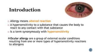
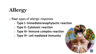
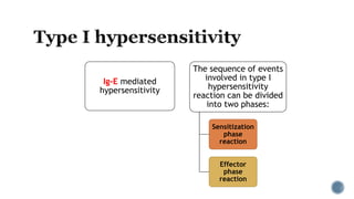
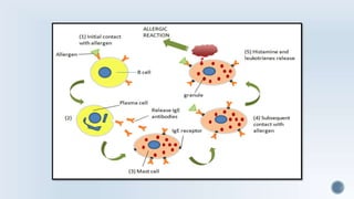
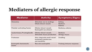
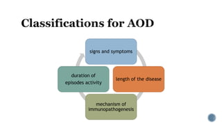
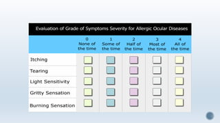
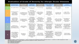
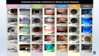
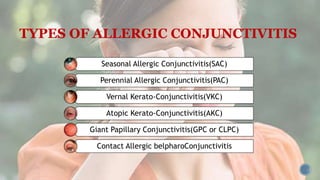
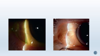
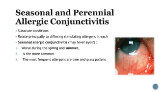
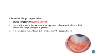
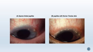
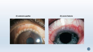
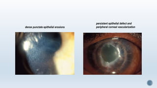
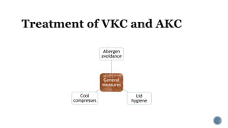
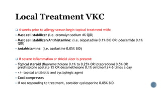
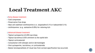
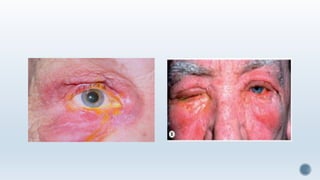
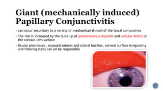
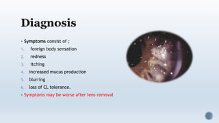
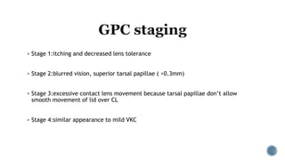
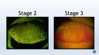
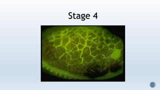
Rasoul Amini Vishte
MSc Student of optometry
IUMS
 Allergy means altered reaction
 A hypersensitivity to a substance that causes the body to
react to any contact with that substance
 Is a term synonymously with hypersensitivity
Ocular allergy are a group of external ocular conditions
resulting from one or more types of hypersensitivity reactions
to allergens
 Approx 20% of the general population have some form of
allergic disorder but about a third of these have
ophthalmic symptoms
Allergic conjunctivitis (AC) is an inflammation of the
conjunctiva secondary to an immune response to external
antigens, usually called allergens.
AC is not a single disease; in fact it is a syndrome affecting
the entire ocular surface, including conjunctiva, lids,
cornea, and tear film
 Four types of allergic responses
 Type I- Immediate/anaphylactic reaction
 Type II- Cytotoxic reaction
 Type III- Immune complex reaction
 Type IV- cell mediated immunity
Ig-E mediated
hypersensitivity
The sequence of events
involved in type I
hypersensitivity
reaction can be divided
into two phases:
Sensitization
phase
reaction
Effector
phase
reaction
Understand the pathophysiology of ocular allergies
Identify the aetiologies and clinical features of allergic conjunctivitis
Recognise the various forms of allergic eye disease
Discuss the various drugs used in the treatment of ocular allergies
Formulate an appropriate management plan Types
signs and symptoms
length of the disease
mechanism of
immunopathogenesis
duration of
episodes activity
TYPES OF ALLERGIC CONJUNCTIVITIS
Seasonal Allergic Conjunctivitis(SAC)
Perennial Allergic Conjunctivitis(PAC)
Vernal Kerato-Conjunctivitis(VKC)
Atopic Kerato-Conjunctivitis(AKC)
Giant Papillary Conjunctivitis(GPC or CLPC)
Contact Allergic belpharoConjunctivitis
Acute Allergic Conjunctivitis
 An acute conjunctival reaction to an environmental allergen, usually pollen
 Typically seen in younger children after playing outside in spring or summer
 Acute itching and watering are common, but the hallmark is chemosis
 Treatment is not usually required, self limited- usually settles within 24hrs
 Subacute conditions
 Relate principally to differing stimulating allergens in each
 Seasonal allergic conjunctivitis (‘hay fever eyes’) :
1. Worse during the spring and summer,
2. is the more common
3. The most frequent allergens are tree and grass pollens
 Perennial allergic conjunctivitis :
1. causes symptoms throughout the year
2. generally worse in the autumn when exposure to house dust mites, animal
dander and fungal allergens is greatest
3. It is less common and tends to be milder than the seasonal form
 Symptoms
 Transient acute or subacute attacks of redness, watering and itching, associated
with sneezing and nasal discharge.
Signs
 Conjunctival hyperaemia with a relatively mild papillary reaction, variable
chemosis and lid oedema.
Investigations
 Are generally not performed although conjunctival scraping in more active cases
may demonstrate the presence of eosinophils. Skin testing for particular allergens is
rarely required.
1. Artificial tears for mild symptoms.
2. Mast cell stabilizers (e.g., nedocromil sodium, lodoxamide) must be used for
a few days before exerting maximal effect, but are suitable (except
lodoxamide) for long-term use if required.
3. Antihistamines can be used for symptomatic exacerbations and are as
effective as mast cell stabilizers
4. Dual action antihistamine and mast cell stabilizers (e.g. azelastine,
ketotifen, olopatadine) act rapidly and are often very effective for
exacerbations
Pathogenesis;
 A recurrent bilateral disorder
 With both IgE- and cell-mediated immune mechanisms
 Primarily affects boys and onset is generally from about the age of 5 years
onwards
 VKC is rare in temperate regions but relatively common in warm dry climates
such as the Mediterranean, sub-Saharan Africa and the Middle East
 VKC often occurs on a seasonal basis, with a peak incidence over late spring and
summer
Palpebral (Tarsal) ;
 Primarily involves the upper tarsal conjunctiva. It may be associated with
significant corneal disease as a result of the close apposition between the inflamed
conjunctiva and the corneal epithelium.
 Typical Maxwell-Lyons sign is recognized for thick strands of mucus over papillaes
 Limbal ;
 Disease typically affects black and Asian patients.
 Mixed ;
 Has features of both palpebral and limbal disease
 Early-mild disease is characterized by conjunctival hyperaemia and diffuse
velvety papillary hypertrophy on the superior tarsal plate
 Macropapillae (<1 mm) have a flat-topped polygonal appearance reminiscent of
cobblestones; focal or diffuse whitish inflammatory infiltrates may be seen in
intense disease
 Progression to giant papillae (>1 mm) can occur, as adjacent smaller lesions
amalgamate when dividing septarupture
 Mucus deposition between giant papillae
 Decreased disease activity is characterized by milder conjunctival injection
and decreased mucus production
(A) Diffuse fine papillary hypertrophy (B) macropapillae with focal inflammatory infiltrates
(C) macropapillae with diffuse infiltrate (D) giant papillae
E) intense disease with mucus (F) milder disease; note mucousdischarge
 Gelatinous limbal conjunctival papillae that may be associated with transient
apically located white cellula rcollections (Horner– Trantas dots )
 gelatinous yellow-gray infiltrates are observed on the limbus
 a peripheral and superficial neovascularization
 In tropical regions, limbal disease may be severe
(A) Sparse limbal papillae (B) papillae with Horner–Trantas dots
(C) extensive papillae (D) severe features
 is more frequent in palpebral disease and may take the following forms;
1. Superior punctate epithelial erosions associated with layers of mucus on the
superior cornea
2. Epithelial macroerosions
3. Plaques and ‘shield’ ulcers may develop in palpebral or mixed disease
4. Subepithelial scars that are typically grey and oval and may affect vision
5. Pseudogerontoxon can develop in recurrent limbal disease
6. Keratoconus and other forms of corneal ectasia are more common in VKC
7. Herpes simplex keratitis is more common than average
(A) Superior punctate erosions and mucus stained with rose Bengal
(B–C) gradual resolution of a macroerosion over months of treatment
(A) Early plaque (B) plaque and shield ulcer
(C) subepithelial scarring following ulceration
 is a rare bilateral disease
 typically develops in adulthood (peak incidence 30–50 years) following a long history
of atopic dermatitis (eczema)
 asthma is also extremely common in these patients
 AKC tends to be chronic and unremitting
 AKC tends to be perennial and is often worse in the winter
 Patients are sensitive to a wide range of airborne environmental allergens
Dennie–Morgan folds: is a fold or line in the skin below the lower eyelid caused by edema in atopic dermatitis
Hertoghe sign(Queen Anne's sign) : absence of the lateral portion of the eyebrows
There may be keratinization of the lid margin
Associated chronic staphylococcal blepharitis and madarosis are common
Skin changes are more prominent than in VKC, and are typically eczematoid: erythema, dryness, scaliness and thickening
Symptoms are similar to those of VKC, but are frequently more severe and unremitting
preferentially inferior palpebral, whereas in VKC it is worse superiorly
Discharge is generally more watery than the stringy mucoid discharge in VKC
Hyperaemia; chemosis is not uncommon during active inflammation
Papillae are initially smaller than in VKC
Diffuse conjunctival infiltration and scarring may give awhitish, featureless appearance
Cicatricial changes can lead to moderate symblepharon formation, forniceal shortening and
keratinization of the caruncle
Limbal involvement similar to that of limbal VKC can be seen, including Horner–Trantas dots
infiltration and scarring of the tarsal conjunctiva Forniceal shortening
keratinization of the caruncle
Punctate
epithelial
erosions over the
inferior third of
the cornea are
common and can
be marked
Persistent
epithelial defects
Plaque formation
may occur
Peripheral
vascularization
and stromal
scarring are more
common than in
VKC
Predisposition to
secondary
bacterial and
fungal infection
Keratoconus is
common (about
15%)
dense punctate epithelial erosions
persistent epithelial defect and
peripheral corneal vascularization
General
measures
Allergen
avoidance
Lid
hygiene
Cool
compresses
 4 weeks prior to allergy season begin topical treatment with:
 Mast cell stabilizer (i.e. cromolyn sodium 4% QID)
 Mast cell stabilizer/Antihistamine: (i.e. olopatadine 0.1% BID OR lodoxamide 0.1%
QID)
 Antahistamine: (i.e. azelastine 0.05% BID)
 If severe inflammation or shield ulcer is present:
 Topical steroid (fluorometholone 0.1% to 0.25% OR lotepredonol 0.5% OR
prednisolone acetate 1% OR dexamethesone 0.1% ointment) 4-6 times a day
 +/- topical antibiotic and cycloplegic agent
 Cool compresses
 If not responding to treatment, consider cyclosporine 0.05% BID
Early disease treatment
 Cold compresses
 Preservative free drops
 Mast cell stabilizer/antihistamine (i.e. olopatadine 0.1% or lodoxamide 0.1%)
 Antihistamine: (e.g. azelastine 0.05%) for relieving itch
Advanced disease treatment
 Topical cyclosporine A 0.05% eye drops
 Topical tacrolimus 0.03% ointment to the eyelid skin
 Topical corticosteroid
 Consultation with allergist and/or dermatologist
 Oral cyclosporine, tacrolimus, or corticosteroids
 Boston keratoprosthesis (if visual loss from corneal opacification has occurred)
Oral antihistamines help itching, promote sleep and reduce
nocturnal eye rubbing.
Antibiotics (e.g. doxycycline 50–100 mg daily for 6 weeks,
azithromycin 500 mg once daily for 3 days) may be given to
reduce blepharitis-aggravated inflammation, usually in AKC
Immunosuppressive agents (e.g. steroids, ciclosporin) may
be effective at relatively low doses in AKC unresponsive to
other measures
Superficial keratectomy may be required to remove plaques or
debride shield ulcers and allow epithelialization
Surface maintenance/restoration surgery such as amniotic
membrane overlay grafting or lamellar keratoplasty, or eyelid
procedures such as botulinum toxin-induced ptosis or lateral
tarsorrhaphy, may be required for severe persistent epithelial defects
or ulceration
 Refers to the acute or subacute T-cell-mediated delayed hypersensitivity reaction
 An allergy that develops over a 48 hr period following exposure to which there has
been previous sensitization
 Contact sensitizing substances include ;
1. locally applied drugs
2. EDTA and preservatives in ophthalmic preparations
3. antimicrobial agents in lotions, creams and cosmetics
4. p-phenylenediamine (hair sprays, fabrics and shoes)
 Involves acute and chronic lesions of the lids, conjunctiva and cornea
 Itching is a prominent symptom
 Acute response comprises injection and chemosis of lower conjunctiva with the
serous or mucoid discharge
 Papillae develop over the entire conjunctiva
 In chronic cases the lid thickens and exhibit dryness, fissuring and crusting
 can occur secondary to a variety of mechanical stimuli of the tarsal conjunctiva
 The risk is increased by the build-up of proteinaceous deposits and cellular debris on
the contact lens surface
 Ocular prostheses , exposed sutures and scleral buckles, corneal surface irregularity
and filtering blebs can all be responsible
 Symptoms consist of ;
1. foreign body sensation
2. redness
3. itching
4. increased mucus production
5. blurring
6. loss of CL tolerance.
 Symptoms may be worse after lens removal
Ptosis may occur, mainly as a result of irritative spasm and tissue laxity secondary to chronic inflammation
Focal apical ulceration and whitish scarring may develop on larger papillae
Superior tarsal hyperaemia and papillae
Excessive CL mobility due to upper lid capture.
Substantial CL protein deposits may be present.
Variable mucous discharge.
 Stage 1:itching and decreased lens tolerance
 Stage 2:blurred vision, superior tarsal papillae ( >0.3mm)
 Stage 3:excessive contact lens movement because tarsal papillae don’t allow
smooth movement of lid over CL
 Stage 4:similar appearance to mild VKC
Stage 2 Stage 3
Stage 4
Topical
Ensure
effective
cleaning of
CL or
prosthesis
Removal of
the
stimulus
 In SEVERE GPC start with
 mast cell stabilizer (pemirolast q.i.d , nedocromil b.i.d ,lodoxamide q.i.d,cromolyn
sodium q.i.d)
 Mast cell stabilizer/antihistamine (olopatadine b.i.d , epinastine b.i.d)
 In VERY SEVERE GPC ;
 Low dose Topical steroid (loteprednole 0.2% ,qid)
 Kanski’s Clinical Ophthalmology , 8th Edition,2015
 Conjunctivitis – A Complex and Multifaceted Disorder , Edited by Zdenek
Pelikan , First published November, 2011
Ocular allergy rasoul amini

Ocular allergy rasoul amini

  • 1.
    Rasoul Amini Vishte MScStudent of optometry IUMS
  • 2.
     Allergy meansaltered reaction  A hypersensitivity to a substance that causes the body to react to any contact with that substance  Is a term synonymously with hypersensitivity Ocular allergy are a group of external ocular conditions resulting from one or more types of hypersensitivity reactions to allergens
  • 3.
     Approx 20%of the general population have some form of allergic disorder but about a third of these have ophthalmic symptoms Allergic conjunctivitis (AC) is an inflammation of the conjunctiva secondary to an immune response to external antigens, usually called allergens. AC is not a single disease; in fact it is a syndrome affecting the entire ocular surface, including conjunctiva, lids, cornea, and tear film
  • 4.
     Four typesof allergic responses  Type I- Immediate/anaphylactic reaction  Type II- Cytotoxic reaction  Type III- Immune complex reaction  Type IV- cell mediated immunity
  • 5.
    Ig-E mediated hypersensitivity The sequenceof events involved in type I hypersensitivity reaction can be divided into two phases: Sensitization phase reaction Effector phase reaction
  • 8.
    Understand the pathophysiologyof ocular allergies Identify the aetiologies and clinical features of allergic conjunctivitis Recognise the various forms of allergic eye disease Discuss the various drugs used in the treatment of ocular allergies Formulate an appropriate management plan Types
  • 9.
    signs and symptoms lengthof the disease mechanism of immunopathogenesis duration of episodes activity
  • 13.
    TYPES OF ALLERGICCONJUNCTIVITIS Seasonal Allergic Conjunctivitis(SAC) Perennial Allergic Conjunctivitis(PAC) Vernal Kerato-Conjunctivitis(VKC) Atopic Kerato-Conjunctivitis(AKC) Giant Papillary Conjunctivitis(GPC or CLPC) Contact Allergic belpharoConjunctivitis
  • 14.
    Acute Allergic Conjunctivitis An acute conjunctival reaction to an environmental allergen, usually pollen  Typically seen in younger children after playing outside in spring or summer  Acute itching and watering are common, but the hallmark is chemosis  Treatment is not usually required, self limited- usually settles within 24hrs
  • 16.
     Subacute conditions Relate principally to differing stimulating allergens in each  Seasonal allergic conjunctivitis (‘hay fever eyes’) : 1. Worse during the spring and summer, 2. is the more common 3. The most frequent allergens are tree and grass pollens
  • 17.
     Perennial allergicconjunctivitis : 1. causes symptoms throughout the year 2. generally worse in the autumn when exposure to house dust mites, animal dander and fungal allergens is greatest 3. It is less common and tends to be milder than the seasonal form
  • 18.
     Symptoms  Transientacute or subacute attacks of redness, watering and itching, associated with sneezing and nasal discharge. Signs  Conjunctival hyperaemia with a relatively mild papillary reaction, variable chemosis and lid oedema. Investigations  Are generally not performed although conjunctival scraping in more active cases may demonstrate the presence of eosinophils. Skin testing for particular allergens is rarely required.
  • 19.
    1. Artificial tearsfor mild symptoms. 2. Mast cell stabilizers (e.g., nedocromil sodium, lodoxamide) must be used for a few days before exerting maximal effect, but are suitable (except lodoxamide) for long-term use if required. 3. Antihistamines can be used for symptomatic exacerbations and are as effective as mast cell stabilizers 4. Dual action antihistamine and mast cell stabilizers (e.g. azelastine, ketotifen, olopatadine) act rapidly and are often very effective for exacerbations
  • 20.
    Pathogenesis;  A recurrentbilateral disorder  With both IgE- and cell-mediated immune mechanisms  Primarily affects boys and onset is generally from about the age of 5 years onwards  VKC is rare in temperate regions but relatively common in warm dry climates such as the Mediterranean, sub-Saharan Africa and the Middle East  VKC often occurs on a seasonal basis, with a peak incidence over late spring and summer
  • 21.
    Palpebral (Tarsal) ; Primarily involves the upper tarsal conjunctiva. It may be associated with significant corneal disease as a result of the close apposition between the inflamed conjunctiva and the corneal epithelium.  Typical Maxwell-Lyons sign is recognized for thick strands of mucus over papillaes  Limbal ;  Disease typically affects black and Asian patients.  Mixed ;  Has features of both palpebral and limbal disease
  • 22.
     Early-mild diseaseis characterized by conjunctival hyperaemia and diffuse velvety papillary hypertrophy on the superior tarsal plate  Macropapillae (<1 mm) have a flat-topped polygonal appearance reminiscent of cobblestones; focal or diffuse whitish inflammatory infiltrates may be seen in intense disease  Progression to giant papillae (>1 mm) can occur, as adjacent smaller lesions amalgamate when dividing septarupture  Mucus deposition between giant papillae  Decreased disease activity is characterized by milder conjunctival injection and decreased mucus production
  • 23.
    (A) Diffuse finepapillary hypertrophy (B) macropapillae with focal inflammatory infiltrates
  • 24.
    (C) macropapillae withdiffuse infiltrate (D) giant papillae
  • 25.
    E) intense diseasewith mucus (F) milder disease; note mucousdischarge
  • 26.
     Gelatinous limbalconjunctival papillae that may be associated with transient apically located white cellula rcollections (Horner– Trantas dots )  gelatinous yellow-gray infiltrates are observed on the limbus  a peripheral and superficial neovascularization  In tropical regions, limbal disease may be severe
  • 27.
    (A) Sparse limbalpapillae (B) papillae with Horner–Trantas dots
  • 28.
    (C) extensive papillae(D) severe features
  • 29.
     is morefrequent in palpebral disease and may take the following forms; 1. Superior punctate epithelial erosions associated with layers of mucus on the superior cornea 2. Epithelial macroerosions 3. Plaques and ‘shield’ ulcers may develop in palpebral or mixed disease 4. Subepithelial scars that are typically grey and oval and may affect vision 5. Pseudogerontoxon can develop in recurrent limbal disease 6. Keratoconus and other forms of corneal ectasia are more common in VKC 7. Herpes simplex keratitis is more common than average
  • 30.
    (A) Superior punctateerosions and mucus stained with rose Bengal
  • 31.
    (B–C) gradual resolutionof a macroerosion over months of treatment
  • 32.
    (A) Early plaque(B) plaque and shield ulcer
  • 33.
    (C) subepithelial scarringfollowing ulceration
  • 34.
     is arare bilateral disease  typically develops in adulthood (peak incidence 30–50 years) following a long history of atopic dermatitis (eczema)  asthma is also extremely common in these patients  AKC tends to be chronic and unremitting  AKC tends to be perennial and is often worse in the winter  Patients are sensitive to a wide range of airborne environmental allergens
  • 35.
    Dennie–Morgan folds: isa fold or line in the skin below the lower eyelid caused by edema in atopic dermatitis Hertoghe sign(Queen Anne's sign) : absence of the lateral portion of the eyebrows There may be keratinization of the lid margin Associated chronic staphylococcal blepharitis and madarosis are common Skin changes are more prominent than in VKC, and are typically eczematoid: erythema, dryness, scaliness and thickening Symptoms are similar to those of VKC, but are frequently more severe and unremitting
  • 38.
    preferentially inferior palpebral,whereas in VKC it is worse superiorly Discharge is generally more watery than the stringy mucoid discharge in VKC Hyperaemia; chemosis is not uncommon during active inflammation Papillae are initially smaller than in VKC Diffuse conjunctival infiltration and scarring may give awhitish, featureless appearance Cicatricial changes can lead to moderate symblepharon formation, forniceal shortening and keratinization of the caruncle Limbal involvement similar to that of limbal VKC can be seen, including Horner–Trantas dots
  • 39.
    infiltration and scarringof the tarsal conjunctiva Forniceal shortening
  • 40.
  • 41.
    Punctate epithelial erosions over the inferiorthird of the cornea are common and can be marked Persistent epithelial defects Plaque formation may occur Peripheral vascularization and stromal scarring are more common than in VKC Predisposition to secondary bacterial and fungal infection Keratoconus is common (about 15%)
  • 42.
    dense punctate epithelialerosions persistent epithelial defect and peripheral corneal vascularization
  • 43.
  • 44.
     4 weeksprior to allergy season begin topical treatment with:  Mast cell stabilizer (i.e. cromolyn sodium 4% QID)  Mast cell stabilizer/Antihistamine: (i.e. olopatadine 0.1% BID OR lodoxamide 0.1% QID)  Antahistamine: (i.e. azelastine 0.05% BID)  If severe inflammation or shield ulcer is present:  Topical steroid (fluorometholone 0.1% to 0.25% OR lotepredonol 0.5% OR prednisolone acetate 1% OR dexamethesone 0.1% ointment) 4-6 times a day  +/- topical antibiotic and cycloplegic agent  Cool compresses  If not responding to treatment, consider cyclosporine 0.05% BID
  • 45.
    Early disease treatment Cold compresses  Preservative free drops  Mast cell stabilizer/antihistamine (i.e. olopatadine 0.1% or lodoxamide 0.1%)  Antihistamine: (e.g. azelastine 0.05%) for relieving itch Advanced disease treatment  Topical cyclosporine A 0.05% eye drops  Topical tacrolimus 0.03% ointment to the eyelid skin  Topical corticosteroid  Consultation with allergist and/or dermatologist  Oral cyclosporine, tacrolimus, or corticosteroids  Boston keratoprosthesis (if visual loss from corneal opacification has occurred)
  • 46.
    Oral antihistamines helpitching, promote sleep and reduce nocturnal eye rubbing. Antibiotics (e.g. doxycycline 50–100 mg daily for 6 weeks, azithromycin 500 mg once daily for 3 days) may be given to reduce blepharitis-aggravated inflammation, usually in AKC Immunosuppressive agents (e.g. steroids, ciclosporin) may be effective at relatively low doses in AKC unresponsive to other measures
  • 47.
    Superficial keratectomy maybe required to remove plaques or debride shield ulcers and allow epithelialization Surface maintenance/restoration surgery such as amniotic membrane overlay grafting or lamellar keratoplasty, or eyelid procedures such as botulinum toxin-induced ptosis or lateral tarsorrhaphy, may be required for severe persistent epithelial defects or ulceration
  • 48.
     Refers tothe acute or subacute T-cell-mediated delayed hypersensitivity reaction  An allergy that develops over a 48 hr period following exposure to which there has been previous sensitization  Contact sensitizing substances include ; 1. locally applied drugs 2. EDTA and preservatives in ophthalmic preparations 3. antimicrobial agents in lotions, creams and cosmetics 4. p-phenylenediamine (hair sprays, fabrics and shoes)
  • 49.
     Involves acuteand chronic lesions of the lids, conjunctiva and cornea  Itching is a prominent symptom  Acute response comprises injection and chemosis of lower conjunctiva with the serous or mucoid discharge  Papillae develop over the entire conjunctiva  In chronic cases the lid thickens and exhibit dryness, fissuring and crusting
  • 51.
     can occursecondary to a variety of mechanical stimuli of the tarsal conjunctiva  The risk is increased by the build-up of proteinaceous deposits and cellular debris on the contact lens surface  Ocular prostheses , exposed sutures and scleral buckles, corneal surface irregularity and filtering blebs can all be responsible
  • 52.
     Symptoms consistof ; 1. foreign body sensation 2. redness 3. itching 4. increased mucus production 5. blurring 6. loss of CL tolerance.  Symptoms may be worse after lens removal
  • 53.
    Ptosis may occur,mainly as a result of irritative spasm and tissue laxity secondary to chronic inflammation Focal apical ulceration and whitish scarring may develop on larger papillae Superior tarsal hyperaemia and papillae Excessive CL mobility due to upper lid capture. Substantial CL protein deposits may be present. Variable mucous discharge.
  • 54.
     Stage 1:itchingand decreased lens tolerance  Stage 2:blurred vision, superior tarsal papillae ( >0.3mm)  Stage 3:excessive contact lens movement because tarsal papillae don’t allow smooth movement of lid over CL  Stage 4:similar appearance to mild VKC
  • 55.
  • 56.
  • 58.
  • 59.
     In SEVEREGPC start with  mast cell stabilizer (pemirolast q.i.d , nedocromil b.i.d ,lodoxamide q.i.d,cromolyn sodium q.i.d)  Mast cell stabilizer/antihistamine (olopatadine b.i.d , epinastine b.i.d)  In VERY SEVERE GPC ;  Low dose Topical steroid (loteprednole 0.2% ,qid)
  • 60.
     Kanski’s ClinicalOphthalmology , 8th Edition,2015  Conjunctivitis – A Complex and Multifaceted Disorder , Edited by Zdenek Pelikan , First published November, 2011