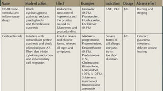
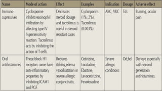
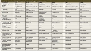
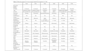
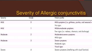
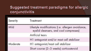
PRESENTER – DR ARNAV SAROYA
INTRODUCTION
 Ocular allergy is estimated to affect 5 – 22% of the
population depending on the geographical setting and
the age of the population.
 It is also often under diagnosed and undertreated.
 Definitive etiology is not known but genetic
predisposition, air pollution in urban area and exposure
to pets have been associated.
 Clinical manifestations include the various forms of allergic
conjunctivitis, as well as hay fever (seasonal allergic rhinitis),
asthma and eczema.
 Allergic conjunctivitis is a Type I (immediate) hypersensitivity
reaction, mediated by degranulation of mast cells in
response to the action of IgE.
 There is evidence of Type IV hypersensitivity in at least some
forms.
TYPES OF ALLERGIC
CONJUNCTIVITIS
 SEASONAL ALLERGIC CONJUNCTIVITIS
(SAC)
 PERINEAL ALLERGIC CONJUNCTIVITIS
(PAC) - ARE THE MOST COMMONLY
SEEN ALLERGIC CONJUNCTIVITIS.
 VERNAL KERATOCONJUNCTIVITIS (VKC)
 ATOPIC KERATOCONJUNCTIVITIS (AKC)
 GIANT PAPILLARY CONJUNCTIVITIS
(GPC) -
SEASONAL ALLERGIC CONJUNCTIVITIS
AND PERINEAL ALLERGIC
CONJUNCTIVITIS
 SAC is the most common form of all ocular allergic
diseases and is triggered by exposure to pollen. It usually
occurs in spring.
 PAC is induced by exposure to dust mites, fungi, animal
epithelial and/or occupational allergens. The affected
patients can show symptoms throughout the year with
acute seasonal exacerbations.
PATHOPHYSIOLOGY
 The response to allergen - due to its interaction with prostaglandins and
leukotrienes.
 It is a type I hypersensitivity reaction and the early response lasts clinically for 20 -
30 minutes.
 The late phase reaction is due to the presence of inflammatory cells in the
conjunctival mucosa.
 Due to activation of vascular endothelial cells, which in turn expresses chemokines,
adhesion molecules such as such as intercellular adhesion molecule (ICAM) and
vascular cell adhesion molecule (VCAM). Other chemokines secreted include
regulated upon activation normal T cell expressed and secreted (RANTES)
chemokines, monocyte chemo attractant protein (MCP), interleukin (IL)-8, eotaxin
and macrophage inflammatory protein (MIP)-1 alpha.
CLINICAL FEATURES
The clinical presentation in both the conditions is similar.
 Bilateral itching, tearing and burning sensation.
 Blurred vision and photophobia may be due to an alteration in the
composition and instability of the tear film.
 PAC affects young adults between 20-40 years of age with no
gender predilection while SAC has no age or gender predilection.
 Both are frequently associated with allergic rhinitis and other
allergic disorder (slightly more with SAC).
 On examination –
mild to moderate conjunctival hyperemia with an
edematous conjunctival surface is seen.
The palpebral conjunctiva appears pale pink in color with
diffuse areas of slightly hypertrophic papillae predominantly
located in the upper tarsal conjunctiva .
 The cornea is rarely affected.
CONJUNCTIVAL HYPERAEMIA
WITH OEDEMA PAPILLARY HYPERTROPHY
Diagnostic features of SAC and PAC
 Itching, redness, and swelling of the conjunctiva.
 Family or personal history of atopy and skin test
 Response to antiallergic treatment
 Serum IgE elevation (found in 78% of all patients with SAC–
69% being specific of pollen)
 Lacrimal IgE elevation
 Increased mast cell infiltration of the conjunctiva
 Increase in type T mast cells with tryptase release in tears.
VERNAL KERATOCONJUNCTIVITIS
 Self-limiting, bilateral chronic inflammation
 Leaves sequelae or permanent alterations in visual acuity in about
5-6% of the patients.
 It is more frequent in young males with an increased incidence
between 11-13 years of age.
 Vkc is rare in adults.
 Pathophysiology is not precisely known, though two
hypersensitivity mechanisms seem to be involved (type I and type
IV).
 Mucosal secretion in these individuals due to increased presence
of goblet cells in the conjunctiva.
Clinical Features -
 itching, redness, photophobia and watering.
 The symptoms may be seasonal or perennial with
exacerbations in heat, dust, wind, bright light.
 Two forms of conjunctival involvement is known:
 palpebral, with giant subtarsal papillae (>1 mm in
diameter) showing a typical cobblestone pattern
with profuse mucus secretion
 limbal with Horner-Trantas dots that appear as
small gelatinous nodules at limbus and are typical
of the active phase of the disease
 Mixed – features of palpebral + limbal
 Corneal involvement can manifest as micropannus
 Superficial punctate keratopathy (normally located in the
upper half of the cornea)
 Corneal macro erosions
 Shield ulcerations covered with mucus and fibrin plaques
 Subepithelial scarring and pseudogerontoxon (paralimbal
band of superficial scarring resembling arcus senilis adjacent
to a previously inflamed segment of the limbus.
 Vkc is strongly associated with keratoconus, hydrops and
may cause dry eye.
SHILED ULCER PSEUDOGERONTOXON SUPERIOR PUNCTATE EROSIONS
Diagnosis
 Large papillae in the upper tarsal conjunctiva and corneoscleral
junction are hallmark of VKC.
 Conjunctival biopsy reveals an increase in basophils, eosinophils,
mast cells, plasma cells and lymphocytes that also appear in the
smears.
 The tears show very high levels of histamine, tryptase, eotaxin and
eosinophil cationic protein and increased adhesion molecules
(VCAM-1) and leukotrienes (LTB4, LTC4).
ATOPIC KERATOCONJUNCTIVITIS
 Bilateral chronic inflammatory disease
affecting the eyelids and ocular surface in
young males with atopic disease.
 Pathophysiology -
It involves type I and type IV hypersensitivity
reaction with activation of Th1 and Th2
lymphocytes and reduction of MU5CAC
secreting goblet cells.
Lower tarsal conjunctiva in AKC.
Note the fornix
foreshortening and pale edema.
Clinical features
 Itching, redness, swelling and eczema of
eyelids.
 Chronic edema of lower lid causes “Dennie
– Morgan fold” at infraorbital area
 Loss of lashes due to scratching results in
“Hertoghe sign”.
 Lid margins may have staphylococcal
blephritis, mebomitis, trichiasis, entropion
and ectropion.
 Papillary hypertrophy is predominantly
seen in lower palpebral conjunctiva and
symblephron formation and conjunctival
scarring occurs in chronic cases.
Dennie – Morgan fold
 Corneal involvement is in the form of superficial
puctate keratitis, ulcer formation,
neovascularisation, pannus formation and rarely
Horner Trantas dot.
 These eyes are prone to herpetic and fungal
keratitis.
 These patients also develop atopic cataracts
which are anterior shield like cataracts
 There is increased association of keratoconus
and retinal detachment with AKC.
Atopic Cataract
Diagnosis
It is done mainly by history and clinical
examination.
A conjunctival biopsy is sometimes
needed to differentiate it from other forms
of cicatrizing conjunctivitis.
GIANT PAPILLARY
CONJUNCTIVITIS
 GPC is non-infectious inflammatory disorder of the surface of
the eye
 Related to the wearing of contact lenses, ocular prostheses,
sutures and even limbal dermoids.
 Affects 5-10% of all contact lens wearers and is more
common among individuals wearing soft lenses.
 Any age and shows no race or gender predilection.
 It can affect both atopic and non-atopic individuals, though
the signs and symptoms are more severe in atopic form.
Etiopathogenesis
 Mechanical trauma to the conjunctiva and
cornea causes release of local inflammatory
mediators such as IL-8 and the recruitment of
dendritic cells which increase antigen
presentation to the cells.
 Bacterial products, lubricating eyedrops,
preservative solutions, disinfecting solutions,
and even the contact lens material -antigenic
stimuli.
 Increased contact lens water content and
thicker and more irregular lens margins are
correlated to an increased frequency of the
GPC
Clinical Features -
 Any age with no race or gender predilection.
 Exacerbations -usually during the spring season.
 Dry eye and genetic predisposition may be contributary.
 Examination of the superior tarsal conjunctiva reveals giant papillae
measuring over 1 mm in size.
 Corneal involvement can be in the form of punctate keratitis,
peripheral infiltrates and corneal neovascularisation.
 Patients present with increase in contact lens soilage, ocular itching and
mucous discharge in tears as well as blurred vision and conjunctival
injection. This can be accompanied by decreased contact lens tolerance and
mechanical stability.
CONTACT DERMATITIS ALLERGY
It is a type IV mediated hypersensitivity
response that involves interaction of
antigen with Th1 and Th2 cells resulting
in release of cytokines.
Pathophysiology
 Typically, it consists of two phases:
 sensitization and inflammatory response.
 In the sensitization phase antigen presenting cells processed antigen - MHC class II complex
interacts with T-lymphocytes, resulting in the differentiation of CD4+ T-lymphocyte into memory T-
lymphocyte.
 In the second phase, the interaction between the antigen- MHC-II complex and memory T-cells
stimulates the proliferation of T-cells.
 The memory T-lymphocytes during proliferation release cytokines.
 In sensitized individuals, the immune response takes 48-72 hours to develop. Some of the products
that can act as antigens are: mydriatic drugs (atropine, homatropine, tropicamide, phenyephrine);
antiglaucoma drugs (Brimonidine, apraclonidine dorzolamide); preservatives (thiomersal,
benzalkoniumchloride, chlorhexidine EDTA); antibiotics (aminoglycosides, sulfamides, polymyxin);
antiviral drugs (idoxuridine, trifuridine, viderabine); anaesthetic agents (procaine, tetracaine);
cosmetics, soaps and detergents.
Clinical features
 The lower palpebral conjunctiva is involved first followed by rest of the
conjunctiva.
 The eyelids may show blephritis, eczematous dermatitis which later resulting
in folds, crusts, fissures, and skin thickening.
 The conjunctiva shows follicles, papillae, psedopemphigoid lesions. Corneal
involvement may be in the form of superficial punctuate keratitis, marginal
infiltrate, ulcer and stromal edema.
 Diagnosis is by history and clinical examination
Treatment of allergic
conjunctivitis
A. Medicals
 Anti allergic drugs- antihistamine, vasoconstrictors, mast cell
stabilisers, dual mode action drugs, corticosteroids and
immunosuppressives.
 The regimen that is usually followed is as follows:
1. In mild cases, antihistamines or vasoconstrictors suffice.
2. In moderate cases, NSAID is added along with dual action drugs.
3. In severe cases, steroid drop are given for a short duration of upto
2 weeks followed by dual action drugs.
 Duration of treatment depends on patients response and
the nature of the allergy.
 Mast cell stabilisers are given prior to the onset of
symptoms in susceptible cases.
 Cyclosporins and tacrolimus is used in severe cases of
VKC and AKC.
 Allergen-specific immunotherapy is an effective treatment
for patients with allergic rhinoconjunctivitis.
B. Supportive
 Supportive therapy includes artificial tears, goggles and
cold eye pads.
 Artificial tears dilulte the concentration of allergens and
inflammatory mediators and flush the ocular surface of
these agents.
 Goggles decrease the amount of allergen reaching the
ocular surface while cold eye pads offer for symptomatic
relief.
 Avoidance of allergen should be tried wherever possible.
C.Surgical
 Surgical treatment may be required in severe cases of AKC
and VKC.
 Surgical excision, cryocoagulation, excision with Mitomycin C
0.02% or CO2 laser is used for papillary hypertrophy.
 Debridement of ulcer base/surgical or excimer laser
keratectomy, amniotic membrane graft/free autologous
conjunctival graft are the various options for managing non
healing shield ulcer.
 Tarsal plate resection is done for mechanical ptosis.
Allergic Conjunctivitis - Dr Arnav Saroya

Allergic Conjunctivitis - Dr Arnav Saroya

  • 1.
    PRESENTER – DRARNAV SAROYA
  • 2.
    INTRODUCTION  Ocular allergyis estimated to affect 5 – 22% of the population depending on the geographical setting and the age of the population.  It is also often under diagnosed and undertreated.  Definitive etiology is not known but genetic predisposition, air pollution in urban area and exposure to pets have been associated.
  • 3.
     Clinical manifestationsinclude the various forms of allergic conjunctivitis, as well as hay fever (seasonal allergic rhinitis), asthma and eczema.  Allergic conjunctivitis is a Type I (immediate) hypersensitivity reaction, mediated by degranulation of mast cells in response to the action of IgE.  There is evidence of Type IV hypersensitivity in at least some forms.
  • 4.
    TYPES OF ALLERGIC CONJUNCTIVITIS SEASONAL ALLERGIC CONJUNCTIVITIS (SAC)  PERINEAL ALLERGIC CONJUNCTIVITIS (PAC) - ARE THE MOST COMMONLY SEEN ALLERGIC CONJUNCTIVITIS.  VERNAL KERATOCONJUNCTIVITIS (VKC)  ATOPIC KERATOCONJUNCTIVITIS (AKC)  GIANT PAPILLARY CONJUNCTIVITIS (GPC) -
  • 5.
    SEASONAL ALLERGIC CONJUNCTIVITIS ANDPERINEAL ALLERGIC CONJUNCTIVITIS  SAC is the most common form of all ocular allergic diseases and is triggered by exposure to pollen. It usually occurs in spring.  PAC is induced by exposure to dust mites, fungi, animal epithelial and/or occupational allergens. The affected patients can show symptoms throughout the year with acute seasonal exacerbations.
  • 6.
    PATHOPHYSIOLOGY  The responseto allergen - due to its interaction with prostaglandins and leukotrienes.  It is a type I hypersensitivity reaction and the early response lasts clinically for 20 - 30 minutes.  The late phase reaction is due to the presence of inflammatory cells in the conjunctival mucosa.  Due to activation of vascular endothelial cells, which in turn expresses chemokines, adhesion molecules such as such as intercellular adhesion molecule (ICAM) and vascular cell adhesion molecule (VCAM). Other chemokines secreted include regulated upon activation normal T cell expressed and secreted (RANTES) chemokines, monocyte chemo attractant protein (MCP), interleukin (IL)-8, eotaxin and macrophage inflammatory protein (MIP)-1 alpha.
  • 7.
    CLINICAL FEATURES The clinicalpresentation in both the conditions is similar.  Bilateral itching, tearing and burning sensation.  Blurred vision and photophobia may be due to an alteration in the composition and instability of the tear film.  PAC affects young adults between 20-40 years of age with no gender predilection while SAC has no age or gender predilection.  Both are frequently associated with allergic rhinitis and other allergic disorder (slightly more with SAC).
  • 8.
     On examination– mild to moderate conjunctival hyperemia with an edematous conjunctival surface is seen. The palpebral conjunctiva appears pale pink in color with diffuse areas of slightly hypertrophic papillae predominantly located in the upper tarsal conjunctiva .  The cornea is rarely affected.
  • 9.
  • 10.
    Diagnostic features ofSAC and PAC  Itching, redness, and swelling of the conjunctiva.  Family or personal history of atopy and skin test  Response to antiallergic treatment  Serum IgE elevation (found in 78% of all patients with SAC– 69% being specific of pollen)  Lacrimal IgE elevation  Increased mast cell infiltration of the conjunctiva  Increase in type T mast cells with tryptase release in tears.
  • 11.
    VERNAL KERATOCONJUNCTIVITIS  Self-limiting,bilateral chronic inflammation  Leaves sequelae or permanent alterations in visual acuity in about 5-6% of the patients.  It is more frequent in young males with an increased incidence between 11-13 years of age.  Vkc is rare in adults.  Pathophysiology is not precisely known, though two hypersensitivity mechanisms seem to be involved (type I and type IV).  Mucosal secretion in these individuals due to increased presence of goblet cells in the conjunctiva.
  • 12.
    Clinical Features - itching, redness, photophobia and watering.  The symptoms may be seasonal or perennial with exacerbations in heat, dust, wind, bright light.  Two forms of conjunctival involvement is known:  palpebral, with giant subtarsal papillae (>1 mm in diameter) showing a typical cobblestone pattern with profuse mucus secretion  limbal with Horner-Trantas dots that appear as small gelatinous nodules at limbus and are typical of the active phase of the disease  Mixed – features of palpebral + limbal
  • 13.
     Corneal involvementcan manifest as micropannus  Superficial punctate keratopathy (normally located in the upper half of the cornea)  Corneal macro erosions  Shield ulcerations covered with mucus and fibrin plaques  Subepithelial scarring and pseudogerontoxon (paralimbal band of superficial scarring resembling arcus senilis adjacent to a previously inflamed segment of the limbus.  Vkc is strongly associated with keratoconus, hydrops and may cause dry eye.
  • 14.
    SHILED ULCER PSEUDOGERONTOXONSUPERIOR PUNCTATE EROSIONS
  • 15.
    Diagnosis  Large papillaein the upper tarsal conjunctiva and corneoscleral junction are hallmark of VKC.  Conjunctival biopsy reveals an increase in basophils, eosinophils, mast cells, plasma cells and lymphocytes that also appear in the smears.  The tears show very high levels of histamine, tryptase, eotaxin and eosinophil cationic protein and increased adhesion molecules (VCAM-1) and leukotrienes (LTB4, LTC4).
  • 16.
    ATOPIC KERATOCONJUNCTIVITIS  Bilateralchronic inflammatory disease affecting the eyelids and ocular surface in young males with atopic disease.  Pathophysiology - It involves type I and type IV hypersensitivity reaction with activation of Th1 and Th2 lymphocytes and reduction of MU5CAC secreting goblet cells. Lower tarsal conjunctiva in AKC. Note the fornix foreshortening and pale edema.
  • 17.
    Clinical features  Itching,redness, swelling and eczema of eyelids.  Chronic edema of lower lid causes “Dennie – Morgan fold” at infraorbital area  Loss of lashes due to scratching results in “Hertoghe sign”.  Lid margins may have staphylococcal blephritis, mebomitis, trichiasis, entropion and ectropion.  Papillary hypertrophy is predominantly seen in lower palpebral conjunctiva and symblephron formation and conjunctival scarring occurs in chronic cases. Dennie – Morgan fold
  • 18.
     Corneal involvementis in the form of superficial puctate keratitis, ulcer formation, neovascularisation, pannus formation and rarely Horner Trantas dot.  These eyes are prone to herpetic and fungal keratitis.  These patients also develop atopic cataracts which are anterior shield like cataracts  There is increased association of keratoconus and retinal detachment with AKC. Atopic Cataract
  • 19.
    Diagnosis It is donemainly by history and clinical examination. A conjunctival biopsy is sometimes needed to differentiate it from other forms of cicatrizing conjunctivitis.
  • 20.
    GIANT PAPILLARY CONJUNCTIVITIS  GPCis non-infectious inflammatory disorder of the surface of the eye  Related to the wearing of contact lenses, ocular prostheses, sutures and even limbal dermoids.  Affects 5-10% of all contact lens wearers and is more common among individuals wearing soft lenses.  Any age and shows no race or gender predilection.  It can affect both atopic and non-atopic individuals, though the signs and symptoms are more severe in atopic form.
  • 21.
    Etiopathogenesis  Mechanical traumato the conjunctiva and cornea causes release of local inflammatory mediators such as IL-8 and the recruitment of dendritic cells which increase antigen presentation to the cells.  Bacterial products, lubricating eyedrops, preservative solutions, disinfecting solutions, and even the contact lens material -antigenic stimuli.  Increased contact lens water content and thicker and more irregular lens margins are correlated to an increased frequency of the GPC
  • 22.
    Clinical Features - Any age with no race or gender predilection.  Exacerbations -usually during the spring season.  Dry eye and genetic predisposition may be contributary.  Examination of the superior tarsal conjunctiva reveals giant papillae measuring over 1 mm in size.  Corneal involvement can be in the form of punctate keratitis, peripheral infiltrates and corneal neovascularisation.  Patients present with increase in contact lens soilage, ocular itching and mucous discharge in tears as well as blurred vision and conjunctival injection. This can be accompanied by decreased contact lens tolerance and mechanical stability.
  • 23.
    CONTACT DERMATITIS ALLERGY Itis a type IV mediated hypersensitivity response that involves interaction of antigen with Th1 and Th2 cells resulting in release of cytokines.
  • 24.
    Pathophysiology  Typically, itconsists of two phases:  sensitization and inflammatory response.  In the sensitization phase antigen presenting cells processed antigen - MHC class II complex interacts with T-lymphocytes, resulting in the differentiation of CD4+ T-lymphocyte into memory T- lymphocyte.  In the second phase, the interaction between the antigen- MHC-II complex and memory T-cells stimulates the proliferation of T-cells.  The memory T-lymphocytes during proliferation release cytokines.  In sensitized individuals, the immune response takes 48-72 hours to develop. Some of the products that can act as antigens are: mydriatic drugs (atropine, homatropine, tropicamide, phenyephrine); antiglaucoma drugs (Brimonidine, apraclonidine dorzolamide); preservatives (thiomersal, benzalkoniumchloride, chlorhexidine EDTA); antibiotics (aminoglycosides, sulfamides, polymyxin); antiviral drugs (idoxuridine, trifuridine, viderabine); anaesthetic agents (procaine, tetracaine); cosmetics, soaps and detergents.
  • 25.
    Clinical features  Thelower palpebral conjunctiva is involved first followed by rest of the conjunctiva.  The eyelids may show blephritis, eczematous dermatitis which later resulting in folds, crusts, fissures, and skin thickening.  The conjunctiva shows follicles, papillae, psedopemphigoid lesions. Corneal involvement may be in the form of superficial punctuate keratitis, marginal infiltrate, ulcer and stromal edema.  Diagnosis is by history and clinical examination
  • 26.
    Treatment of allergic conjunctivitis A.Medicals  Anti allergic drugs- antihistamine, vasoconstrictors, mast cell stabilisers, dual mode action drugs, corticosteroids and immunosuppressives.  The regimen that is usually followed is as follows: 1. In mild cases, antihistamines or vasoconstrictors suffice. 2. In moderate cases, NSAID is added along with dual action drugs. 3. In severe cases, steroid drop are given for a short duration of upto 2 weeks followed by dual action drugs.
  • 27.
     Duration oftreatment depends on patients response and the nature of the allergy.  Mast cell stabilisers are given prior to the onset of symptoms in susceptible cases.  Cyclosporins and tacrolimus is used in severe cases of VKC and AKC.  Allergen-specific immunotherapy is an effective treatment for patients with allergic rhinoconjunctivitis.
  • 28.
    B. Supportive  Supportivetherapy includes artificial tears, goggles and cold eye pads.  Artificial tears dilulte the concentration of allergens and inflammatory mediators and flush the ocular surface of these agents.  Goggles decrease the amount of allergen reaching the ocular surface while cold eye pads offer for symptomatic relief.  Avoidance of allergen should be tried wherever possible.
  • 29.
    C.Surgical  Surgical treatmentmay be required in severe cases of AKC and VKC.  Surgical excision, cryocoagulation, excision with Mitomycin C 0.02% or CO2 laser is used for papillary hypertrophy.  Debridement of ulcer base/surgical or excimer laser keratectomy, amniotic membrane graft/free autologous conjunctival graft are the various options for managing non healing shield ulcer.  Tarsal plate resection is done for mechanical ptosis.