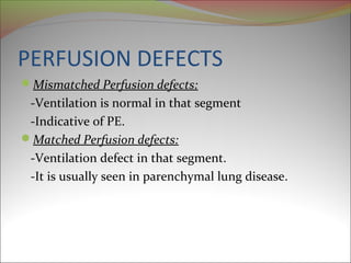
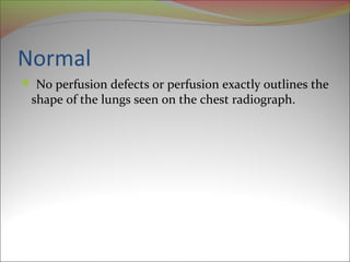
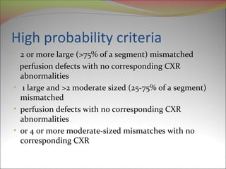
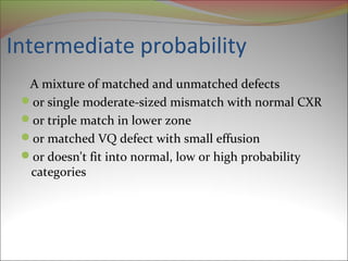
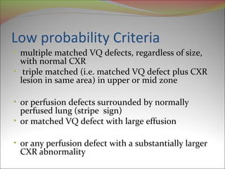
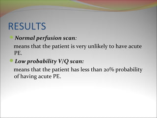
V/Q SCAN 
A type of medical imaging using scintigraphy to 
evaluate the circulation of air and blood within a 
patient's lungs . 
 The ventilation part of the test looks at the ability of 
air to reach all parts of the lungs 
The perfusion part evaluates how well blood 
circulates within the lungs.
INDICATIONS 
 Most commonly done to check the presence of a 
blood clot or abnormal blood flow inside the lungs. 
PE 
COPD 
Pneumonia 
Post lobectomy
Pulmonary embolism 
Pathophysiology 
Rudolph Virchow, 1858 
Triad: 
 Hypercoagulability 
 Stasis to flow 
 Vessel injury
Risk Factors 
Hypercoagulability 
Malignancy 
Pregnancy 
Postpartum status (<4wk) 
Estrogen/ OCP’s 
Venous Stasis 
Bed rest > 24 hr 
Recent cast or external fixator 
Long-distance travel or prolong automobile travel 
Venous Injury 
surgery 
trauma (especially the lower extremities and pelvis)
Presentation 
Dyspnea 
Pleuritic chest pain 
Low-grade fever 
Tachycardia
RADIOGRAPHIC EVALUATION 
• CXR 
• V/Q Scan 
• Spiral CT with contrast 
• Angiogram
CXR 
Initial CXR usually normal. 
May progress to show 
 atelactasis 
 pleural effusion 
 elevated hemi diaphragm.
CXR 
Chest X-Ray Myth: 
“You have to do a chest x-ray so you can find 
Hampton’s hump or a Westermarck sign.” 
Reality: 
Most chest x-rays in patients with PE are 
nonspecific and insensitive 
9
Chest X-ray Eponyms of PE 
Westermarck's sign 
A dilation of the pulmonary vessels proximal to the 
embolism along with collapse of distal vessels, 
sometimes with a sharp cutoff. 
Hampton’s Hump 
A triangular or rounded pleural-based infiltrate or 
consolidation with the apex toward the hilum.
Westermark’s 
Sign 
Hampton’s Hump

CXR
Ventilation/Perfusion Scan 
- “V/Q Scan” 
A common modality to image the lung and its use 
still stems from the PIOPED study. 
Relatively noninvasive 
In many centers remains the initial test of choice 
Preferred test in pregnant patients 
 50 mrem vs 800mrem (with spiral CT)
Procedure 
• Ventilation phase 
a gaseous radionuclide such Krypton ,Xenon or 
technetium DTPA in an aerosol form is inhaled by the 
patient through a mask. 
• The perfusion phase involves the I/V inj of radioactive 
technetium macro aggregated albumin (Tc99m- 
MAA). 
• A gamma camera acquires the images for both phases 
of the study.
The ventilation and perfusion phases of a V/Q lung 
scan are performed together along with a chest x-ray 
for comparison or to look for other causes of lung 
disease
NORMAL PERFUSION IMAGES
NORMAL VENTILATION 
IMAGES
PIOPED STUDY 
PIOPED STUDY The Prospective Investigation of 
Pulmonary Embolism Diagnosis (PIOPED) was a 
multicenter, collaborative effort designed to 
determine the sensitivity and specificity of the V/Q 
scan in patients with suspected acute PE
Defect Descriptors, as Used in 
PIOPED 
 Small defect (small subsegmental): Less than 25% of 
a segment. 
 Moderate defect (moderate subsegmental): > 25%, 
but < 75% of a segment. 
 Large defect (segmental): Greater than 75% of a 
segment.
PERFUSION DEFECTS 
Mismatched Perfusion defects: 
-Ventilation is normal in that segment 
-Indicative of PE. 
Matched Perfusion defects: 
-Ventilation defect in that segment. 
-It is usually seen in parenchymal lung disease.
Normal 
 No perfusion defects or perfusion exactly outlines the 
shape of the lungs seen on the chest radiograph.
High probability criteria 
• 2 or more large (>75% of a segment) mismatched 
perfusion defects with no corresponding CXR 
abnormalities 
• 1 large and >2 moderate sized (25-75% of a segment) 
mismatched 
• perfusion defects with no corresponding CXR 
abnormalities 
• or 4 or more moderate-sized mismatches with no 
corresponding CXR
Intermediate probability 
A mixture of matched and unmatched defects 
or single moderate-sized mismatch with normal CXR 
or triple match in lower zone 
or matched VQ defect with small effusion 
or doesn't fit into normal, low or high probability 
categories
Low probability Criteria 
• multiple matched VQ defects, regardless of size, 
with normal CXR 
• triple matched (i.e. matched VQ defect plus CXR 
lesion in same area) in upper or mid zone 
• or perfusion defects surrounded by normally 
perfused lung (stripe sign) 
• or matched VQ defect with large effusion 
• or any perfusion defect with a substantially larger 
CXR abnormality
RESULTS 
Normal perfusion scan: 
means that the patient is very unlikely to have acute 
PE. 
Low probability V/Q scan: 
means that the patient has less than 20% probability 
of having acute PE.
High probability V/Q scan: 
means that the patient has greater than 80% 
probability of having acute PE. 
Intermediate probability V/Q scan: 
means that the patient has between 20 – 80% 
probabilities of having acute PE.
Visible renal uptake indicates a right to left shunt 
 faulty radiopharmaceutical preparation 
 Thyroid activity usually indicates the presence of 
unbound per technetate in injected dose.
Artefacts may result from clumping of inhaled 
particles with severe airways disease 
OR 
where errors occurred in the preparation or 
administration of the particles
CONTRAINDICATIONS 
No absolute CI 
RELATIVE CI 
Pulmonary Hypertension 
Right to Left shunts e.g. VSD.
Spiral (Helical) Chest CT 
Advantages 
Rapid 
Alternative Diagnosis 
Disadvantages 
Costly 
Risk to patients with borderline renal function 
Hard to detect subsegmental pulmonary emboli
CT Angiogram
Pulmonary angiogram 
Gold Standard. 
Positive angiogram provides 100% certainty that an 
obstruction exists in the pulmonary artery. 
Negative angiogram provides > 90% certainty in the 
exclusion of PE.
PULMONARY ANGIOGRAPHY
Summary 
Plain chest radiograph – Usually normal and non-specific 
signs. 
Radionuclide ventilation-perfusion lung scan. 
CT Angiography of the pulmonary arteries – Quickly 
becoming method of choice. 
Pulmonary angiography – Gold standard but invasive.
QUESTION 
A 52 yr old male presented with SOB and his V/Q 
scan showed multiple matched V/Q defects and his 
CXR was normal. 
 Where will you place the pt in terms of probability. 
 Describe the scheme for interpretation of V/Q scan. 
Describe the different investigation for diagnosis of 
PE and its plain CXR findings.
THANK YOU

VQ scan of lung

  • 2.
    V/Q SCAN Atype of medical imaging using scintigraphy to evaluate the circulation of air and blood within a patient's lungs .  The ventilation part of the test looks at the ability of air to reach all parts of the lungs The perfusion part evaluates how well blood circulates within the lungs.
  • 3.
    INDICATIONS  Mostcommonly done to check the presence of a blood clot or abnormal blood flow inside the lungs. PE COPD Pneumonia Post lobectomy
  • 4.
    Pulmonary embolism Pathophysiology Rudolph Virchow, 1858 Triad:  Hypercoagulability  Stasis to flow  Vessel injury
  • 5.
    Risk Factors Hypercoagulability Malignancy Pregnancy Postpartum status (<4wk) Estrogen/ OCP’s Venous Stasis Bed rest > 24 hr Recent cast or external fixator Long-distance travel or prolong automobile travel Venous Injury surgery trauma (especially the lower extremities and pelvis)
  • 6.
    Presentation Dyspnea Pleuriticchest pain Low-grade fever Tachycardia
  • 7.
    RADIOGRAPHIC EVALUATION •CXR • V/Q Scan • Spiral CT with contrast • Angiogram
  • 8.
    CXR Initial CXRusually normal. May progress to show  atelactasis  pleural effusion  elevated hemi diaphragm.
  • 9.
    CXR Chest X-RayMyth: “You have to do a chest x-ray so you can find Hampton’s hump or a Westermarck sign.” Reality: Most chest x-rays in patients with PE are nonspecific and insensitive 9
  • 10.
    Chest X-ray Eponymsof PE Westermarck's sign A dilation of the pulmonary vessels proximal to the embolism along with collapse of distal vessels, sometimes with a sharp cutoff. Hampton’s Hump A triangular or rounded pleural-based infiltrate or consolidation with the apex toward the hilum.
  • 11.
  • 12.
  • 13.
  • 14.
    Ventilation/Perfusion Scan -“V/Q Scan” A common modality to image the lung and its use still stems from the PIOPED study. Relatively noninvasive In many centers remains the initial test of choice Preferred test in pregnant patients  50 mrem vs 800mrem (with spiral CT)
  • 15.
    Procedure • Ventilationphase a gaseous radionuclide such Krypton ,Xenon or technetium DTPA in an aerosol form is inhaled by the patient through a mask. • The perfusion phase involves the I/V inj of radioactive technetium macro aggregated albumin (Tc99m- MAA). • A gamma camera acquires the images for both phases of the study.
  • 16.
    The ventilation andperfusion phases of a V/Q lung scan are performed together along with a chest x-ray for comparison or to look for other causes of lung disease
  • 17.
  • 18.
  • 19.
    PIOPED STUDY PIOPEDSTUDY The Prospective Investigation of Pulmonary Embolism Diagnosis (PIOPED) was a multicenter, collaborative effort designed to determine the sensitivity and specificity of the V/Q scan in patients with suspected acute PE
  • 20.
    Defect Descriptors, asUsed in PIOPED  Small defect (small subsegmental): Less than 25% of a segment.  Moderate defect (moderate subsegmental): > 25%, but < 75% of a segment.  Large defect (segmental): Greater than 75% of a segment.
  • 21.
    PERFUSION DEFECTS MismatchedPerfusion defects: -Ventilation is normal in that segment -Indicative of PE. Matched Perfusion defects: -Ventilation defect in that segment. -It is usually seen in parenchymal lung disease.
  • 22.
    Normal  Noperfusion defects or perfusion exactly outlines the shape of the lungs seen on the chest radiograph.
  • 24.
    High probability criteria • 2 or more large (>75% of a segment) mismatched perfusion defects with no corresponding CXR abnormalities • 1 large and >2 moderate sized (25-75% of a segment) mismatched • perfusion defects with no corresponding CXR abnormalities • or 4 or more moderate-sized mismatches with no corresponding CXR
  • 25.
    Intermediate probability Amixture of matched and unmatched defects or single moderate-sized mismatch with normal CXR or triple match in lower zone or matched VQ defect with small effusion or doesn't fit into normal, low or high probability categories
  • 26.
    Low probability Criteria • multiple matched VQ defects, regardless of size, with normal CXR • triple matched (i.e. matched VQ defect plus CXR lesion in same area) in upper or mid zone • or perfusion defects surrounded by normally perfused lung (stripe sign) • or matched VQ defect with large effusion • or any perfusion defect with a substantially larger CXR abnormality
  • 29.
    RESULTS Normal perfusionscan: means that the patient is very unlikely to have acute PE. Low probability V/Q scan: means that the patient has less than 20% probability of having acute PE.
  • 30.
    High probability V/Qscan: means that the patient has greater than 80% probability of having acute PE. Intermediate probability V/Q scan: means that the patient has between 20 – 80% probabilities of having acute PE.
  • 31.
    Visible renal uptakeindicates a right to left shunt  faulty radiopharmaceutical preparation  Thyroid activity usually indicates the presence of unbound per technetate in injected dose.
  • 33.
    Artefacts may resultfrom clumping of inhaled particles with severe airways disease OR where errors occurred in the preparation or administration of the particles
  • 35.
    CONTRAINDICATIONS No absoluteCI RELATIVE CI Pulmonary Hypertension Right to Left shunts e.g. VSD.
  • 36.
    Spiral (Helical) ChestCT Advantages Rapid Alternative Diagnosis Disadvantages Costly Risk to patients with borderline renal function Hard to detect subsegmental pulmonary emboli
  • 37.
  • 38.
    Pulmonary angiogram GoldStandard. Positive angiogram provides 100% certainty that an obstruction exists in the pulmonary artery. Negative angiogram provides > 90% certainty in the exclusion of PE.
  • 39.
  • 40.
    Summary Plain chestradiograph – Usually normal and non-specific signs. Radionuclide ventilation-perfusion lung scan. CT Angiography of the pulmonary arteries – Quickly becoming method of choice. Pulmonary angiography – Gold standard but invasive.
  • 41.
    QUESTION A 52yr old male presented with SOB and his V/Q scan showed multiple matched V/Q defects and his CXR was normal.  Where will you place the pt in terms of probability.  Describe the scheme for interpretation of V/Q scan. Describe the different investigation for diagnosis of PE and its plain CXR findings.
  • 42.

Editor's Notes

  • #12 Here we see the dilated vessels and oligemia of westermark’s sign And below Hampton’s Hump
  • #37 The entire lung can be scanned while the patient holds there breath. Advantages: CT most useful benefit is in providing evidence for an alternative diagnosis or excluding it entirely. Disadvantages: The clinical significance for subsegmental PE are not well known, but may be a marker for a larger PE Given that the majority of V/Q studies are non-diagnostic, I prefer the CT as the initial test of choice in place of V/Q scan.