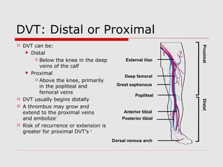
This document discusses the diagnostic and treatment approaches to venous thromboembolism (VTE), which includes deep vein thrombosis (DVT) and pulmonary embolism (PE). It provides details on evaluating patients using Wells criteria and D-dimer testing to determine pre-test probability and decide between imaging with CT pulmonary angiogram or VQ scan. For confirmed VTE, treatment options include warfarin, novel oral anticoagulants (NOACs), inferior vena cava filters or thrombolytics. The document reviews best practices for treating isolated distal DVT, catheter-related thrombosis, and selecting appropriate long-term anticoagulation therapy.
A lecture highlighting the role of Echocardiography as a major hemodynamic monitoring tool in the Intensive Care settings and the assessment of loading conditions.
various trial about on pump vs off pump and pci vs CABG
coronary trial
ROOBY TRAIL
DOORS TRAIL
GOPCABE TRAIL
PRAGUE 6 TRAIL
SMART STUDY
PROMOTE PATENCY TRIAL
AV nodal reentrant tachycardia (AVNRT), or atrioventricular nodal reentrant tachycardia, is a type of tachycardia (fast rhythm) of the heart. It is a type of supraventricular tachycardia (SVT), meaning that it originates from a location within the heart above the bundle of His. AV nodal reentrant tachycardia is the most common regular supraventricular tachycardia. It is more common in women than men (approximately 75% of cases occur in females). The main symptom is palpitations. Treatment may be with specific physical maneuvers, medication, or, rarely, synchronized cardioversion. Frequent attacks may require radiofrequency ablation, in which the abnormally conducting tissue in the heart is destroyed.
AVNRT occurs when a reentry circuit forms within or just next to the atrioventricular node. The circuit usually involves two anatomical pathways: the fast pathway and the slow pathway, which are both in the right atrium. The slow pathway (which is usually targeted for ablation) is located inferior and slightly posterior to the AV node, often following the anterior margin of the coronary sinus. The fast pathway is usually located just superior and posterior to the AV node. These pathways are formed from tissue that behaves very much like the AV node, and some authors regard them as part of the AV node.
The fast and slow pathways should not be confused with the accessory pathways that give rise to Wolff-Parkinson-White syndrome (WPW syndrome) or atrioventricular reciprocating tachycardia (AVRT). In AVNRT, the fast and slow pathways are located within the right atrium close to or within the AV node and exhibit electrophysiologic properties similar to AV nodal tissue. Accessory pathways that give rise to WPW syndrome and AVRT are located in the atrioventricular valvular rings. They provide a direct connection between the atria and ventricles, and have electrophysiologic properties similar to ventricular myocardium.
Our concepts of heart disease are based on the enormous reservoir of physiologic and anatomic knowledge derived from the past 70 years' of experience in the cardiac catheterization laboratory.
As Andre Cournand remarked in his Nobel lecture of December 11, 1956, the cardiac catheter was the key in the lock.
By turning this key, Cournand and his colleagues led us into a new era in the understanding of normal and disordered cardiac function in huma
Endovascular and surgical treatment of pulmonary embolism 26.11.17Ivo Petrov
Interventional treatment (thrombus fragmentation and supraselective fibrinolysis) of high and intermediate risk patients with pulmonary embolism.
Protocols of intervention, results, clinical cases provided
Physician should have a high suspicion to diagnose patient with pulmonary Embolism, this slides will give you precise Diagnosis, Investigation and guideline directed Treatment.
Defecation
Normal defecation begins with movement in the left colon, moving stool toward the anus. When stool reaches the rectum, the distention causes relaxation of the internal sphincter and an awareness of the need to defecate. At the time of defecation, the external sphincter relaxes, and abdominal muscles contract, increasing intrarectal pressure and forcing the stool out
The Valsalva maneuver exerts pressure to expel faeces through a voluntary contraction of the abdominal muscles while maintaining forced expiration against a closed airway. Patients with cardiovascular disease, glaucoma, increased intracranial pressure, or a new surgical wound are at greater risk for cardiac dysrhythmias and elevated blood pressure with the Valsalva maneuver and need to avoid straining to pass the stool.
Normal defecation is painless, resulting in passage of soft, formed stool
CONSTIPATION
Constipation is a symptom, not a disease. Improper diet, reduced fluid intake, lack of exercise, and certain medications can cause constipation. For example, patients receiving opiates for pain after surgery often require a stool softener or laxative to prevent constipation. The signs of constipation include infrequent bowel movements (less than every 3 days), difficulty passing stools, excessive straining, inability to defecate at will, and hard feaces
IMPACTION
Fecal impaction results from unrelieved constipation. It is a collection of hardened feces wedged in the rectum that a person cannot expel. In cases of severe impaction the mass extends up into the sigmoid colon.
DIARRHEA
Diarrhea is an increase in the number of stools and the passage of liquid, unformed feces. It is associated with disorders affecting digestion, absorption, and secretion in the GI tract. Intestinal contents pass through the small and large intestine too quickly to allow for the usual absorption of fluid and nutrients. Irritation within the colon results in increased mucus secretion. As a result, feces become watery, and the patient is unable to control the urge to defecate. Normally an anal bag is safe and effective in long-term treatment of patients with fecal incontinence at home, in hospice, or in the hospital. Fecal incontinence is expensive and a potentially dangerous condition in terms of contamination and risk of skin ulceration
HEMORRHOIDS
Hemorrhoids are dilated, engorged veins in the lining of the rectum. They are either external or internal.
FLATULENCE
As gas accumulates in the lumen of the intestines, the bowel wall stretches and distends (flatulence). It is a common cause of abdominal fullness, pain, and cramping. Normally intestinal gas escapes through the mouth (belching) or the anus (passing of flatus)
FECAL INCONTINENCE
Fecal incontinence is the inability to control passage of feces and gas from the anus. Incontinence harms a patient’s body image
PREPARATION AND GIVING OF LAXATIVESACCORDING TO POTTER AND PERRY,
An enema is the instillation of a solution into the rectum and sig
CRISPR-Cas9, a revolutionary gene-editing tool, holds immense potential to reshape medicine, agriculture, and our understanding of life. But like any powerful tool, it comes with ethical considerations.
Unveiling CRISPR: This naturally occurring bacterial defense system (crRNA & Cas9 protein) fights viruses. Scientists repurposed it for precise gene editing (correction, deletion, insertion) by targeting specific DNA sequences.
The Promise: CRISPR offers exciting possibilities:
Gene Therapy: Correcting genetic diseases like cystic fibrosis.
Agriculture: Engineering crops resistant to pests and harsh environments.
Research: Studying gene function to unlock new knowledge.
The Peril: Ethical concerns demand attention:
Off-target Effects: Unintended DNA edits can have unforeseen consequences.
Eugenics: Misusing CRISPR for designer babies raises social and ethical questions.
Equity: High costs could limit access to this potentially life-saving technology.
The Path Forward: Responsible development is crucial:
International Collaboration: Clear guidelines are needed for research and human trials.
Public Education: Open discussions ensure informed decisions about CRISPR.
Prioritize Safety and Ethics: Safety and ethical principles must be paramount.
CRISPR offers a powerful tool for a better future, but responsible development and addressing ethical concerns are essential. By prioritizing safety, fostering open dialogue, and ensuring equitable access, we can harness CRISPR's power for the benefit of all. (2998 characters)
Global launch of the Healthy Ageing and Prevention Index 2nd wave – alongside...ILC- UK
The Healthy Ageing and Prevention Index is an online tool created by ILC that ranks countries on six metrics including, life span, health span, work span, income, environmental performance, and happiness. The Index helps us understand how well countries have adapted to longevity and inform decision makers on what must be done to maximise the economic benefits that comes with living well for longer.
Alongside the 77th World Health Assembly in Geneva on 28 May 2024, we launched the second version of our Index, allowing us to track progress and give new insights into what needs to be done to keep populations healthier for longer.
The speakers included:
Professor Orazio Schillaci, Minister of Health, Italy
Dr Hans Groth, Chairman of the Board, World Demographic & Ageing Forum
Professor Ilona Kickbusch, Founder and Chair, Global Health Centre, Geneva Graduate Institute and co-chair, World Health Summit Council
Dr Natasha Azzopardi Muscat, Director, Country Health Policies and Systems Division, World Health Organisation EURO
Dr Marta Lomazzi, Executive Manager, World Federation of Public Health Associations
Dr Shyam Bishen, Head, Centre for Health and Healthcare and Member of the Executive Committee, World Economic Forum
Dr Karin Tegmark Wisell, Director General, Public Health Agency of Sweden
We understand the unique challenges pickleball players face and are committed to helping you stay healthy and active. In this presentation, we’ll explore the three most common pickleball injuries and provide strategies for prevention and treatment.
Welcome to Secret Tantric, London’s finest VIP Massage agency. Since we first opened our doors, we have provided the ultimate erotic massage experience to innumerable clients, each one searching for the very best sensual massage in London. We come by this reputation honestly with a dynamic team of the city’s most beautiful masseuses.
India Clinical Trials Market: Industry Size and Growth Trends [2030] Analyzed...Kumar Satyam
According to TechSci Research report, "India Clinical Trials Market- By Region, Competition, Forecast & Opportunities, 2030F," the India Clinical Trials Market was valued at USD 2.05 billion in 2024 and is projected to grow at a compound annual growth rate (CAGR) of 8.64% through 2030. The market is driven by a variety of factors, making India an attractive destination for pharmaceutical companies and researchers. India's vast and diverse patient population, cost-effective operational environment, and a large pool of skilled medical professionals contribute significantly to the market's growth. Additionally, increasing government support in streamlining regulations and the growing prevalence of lifestyle diseases further propel the clinical trials market.
Growing Prevalence of Lifestyle Diseases
The rising incidence of lifestyle diseases such as diabetes, cardiovascular diseases, and cancer is a major trend driving the clinical trials market in India. These conditions necessitate the development and testing of new treatment methods, creating a robust demand for clinical trials. The increasing burden of these diseases highlights the need for innovative therapies and underscores the importance of India as a key player in global clinical research.
How many patients does case series should have In comparison to case reports.pdfpubrica101
Pubrica’s team of researchers and writers create scientific and medical research articles, which may be important resources for authors and practitioners. Pubrica medical writers assist you in creating and revising the introduction by alerting the reader to gaps in the chosen study subject. Our professionals understand the order in which the hypothesis topic is followed by the broad subject, the issue, and the backdrop.
https://pubrica.com/academy/case-study-or-series/how-many-patients-does-case-series-should-have-in-comparison-to-case-reports/
CHAPTER 1 SEMESTER V - ROLE OF PEADIATRIC NURSE.pdfSachin Sharma
Pediatric nurses play a vital role in the health and well-being of children. Their responsibilities are wide-ranging, and their objectives can be categorized into several key areas:
1. Direct Patient Care:
Objective: Provide comprehensive and compassionate care to infants, children, and adolescents in various healthcare settings (hospitals, clinics, etc.).
This includes tasks like:
Monitoring vital signs and physical condition.
Administering medications and treatments.
Performing procedures as directed by doctors.
Assisting with daily living activities (bathing, feeding).
Providing emotional support and pain management.
2. Health Promotion and Education:
Objective: Promote healthy behaviors and educate children, families, and communities about preventive healthcare.
This includes tasks like:
Administering vaccinations.
Providing education on nutrition, hygiene, and development.
Offering breastfeeding and childbirth support.
Counseling families on safety and injury prevention.
3. Collaboration and Advocacy:
Objective: Collaborate effectively with doctors, social workers, therapists, and other healthcare professionals to ensure coordinated care for children.
Objective: Advocate for the rights and best interests of their patients, especially when children cannot speak for themselves.
This includes tasks like:
Communicating effectively with healthcare teams.
Identifying and addressing potential risks to child welfare.
Educating families about their child's condition and treatment options.
4. Professional Development and Research:
Objective: Stay up-to-date on the latest advancements in pediatric healthcare through continuing education and research.
Objective: Contribute to improving the quality of care for children by participating in research initiatives.
This includes tasks like:
Attending workshops and conferences on pediatric nursing.
Participating in clinical trials related to child health.
Implementing evidence-based practices into their daily routines.
By fulfilling these objectives, pediatric nurses play a crucial role in ensuring the optimal health and well-being of children throughout all stages of their development.
QA Paediatric dentistry department, Hospital Melaka 2020Azreen Aj
QA study - To improve the 6th monthly recall rate post-comprehensive dental treatment under general anaesthesia in paediatric dentistry department, Hospital Melaka
Artificial Intelligence to Optimize Cardiovascular Therapy
Vte 2014
1. Diagnostic and Therapeutic
Approach to Venous
Thromboembolism
Dr. Mark Choi, MD
General Internal Medicine
Abbotsford Regional Hospital and Cancer
Centre
3. Pulmonary Embolism
VTE incidence in total population is
~100/100 000 patients/year
1/3 of VTE cases are PE
Acute PE has an estimated 30-day
mortality ranging from 5 to 30%1
Causes ~300,000 deaths in the
United States and over 500,000
deaths in Europe per year2,3
In the EU, more than twice as
many people die from VTE than
from AIDS, breast cancer, prostate
cancer and transportation
accidents combined3
296,370
543,454
• Régie de l'assurance maladie du Québec
(RAMQ) retrospective health database
review study suggests that the overall
incidence of first definite or probable PE
event is 4.5/10,000 person-years4
• 15,700 annual incident cases of PE
across Canada4
4. DVT: Distal or Proximal
DVT can be:
Distal
Below the knee in the deep
veins of the calf
Proximal
Above the knee, primarily
in the popliteal and
femoral veins
DVT usually begins distally
A thrombus may grow and
extend to the proximal veins
and embolize1
Risk of recurrence or extension is
greater for proximal DVT’s 2
External iliac
Deep femoral
Great saphenous
Popliteal
Anterior tibial
Posterior tibial
Dorsal venous arch
Distal Proximal
5. Venous Thromboembolism
Deep Vein Thrombosis
Diagnostic Algorithm / Wells Criteria for DVT
Are isolated distal DVTs treated the same way as above
knee DVTs?
Are superficial vein thromboses ever treated with OAC?
Iatrogenic venous thrombosis
Treatment and its duration
Congenital workup
Pulmonary Embolism
Diagnostic Algorithm / Wells Criteria for PE
Is CT chest always superior to VQ scan?
What is the VQ criteria?
Treatment Options
Warfarin vs. NOACs (dabigatran and rivaroxaban)
Pros and Cons of NOACs
8. Well’s Criteria for DVT
1 point each for below:
Paralysis, paresis or recent orthopedic casting of lower
extremity
Recently bedridden (> 3 days) or major Sx within 4
weeks
Localized tenderness in deep vein system
Swelling of entire leg
Calf swelling 3cm greater than other leg (measured 10cm
below the tibial tuberosity)
Pitting edema greater in the symptomatic leg
Collateral non-varicosesuperficial veins
Active cancer or cancer treated within 6 months
-2 points for alternative more likely diagnosis (baker’s cyst,
cellulitis, muscle damage, SVT, post-thrombotic syndrome,
inguinal lymphadenopathy, external venous compression)
9. Isolated Distal DVT
Conflicting studies. Extension to proximal veins noted
in 4 to 16% of patients without treatment; 7 to 10% of
PE’s found to have isolated distal DVTs
Issues:
Fewer late sequelae than proximal DVT
Operator dependent
Less sensitive than proximal vein examination
Major risk factor for extension was active cancer
Isolated distal DVT had lower rate of recurrence
whereas bilateral distal DVT had same recurrence as
proximal DVT
Current guideline: treat for 3 months
10. Superficial Vein Thrombosis
Treatment
Elevation of affected limb
Warm and cold compresses
Topical or oral NSAID’s
Encourage ambulation
Treat pts with OAC (3 months) in higher
risk for extension
High risk features:
Affected vein segment > 5cm,
saphenofemoral/saphenopopliteal junction, <
5cm to deep venous system, medical risk factors
Chest. 2008;133(6 Suppl):454S
11. Catheter-related venous
thrombosis
Risk of thrombosis present with central
(CVCs) and peripheral catheters (PICCs)
Most patients are asymptomatic
Higher risks of thrombosis with PICCs (both
peripheral and central venous thrombsis)
RFs for CRVT = mal-positioned catheter,
larger diameter catheters, active cancer,
prior hx of DVT, hormonal therapy and
congenital VTE predisposition
12. Catheter-related venous
thrombosis
Superficial phlebitis
Remove catheter
Elevate affected limb
Warm or cold compresses
Topical or oral NSAIDs
Repeat U/S in 1 week in patients with RF’s
Upper extremity DVT
OAC as long as the catheter is in situ and
consider longer depending on RFs
If OAC is discontinued, repeat U/S to look for
extension in patients with RFs
13. Treatment of proximal/distal DVT
Provoked DVT = treat for 3 months
Unprovoked DVT = treat for 3 months and
consider longer
Rebound DVT = Risk roughly 20% in 2 years
after discontinuation of OAC.
Follow-up Baseline DVT and d-dimer necessary
at the end of treatment with OAC in order to
differentiate between recurrent DVT and post-thrombotic
syndrome if his/her symptoms
return
Congenital workup = reserved for unprovoked
VTE’s in younger patients or strong family
history of VTE
15. Pulmonary Embolism
Diagnostic approach that we are used to:
RYU JH, SWENSEN SJ, OLSON EJ, PELLIKKA PA. DIAGNOSIS OF PULMONARY EMBOLISM WITH
USE OF COMPUTED TOMOGRAPHIC ANGIOGRAPHY. MAYO CLIN PROC 2001;76:63.
16. Wells Criteria for PE
Alternative diagnosis less likely than PE 3.0
Signs or symptoms of DVT 3.0
History of PE or DVT 1.5
Immobilization for at least 3 days or surgery 1.5
in the previous month
Heart rate >100 beats/min 1.5
Hemoptysis 1.0
Active cancer (treatment ongoing, within 1.0
previous 6 months or palliative)
For high sensitivity D-dimer:
Low probability: <2 points
Intermediate probability: 2–6 points
High probability: ≥6 points
17. Is CT chest superior to VQ scan?
Initial VQ study showed good
sensitivity and specificity to the gold
standard (pulmonary angiogram)
CTPA compared to VQ study and
deemed non-inferior to VQ for
diagnosis of PE
18. Pros and Cons of CT Chest
CT chest with IV contrast
Pros
Non-inferior to VQ scan in diagnosis of PE
May catch other pulmonary findings (pneumonia, mass,
emphysema, ILD etc)
Cons
Radiation exposure (ei. Pregnancy)
Risk of contrast-induced nephropathy in kidney dz
May give indeterminate findings
Patient may move during the study
IV contrast timed incorrectly
Incidental findings of isolated subsegmental PE may
lead to over-treatment
Potential risk of allergy to IV contrast dye
19. Pros and Cons of VQ Scan
VQ Scan
Pros
Good sensitivity and specificity in comparison to gold
standard (pulmonary angiogram)
May catch other pulmonary findings (focal
consolidation)
Safe in kidney disease
Relatively small radiation exposure (equiv ~ 5 CXRs)
Cons
May give indeterminate findings
If performed in patients with chronic cardiac or
pulmonary disease
Availability: nuclear medicine open daytime Mon to Fri
in most tertiary centres
20. Radiation Exposure
64-slice CTPA delivers 50-60mSv to
the breast
V/Q Scan delivers only 0.28-0.9mSv
In comparison, a 2-view
mammogram is associated with 3mSv
Radiation exposure to female breast
is 70 to 100-fold greater in CTPA vs.
V/Q scan
Einstein AJ. Estimating risk of cancer associated with radiation exposure from 64-slice CT angiography. JAMA. 2007; 298: 317-323
Parker MS. Female breast radiation exposure during CT pulmonary angiography. Am J Radiol. 2005. 185: 1229-1233
Cook JV. Radiation from CT and perfusion scanning in prengnancy
Task Group on Control of Radiation Dose in CT: a report of the International Commission on Radiological Protection. AnnICRP 2000;30:7-45
21. Radiation Exposure
There is non-negligible increase in
lifetime attributable risk of cancer,
particularly to the breasts of young
women
1 in 143 for a 20-year-old woman
1 in 284 for a 40-year-old woman
There is also increased odds of lung
cancer in both gender
Einstein AJ. Estimating risk of cancer associated with radiation exposure from 64-slice CT angiography. JAMA. 2007; 298: 317-323
22. When do physicians choose VQ
Scan?
In the setting of decreased eGFR
Known IV contrast allergy
When CTPA result is indeterminate
Pregnancy and Post-Partum
Because it causes less radiation
23. So when is V/Q scan more appropriate to
order over a CT scan?
younger population
female population
no other intrathoracic disease is
suspected
decreased eGFR
Pregnancy/Post Partum
Consider in those meeting the “VQ
criteria”
24. The VQ Criteria
There are 2 VQ Criteria
A normal CXR
No history of chronic lung disease or
congestive heart failure
If above 2 criteria are met, it is very
unlikely that the VQ scan will give an
indeterminate finding
25. V/Q vs. CTPE Study
Data on all CT and V/Q scans performed to
assess for PE in St. Michael's Hospital
(SMH) inpatients over 2-year period
collected
Exclusion
CT scans ordered at hours when V/Q scan is
unavailable (weeknights, weekends and
holidays) were excluded
CT scans looking for PE and other diagnoses
were excluded
CT scans ordered on intensive care patients
were excluded.
26. V/Q vs. CTPE Study
29.2% of CTPE studies met the V/Q
criteria
28. V/Q Scan as primary imaging
modality
Only half (14 of 28) of these studies
met the VQ Criteria
29. V/Q vs. CTPE study conclusion
V/Q scan under-utilized in patients
meeting V/Q criteria
Not using the V/Q criteria leads to
many indeterminate findings
30. Preferred Algorithm
Pre-test Probability using Dichotomized Well’s Score
Low (4 or less) High (5 or higher)
D-dimer Normal CXR and no hx of
cardiopulmonary disease
Low (< 500) High (>500) Yes No
PE ruled out
VQ Scan
Renal Dysfunction or
IV contrast allergy
Yes No
CT Scan
If either test indeterminate
37. Treatment Options
Warfarin
vitamin K antagonist
IV heparin
binds AT and inactivates fXa and prevents conversion of
fibrinogen to fibrin
LMWH
binds AT and inactivates fXa
fondaparinux
enhances the anti-Xa activity of AT by 300-fold
NOAC’s
Dabigatran (direct thrombin inhibitor)
Rivaroxaban (direct factor Xa inhibitor)
Apixaban (not approved in Canada for treatment of PE or DVT
yet)
IVC filter
Intravenous thrombolytics
38. Warfarin is Highly Effective for the
Prevention of Stroke in AF or Recurrent VTE
Stroke Prevention in Atrial
Fibrillation
Treatment of Venous
Thromboembolism (VTE)
Standard Care: Warfarin (INR 2.0-3.0) Standard Care: Warfarin (INR 2.0-3.0),
initial parenteral anticoagulation until INR >2
Chance of recurrent VTE in
first 3 months
47%
If proximal DVT inadequately treated3
1
1
2
<2%
If adequate anticoagulant
response is achieved4
1. Hart et al Ann Intern Med. 2007;146:857-867; 2. Connolly et al. Lancet. 2006;367:1903-12; 3. Line B. Semin Nucl Med. 2001;31:90–101 4. Kearon C. Circulation 2003;107:I22–I30.
39. Properties of an Ideal Anticoagulant vs.
Currently Available Agents
IDEAL LMWH UFH VKA NOAC
Oral
No significant food/drug
interactions
Predictable response
No requirement for
coagulation monitoring
Fixed dosing
No HIT
Cost
Reversible
HIT: Heparin induced thrombocytopenia
?
40. Canadian Approvals of New Oral
Anticoagulants
Rivaroxaban
VTE prophylaxis
following THR/TKR
2008 2009 2010 2011
2012 2013
Dabigatran
VTE prophylaxis
following THR/TKR
Health Canada NOC database: http://webprod5.hc-sc.gc.ca/noc-ac/index-eng.jsp
Apixaban
VTE prophylaxis
following THR/TKR
Dabigatran
Stroke prevention
in AF
Rivaroxaban
Stroke prevention
in AF
Rivaroxaban
Treatment and secondary prevention
of DVT (2012) and PE (2013)
New oral anticoagulants
Dabigatran
Treatment and
secondary
prevention of DVT
Apixaban and PE
Stroke prevention
in AF
2014
41. Novel Oral Anticoagulants –
Pharmacological Properties
Characteristic Rivaroxaban1 Dabigatran2 Apixaban3
Target Factor Xa Factor IIa Factor Xa
Dosing OD BID BID
Bioavailability, % 80-100%* 6.5% 50%
Half-life 5-13h 12-14 h 8-15 h
Renal clearance
(unchanged
~33% 85% 27%†
bioavailable drug)
Cmax 2-4 h 1-2 h 3-4 h
Bridging with
No Yes No
LMWH
* When the 15mg and 20mg dose is taken with food
† Factoring in the absolute bioavailability of apixaban, ~ 50 % of the systemically available dose is eliminated in urine
1. Xarelto® PM, June 2013; 2. Pradaxa ® PM November, 2012; 3. Eliquis® PM November, 2012;
4. FDA Eliquis Drug Approval Package, Clinical Pharmacology/Biopharmaceutics Review
42. New Oral Anticoagulants: Total Drug Exposure
(AUC) with Declining Renal Function
Rivaroxaban
(33% cleared renally*)1
Dabigatran
(85% cleared renally)2
Apixaban
(27% cleared renally†)3
* active drug
† Factoring in the absolute bioavailability of apixaban, ~ 50 % of the systemically available dose is eliminated in urine
1. Xarelto® PM, June 2013; 2. Pradaxa ® PM November, 2012; 3. Eliquis® PM November, 2012;
4. FDA Eliquis Drug Approval Package, Clinical Pharmacology/Biopharmaceutics Review
43. Landmark DVT/PE Trials
Phase II Phase III
Rivaroxaban
Oral, direct Factor Xa
inhibitor
EINSTEIN DVT2
Rivaroxaban vs LMWH/UFH
followed by VKA
ODIXa-DVT3
Rivaroxaban vs enoxaparin
followed by VKA
EINSTEIN DVT/PE5,6
Rivaroxaban for 3, 6 or 12 months vs enoxaparin for ≥5 days
followed by VKA for 3, 6, or 12 months
EINSTEIN EXT5
Pre-treatment with rivaroxaban or VKA for 6 or
12 months followed by rivaroxaban or placebo for 6 or 12 months
Dabigatran
Oral, direct thrombin
inhibitor
RE-COVER7 & RE-COVER II8
5–10 days pre-treatment with LMWH bridging to dabigatran or VKA
for 6 months
RE-MEDY9
3–6 months’ treatment with approved anticoagulant; switch to
dabigatran or VKA
RE-SONATE10
6–18 months’ VKA treatment followed by 6 months dabigatran or
placebo
Apixaban
Oral, direct Factor Xa
inhibitor
Botticelli-DVT4
Apixaban vs LMWH or
fondaparinux followed
by VKA
AMPLIFY1
Apixaban 10 mg bid followed by 5 mg bid for 6 months vs.
enoxaparin followed by VKA
AMPLIFY-EXT11
Apixaban 2.5 mg bid or 5 mg bid for extended 12 months period vs
placebo
Bridging with LMWH and duration of treatment are the main differences between the trials
The following slides summarize trial results with NOACs in VTE treatment
Not intended as cross-trial comparison
1. http://clinicaltrials.gov; 2. Buller HR et al. Blood 2008; 3. Agnelli GA, et al. Circulation 2007; 4. Büller HR, et al. J Thromb Haemost 2008; 5. The EINSTEIN Investigators. N Engl
J Med 2010; 6. The EINSTEIN–PE Investigators. N Engl J Med 2012; 7. Schulman S, et al. N Engl J Med 2009; 8. Schulman S, et al. ASH Annual Meeting Abstracts. Blood 2011;
9. Schulmann S et al. ISTH 2011; 10. Schulman S, et al. ISTH 2011; 11. Agnelli G et al. N Engl J Med 2012; 12. Raskob G et al. J Thromb Haemost. 2013.
44. VTE treatment Results comparison: pivotal
phase III clinical trials – Efficacy
Study drug
n (%)
LMWH/
VKA
n (%)
ARD
%
HR 95% CI P
EINSTEIN
Pooled
3, 6, 12 months
86 (2.1) 95
(2.3)
0.2 0.89 0.66-
1.19
N-inf
RE-COVER
Pooled
6 months
(2.7) (2.4) -0.3 1.09 0.77-
1.54
N-inf
AMPLIFY
6 months
59 (2.3) 71
(2.7)
0.4 0.84 0.60–
1.18
N-inf
Recurrent VTE or VTE-related death
Hazard ratio
and 95% CIs
Favours study
drug
Favours
LMWH/VKA
* Event during on-treatment period
Hazard ratio
and 95% CIs
Favours study drug Favours
LMWH/VKA
45. VTE treatment Results comparison: pivotal
phase III clinical trials – Safety
Major Bleeding
Study
drug
n (%)
LMWH/VK
A
n (%)
ARR
%
HR 95% CI P
EINSTEIN
Pooled
3, 6, 12
Months
40 (1.0) 72 (1.7) 0.7 0.54 0.37-0.79 Sup.
RE-COVER
Pooled
6 months
37 (1.4) 51 (2.0) 0.6 0.73 0.48-1.11 N-inf
AMPLIFY
6 months
15 (0.6) 49 (1.8) 1.2 0.31 0.17–0.55 Sup.
Hazard ratio
and 95% CIs
Favours study drug
Favours
LMWH/VKA
46. Summary
Main Phase
Few differences in trial designs
Bridging with LMWH in RE-COVER vs. Single-drug
approach in EINSTEIN and AMPLIFY
Duration of treatment – 6 months in RE-COVER
and AMPLIFY vs. 3, 6 or 12 months in EINSTEIN
OD in EINSTEIN (after initial acute phase for
EINSTEIN) vs. BID in RE-COVER and AMPLIFY
All agents were non-inferior vs. warfarin in
preventing VTE recurrence
Rivaroxaban and apixaban were superior to warfarin
in reducing major bleeding whereas dabigatran was
non-inferior
47. Cost of NOAC’s
May apply for Special Authority for the
treatment of DVT with rivaroxaban. Special
Authority not approved for dabigatran for
treatment of DVT or PE.
If the patient has reached his/her
deductible on Pharmacare, rivaroxaban
does not cost the patient any money
Otherwise, cost of rivaroxaban is ~$3/day
for 20mg po daily. Cost of dabigatran is
~$3.4/day.
48. Rivaroxaban vs. Dabigatran
No bridging with LMWH required with
rivaroxaban
OD vs. BID dosing
Rivaroxaban is superior to warfarin in reducing
major bleeding events whereas dabigatran is
non-inferior
Rivaroxaban is cheaper; rivaroxaban may be free
for patients who have reached their deductible
Less renally cleared
Both irreversible but rivaroxaban has shorter
half-life
49. Rivaroxaban vs. Apixaban
No bridging needed for both drugs
OD vs. BID dosing
Both non-inferior to warfarin in
recurrent VTEs and VTE-related
deaths
Both superior to warfarin in number
of major bleeding events
Both are less renally cleared in
comparison to dabigatran
50. Take Away Points
Not all SVT are treated conservatively
Remember to utilize VQ scans in those who
meet the VQ criteria
Direct Factor Xa inhibitors are superior to
warfarin in reducing major bleeding risk
Bridging not required for rivaroxaban or
apixaban
Your patient’s rivaroxaban cost may be
covered fully in the treatment of DVT.
Please use special authority.