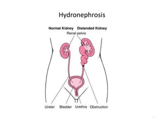
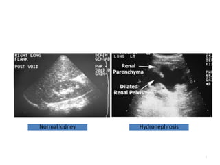
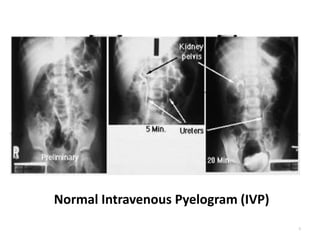
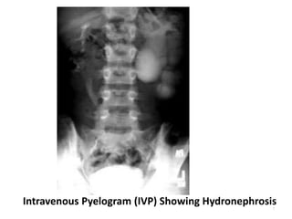
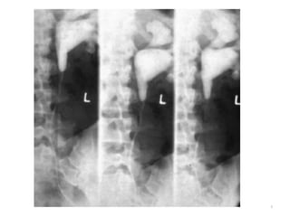
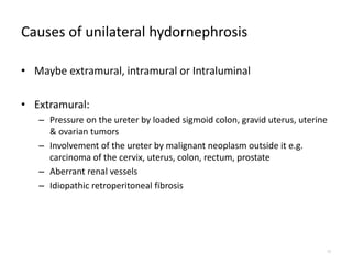
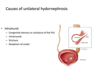
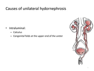
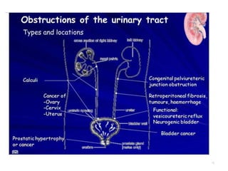
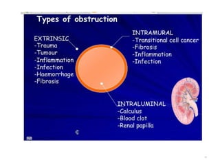
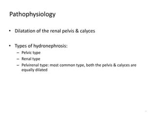
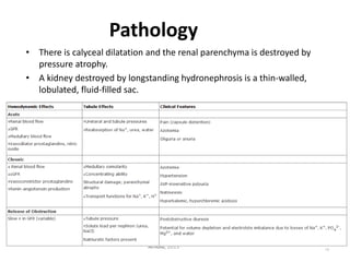
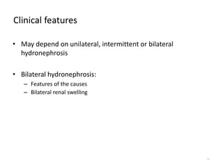
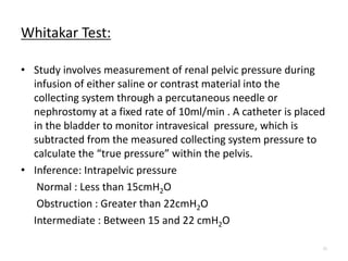
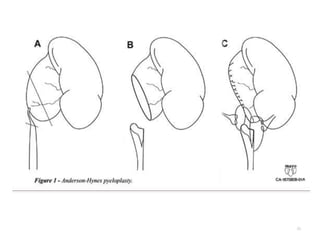
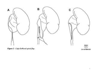
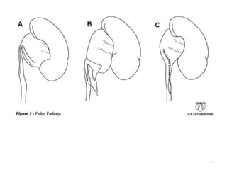
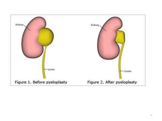
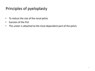
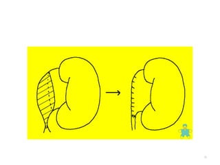
3. DEFINITION
• Aseptic dilatation of renal pelvis & calyces with accompanying
destruction of the kidney parenchyma
• Usually due to partial obstruction to the outflow of urine
• Infected hydronephrosis is bacterial infection in a
hydronephrotic Kidney
• The term pyonephrosis refers to Infected hydronephrosis
associated with suppurative destruction of parenchyma of
kidney, in which there is total or nearly total loss of renal
function
3
9. Etiology
• Primary or secondary
• Primary: idiopathic
• Secondary: maybe unilateral or bilateral
9
10. Causes of unilateral hydornephrosis
• Maybe extramural, intramural or Intraluminal
• Extramural:
– Pressure on the ureter by loaded sigmoid colon, gravid uterus, uterine
& ovarian tumors
– Involvement of the ureter by malignant neoplasm outside it e.g.
carcinoma of the cervix, uterus, colon, rectum, prostate
– Aberrant renal vessels
– Idiopathic retroperitoneal fibrosis
10
11. Causes of unilateral hydornephrosis
• Intramural:
– Congenital stenosis or achalasia of the PUJ
– Ureterocele
– Stricture
– Neoplasm of ureter
11
12. Causes of unilateral hydornephrosis
• Intraluminal:
– Calculus
– Congenital folds at the upper end of the ureter
12
13. Causes of bilateral hydornephrosis
• Due to pathologies in the urethra or the urinary bladder
• Causes in the urethra:
– Pin – hole meatus
– Congenital valves
– BPH & carcinoma of prostate
– Stricture
– Carcinoma of cervix & uterus
13
14. Causes of bilateral hydornephrosis
• Due to pathologies in the urethra or the urinary bladder
• Causes in the bladder:
– Calculus
– Neoplasms
– Sphincter dysfunction
14
17. Pathophysiology
• Dilatation of the renal pelvis & calyces
• Types of hydronephrosis:
– Pelvic type
– Renal type
– Pelvirenal type: most common type, both the pelvis & calyces are
equally dilated
17
19. 19
Pathology
• There is calyceal dilatation and the renal parenchyma is destroyed by
pressure atrophy.
• A kidney destroyed by longstanding hydronephrosis is a thin-walled,
lobulated, fluid-filled sac.
N. S.Williams et al, The Kidney and Ureters ,Bailey & Love's Short Practice of Surgery. 25th Edn USA : Hodder
Arnold, 2013
20. Clinical features
• May depend on unilateral, intermittent or bilateral
hydronephrosis
• Bilateral hydronephrosis:
– Features of the causes
– Bilateral renal swelling
20
21. Clinical features
• May depend on unilateral, intermittent or bilateral
hydronephrosis
• Bilateral hydronephrosis:
– Features of the causes
– Bilateral renal swelling
21
22. Special investigations
• X- ray KUB
• Excretory urography
• Retrograde urography
• Ultrasound
• Nuclear renography
• Computed Tomography
• Magnetic Resonance Imaging
22
23. Whitakar Test:
• Study involves measurement of renal pelvic pressure during
infusion of either saline or contrast material into the
collecting system through a percutaneous needle or
nephrostomy at a fixed rate of 10ml/min . A catheter is placed
in the bladder to monitor intravesical pressure, which is
subtracted from the measured collecting system pressure to
calculate the “true pressure” within the pelvis.
• Inference: Intrapelvic pressure
Normal : Less than 15cmH2O
Obstruction : Greater than 22cmH2O
Intermediate : Between 15 and 22 cmH2O
23
24. Treatment
• Secondary hydronephrosis: treatment of the cause
• Primary hydronephrosis:
– Pyeloplasty:
• Anderson- Hynes
• Culp
• Foley
– Nephrectomy
24
29. Principles of pyeloplasty
• To reduce the size of the renal pelvis
• Excision of the PUJ
• The ureter is attached to the most dependent part of the pelvis
29