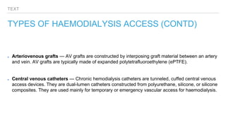
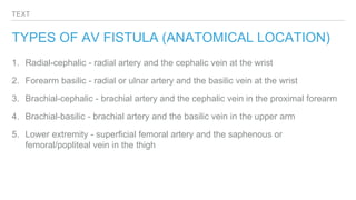
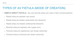
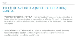
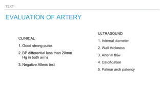
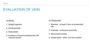
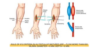
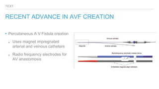
TEXT
NEED FOR AV FISTULA
‣ End stage renal failure needs haemodialysis. For regular haemodialysis, well perfused
venous access is essential. AV fistula is created at different levels to achieve this
hyperdynamic circulation so that regular venous access will not be a problem.
‣ AV fistula creation requires adequate arterial and venous anatomy to support its creation,
and a sufficient time interval to allow the AV fistula to mature prior to its use.
TEXT
NKF Guidelines for AVF creation
‣ Creatinine more than 4mg/dl
‣ Creatinine clearance less than 25ml/min
‣ Within 1 yr of anticipated need
TYPES OF HAEMODIALYSIS ACCESS
Two types of permanent vascular access are available: the arteriovenous (AV) fistula and the AV
graft
Arteriovenous fistulas — An AV fistula is a deliberate connection between a native artery and
vein and is typically constructed with an end-to-side, vein-to-artery anastomosis. The most
commonly used AV fistulas are created by anastomosing the radial artery to the cephalic vein
(radiocephalic fistula) or by anastomosing the brachial artery to the cephalic vein (brachiocephalic
fistula).
TEXT
TYPES OF HAEMODIALYSIS ACCESS (CONTD)
Arteriovenous grafts — AV grafts are constructed by interposing graft material between an artery
and vein. AV grafts are typically made of expanded polytetrafluoroethylene (ePTFE).
Central venous catheters — Chronic hemodialysis catheters are tunneled, cuffed central venous
access devices. They are dual-lumen catheters constructed from polyurethane, silicone, or silicone
composites. They are used mainly for temporary or emergency vascular access for haemodialysis.
TEXT
PRINCIPLES FOR FISTULA CREATION
▸The AV fistula must be accessible with the patient in a comfortable sitting position. The AV fistula must be able to be reliably
cannulated repeatedly
▸Non dominant arm should be used first for fistula creation
▸Fistula should initially be done as distally as possible
▸One should avoid vein punctures/injections on the desired arm at least 3 months prior to fistula creation
▸In the forearm, the AV fistula should be on the volar surface. In the upper arm, the AV fistula should be on the anterior or
lateral surface
▸The AV fistula should be within 5 to 6 mm of the skin surface
▸A relatively straight segment 8 to 10 cm long needs to be available for cannulation
▸Blood flow must be adequate to support the dialysis prescription, generally at least 500 to 700 cc/min. Donor artery should
have an adequate pulsation, volume and devoid of calcification to make successful fistula.
TEXT
TYPES OF AV FISTULA (ANATOMICAL LOCATION)
1. Radial-cephalic - radial artery and the cephalic vein at the wrist
2. Forearm basilic - radial or ulnar artery and the basilic vein at the wrist
3. Brachial-cephalic - brachial artery and the cephalic vein in the proximal forearm
4. Brachial-basilic - brachial artery and the basilic vein in the upper arm
5. Lower extremity - superficial femoral artery and the saphenous or
femoral/popliteal vein in the thigh
TEXT
TYPES OF AV FISTULA (MODE OF CREATION)
‣ SIMPLE DIRECT FISTULA - the vein and the artery are used in their normal positions
Radial artery-to-cephalic vein (wrist)
Radial artery-to-median antecubital vein (forearm)
Radial artery–to-median antebrachial (forearm)
Brachial artery-to-cephalic vein (upper arm)
Femoral artery-to–saphenous vein (lower extremity)
Femoral artery to femoral vein (lower extremity)
TEXT
TYPES OF AV FISTULA (MODE OF CREATION)
CONTD..
‣ VEIN TRANSPOSITION FISTULA - vein is moved or transposed to a position that is
better suited for the construction or cannulation of a fistula. Although the downstream
or proximal end of the vein is left intact, the tributaries to the distal portion are divided
and ligated.
‣ VEIN TRANSLOCATION FISTULA - a vein is removed from its normal anatomic
location to another location and thus requires the creation of a venovenous
anastomosis and venoarterial anastomosis.
TEXT
PREPARING A PATIENT FOR AVF CREATION
▸CLINICAL EXAMINATION
▸DUPLEX ULTRASOUND - for preoperative arterial and veinous mapping, vein
diameter assessment, flow velocity measurement.
▸TIMING - The minimum time for AV fistula maturation is one month, but a lead
time of 6 to 12 months is recommended since intervention may be required to
facilitate maturation of AV fistulas.
▸SITE - distal part of non-dominant hand is the most preferred location. AV grafts
to be only considered when viable sites for fistula creation not available.
TEXT
EVALUATION OF ARTERY
CLINICAL
1. Good strong pulse
2. BP differential less than 20mm
Hg in both arms
3. Negative Allens test
ULTRASOUND
1. Internal diameter
2. Wall thickness
3. Arterial flow
4. Calcification
5. Palmar arch patency
TEXT
EVALUATION OF VEIN
CLINICAL
1. Straight segment
2. Compressibility
3. Distensibility
4. Evidence of frequent phlebotomies OR
vascular access
ULTRASOUND
1. Diameter - at least 2.5mm at anastomotic
site
2. Drainage - continuous proximally
3. Stenosis/thrombosis
4. Vessel depth - within 1cm from surface
TEXT
SURGICAL STRATEGY
▸Local or Regional anaesthesia
▸Preferably with surgical loupes
▸Longitudinal or transverse incision located between target artery
and vein, raising of skin flaps, dissection of artery and vein
along with ligation of small tributary vessels.
▸On table assessment of veinous patency, straightening and
superficialization if needed
▸Heparinisation
▸End-to-end or end-to-side anastomosis of vein to artery
▸Post op on-table assessment of fistula patency/success
Castroveijo needle driver
Vessel loops, forceps, bull-dog clamps
RULE OF 6’S CRITERIA IN AV FISTULA IF MATURED ARE – FLOW MORE THAN 600
ML/MIN; DIAMETER > 6 MM; DEPTH < 6 MM.
TEXT
COMPLICATIONS OF FISTULA PLACEMENT
▸Local complications
▸Bleeding
▸Veinous hypertension - leading to extremity swelling, ischaemic changes of the skin, access dysfunction
▸ Aneurysm or pseudoaneurysm - leading to rupture, bleeding, infection, erosion of overlying skin, canulation
difficulty.
▸Infection
▸Neuropathy - amyloid deposition leading to Carpal Tunnel Syndrome
▸Primary failure - failure to mature
True aneurysm - abnormal focal dilatation of blood vessel that contains all layers of blood vessel wall.
Pseudoaneurysm - focal disruption of vessel wall with blood collected outside , which is contained by fibrous tissue.
TEXT
COMPLICATIONS OF FISTULA PLACEMENT
▸Ischaemia and other systemic problems
▸Dialysis access steal syndrome and Ischaemic Monomelic Neuropathy
▸Coronary steal - in patients with ipsilateral internal mammary as CABG graft
▸Heart failure
▸Pulmonary hypertension
▸Rare complication
▸Malignancy (angiosarcoma)
TEXT
CARE OF THE FISTULA
▸Keep fistula arm raised on a pillow to reduce swelling
▸Isometric arm exercises as soon a surgical pain subsides
▸Do not allow BP measurement, blood drawing or iv injections in fistula arm
▸Self examination regularly for thrill
▸Adopt hygenic and proper canulation techniques
TEXT
RECENT ADVANCE IN AVF CREATION
▸Percutaneous A V Fistula creation
Uses magnet impregnated
arterial and veinous catheters
Radio frequency electrodes for
AV anastomosis
Thank you

A V FISTULA .pptx

  • 2.
    TEXT NEED FOR AVFISTULA ‣ End stage renal failure needs haemodialysis. For regular haemodialysis, well perfused venous access is essential. AV fistula is created at different levels to achieve this hyperdynamic circulation so that regular venous access will not be a problem. ‣ AV fistula creation requires adequate arterial and venous anatomy to support its creation, and a sufficient time interval to allow the AV fistula to mature prior to its use.
  • 3.
    TEXT NKF Guidelines forAVF creation ‣ Creatinine more than 4mg/dl ‣ Creatinine clearance less than 25ml/min ‣ Within 1 yr of anticipated need
  • 4.
    TYPES OF HAEMODIALYSISACCESS Two types of permanent vascular access are available: the arteriovenous (AV) fistula and the AV graft Arteriovenous fistulas — An AV fistula is a deliberate connection between a native artery and vein and is typically constructed with an end-to-side, vein-to-artery anastomosis. The most commonly used AV fistulas are created by anastomosing the radial artery to the cephalic vein (radiocephalic fistula) or by anastomosing the brachial artery to the cephalic vein (brachiocephalic fistula).
  • 5.
    TEXT TYPES OF HAEMODIALYSISACCESS (CONTD) Arteriovenous grafts — AV grafts are constructed by interposing graft material between an artery and vein. AV grafts are typically made of expanded polytetrafluoroethylene (ePTFE). Central venous catheters — Chronic hemodialysis catheters are tunneled, cuffed central venous access devices. They are dual-lumen catheters constructed from polyurethane, silicone, or silicone composites. They are used mainly for temporary or emergency vascular access for haemodialysis.
  • 6.
    TEXT PRINCIPLES FOR FISTULACREATION ▸The AV fistula must be accessible with the patient in a comfortable sitting position. The AV fistula must be able to be reliably cannulated repeatedly ▸Non dominant arm should be used first for fistula creation ▸Fistula should initially be done as distally as possible ▸One should avoid vein punctures/injections on the desired arm at least 3 months prior to fistula creation ▸In the forearm, the AV fistula should be on the volar surface. In the upper arm, the AV fistula should be on the anterior or lateral surface ▸The AV fistula should be within 5 to 6 mm of the skin surface ▸A relatively straight segment 8 to 10 cm long needs to be available for cannulation ▸Blood flow must be adequate to support the dialysis prescription, generally at least 500 to 700 cc/min. Donor artery should have an adequate pulsation, volume and devoid of calcification to make successful fistula.
  • 7.
    TEXT TYPES OF AVFISTULA (ANATOMICAL LOCATION) 1. Radial-cephalic - radial artery and the cephalic vein at the wrist 2. Forearm basilic - radial or ulnar artery and the basilic vein at the wrist 3. Brachial-cephalic - brachial artery and the cephalic vein in the proximal forearm 4. Brachial-basilic - brachial artery and the basilic vein in the upper arm 5. Lower extremity - superficial femoral artery and the saphenous or femoral/popliteal vein in the thigh
  • 8.
    TEXT TYPES OF AVFISTULA (MODE OF CREATION) ‣ SIMPLE DIRECT FISTULA - the vein and the artery are used in their normal positions Radial artery-to-cephalic vein (wrist) Radial artery-to-median antecubital vein (forearm) Radial artery–to-median antebrachial (forearm) Brachial artery-to-cephalic vein (upper arm) Femoral artery-to–saphenous vein (lower extremity) Femoral artery to femoral vein (lower extremity)
  • 9.
    TEXT TYPES OF AVFISTULA (MODE OF CREATION) CONTD.. ‣ VEIN TRANSPOSITION FISTULA - vein is moved or transposed to a position that is better suited for the construction or cannulation of a fistula. Although the downstream or proximal end of the vein is left intact, the tributaries to the distal portion are divided and ligated. ‣ VEIN TRANSLOCATION FISTULA - a vein is removed from its normal anatomic location to another location and thus requires the creation of a venovenous anastomosis and venoarterial anastomosis.
  • 10.
    TEXT PREPARING A PATIENTFOR AVF CREATION ▸CLINICAL EXAMINATION ▸DUPLEX ULTRASOUND - for preoperative arterial and veinous mapping, vein diameter assessment, flow velocity measurement. ▸TIMING - The minimum time for AV fistula maturation is one month, but a lead time of 6 to 12 months is recommended since intervention may be required to facilitate maturation of AV fistulas. ▸SITE - distal part of non-dominant hand is the most preferred location. AV grafts to be only considered when viable sites for fistula creation not available.
  • 11.
    TEXT EVALUATION OF ARTERY CLINICAL 1.Good strong pulse 2. BP differential less than 20mm Hg in both arms 3. Negative Allens test ULTRASOUND 1. Internal diameter 2. Wall thickness 3. Arterial flow 4. Calcification 5. Palmar arch patency
  • 12.
    TEXT EVALUATION OF VEIN CLINICAL 1.Straight segment 2. Compressibility 3. Distensibility 4. Evidence of frequent phlebotomies OR vascular access ULTRASOUND 1. Diameter - at least 2.5mm at anastomotic site 2. Drainage - continuous proximally 3. Stenosis/thrombosis 4. Vessel depth - within 1cm from surface
  • 13.
    TEXT SURGICAL STRATEGY ▸Local orRegional anaesthesia ▸Preferably with surgical loupes ▸Longitudinal or transverse incision located between target artery and vein, raising of skin flaps, dissection of artery and vein along with ligation of small tributary vessels. ▸On table assessment of veinous patency, straightening and superficialization if needed ▸Heparinisation ▸End-to-end or end-to-side anastomosis of vein to artery ▸Post op on-table assessment of fistula patency/success Castroveijo needle driver Vessel loops, forceps, bull-dog clamps
  • 14.
    RULE OF 6’SCRITERIA IN AV FISTULA IF MATURED ARE – FLOW MORE THAN 600 ML/MIN; DIAMETER > 6 MM; DEPTH < 6 MM.
  • 15.
    TEXT COMPLICATIONS OF FISTULAPLACEMENT ▸Local complications ▸Bleeding ▸Veinous hypertension - leading to extremity swelling, ischaemic changes of the skin, access dysfunction ▸ Aneurysm or pseudoaneurysm - leading to rupture, bleeding, infection, erosion of overlying skin, canulation difficulty. ▸Infection ▸Neuropathy - amyloid deposition leading to Carpal Tunnel Syndrome ▸Primary failure - failure to mature True aneurysm - abnormal focal dilatation of blood vessel that contains all layers of blood vessel wall. Pseudoaneurysm - focal disruption of vessel wall with blood collected outside , which is contained by fibrous tissue.
  • 16.
    TEXT COMPLICATIONS OF FISTULAPLACEMENT ▸Ischaemia and other systemic problems ▸Dialysis access steal syndrome and Ischaemic Monomelic Neuropathy ▸Coronary steal - in patients with ipsilateral internal mammary as CABG graft ▸Heart failure ▸Pulmonary hypertension ▸Rare complication ▸Malignancy (angiosarcoma)
  • 17.
    TEXT CARE OF THEFISTULA ▸Keep fistula arm raised on a pillow to reduce swelling ▸Isometric arm exercises as soon a surgical pain subsides ▸Do not allow BP measurement, blood drawing or iv injections in fistula arm ▸Self examination regularly for thrill ▸Adopt hygenic and proper canulation techniques
  • 18.
    TEXT RECENT ADVANCE INAVF CREATION ▸Percutaneous A V Fistula creation Uses magnet impregnated arterial and veinous catheters Radio frequency electrodes for AV anastomosis
  • 19.