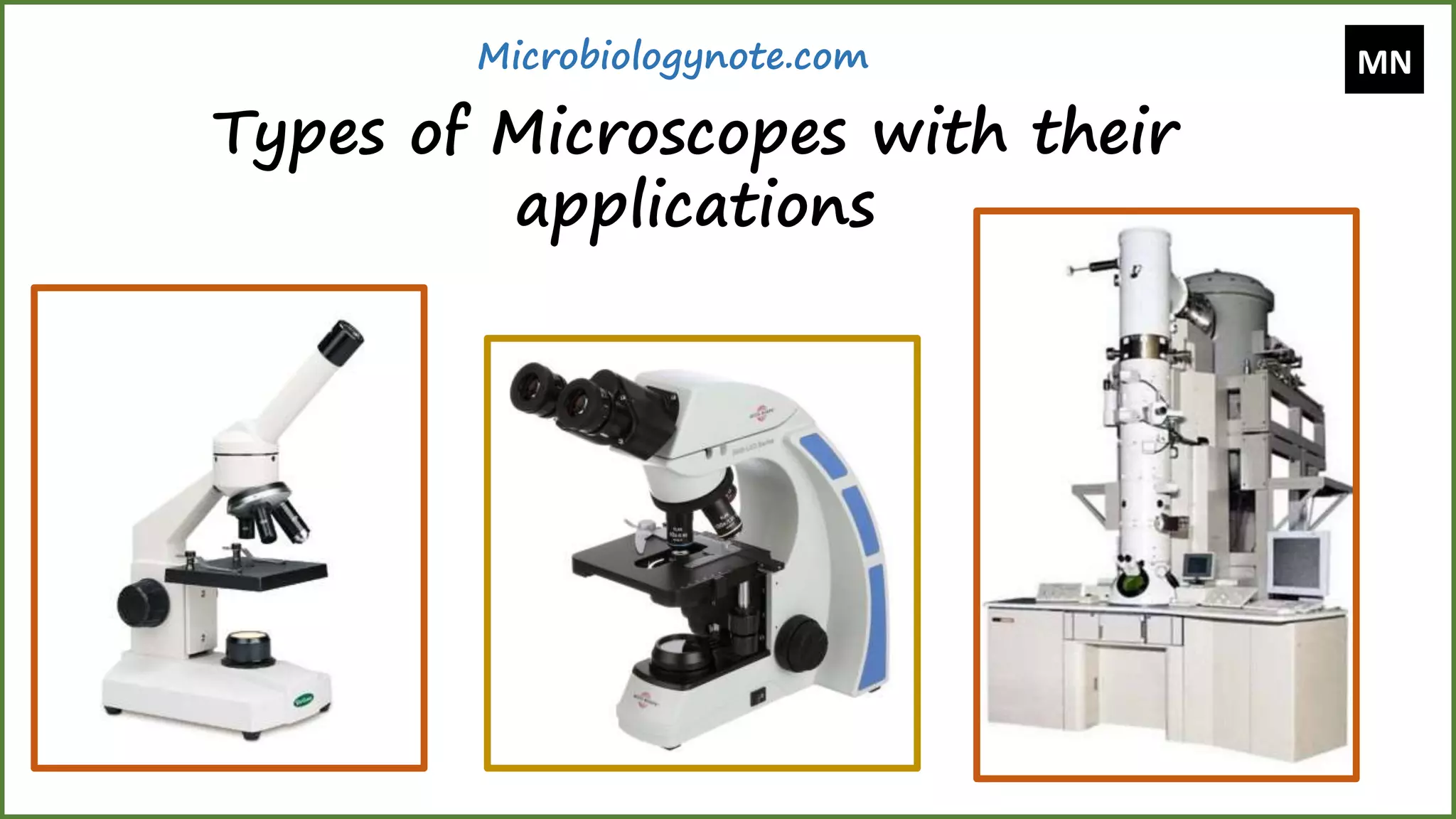
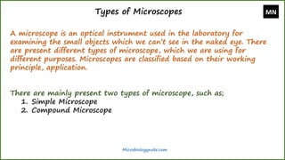
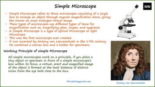
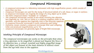
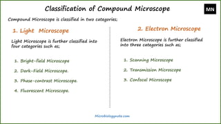
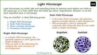
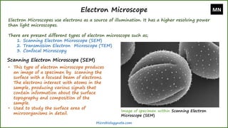
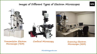
Types of Microscopes with their applications - Microbiologynote.com
https://microbiologynote.com/types-of-microscopes-with-their-applications/
Youtube Lecture Video:
https://www.youtube.com/watch?v=nuJZtXohFFQ&ab_channel=MicrobiologyNote
2. Types of Microscopes
A microscope is an optical instrument used in the laboratory for
examining the small objects which we can’t see in the naked eye. There
are present different types of microscope, which we are using for
different purposes. Microscopes are classified based on their working
principle, application.
There are mainly present two types of microscope, such as;
1. Simple Microscope
2. Compound Microscope
MN
Microbiologynote.com
3. Simple Microscope
• Simple Microscope refers to those microscopes consisting of a single
lens to enlarge an object through angular magnification alone, giving
the viewer an erect enlarged virtual image.
• These types of microscopes use different types of lense for
magnification such as; magnifying glass, loupes, and eyepieces.
• A Simple Microscope is a type of optical Microscope or light
Microscope.
• This was the first microscope ever created.
• It was invented by Antony van Leeuwenhoek in the 17th century.
He combined a convex lens and a holder for specimens.
Working Principle of simple Microscope
All simple microscopes work on a principle, if you place a
tiny object or specimen in front of a simple microscope’s
lens within its focus, a virtual, erect and magnified image
of the object is formed at the least distance of distinct
vision from the eye held close to the lens.
MN
Microbiologynote.com
Antony van leeuwenhoek
4. Compound Microscope
• A compound microscope is a laboratory instrument with high magnification power, which consists of
more than one lens.
• Compound Microscopes are used for the study of structural details of a cell, tissue, or organ in sections.
• A compound microscope can magnify the image of a tiny object up to 1000.
• The term compound means “multiple” or “complex”.
Working Principle of Compound Microscope
The compound microscopes are works on the principle that when
a tiny specimen to be magnified is placed just beyond the focus of
its objective lens, a virtual, inverted and highly magnified image
of the object are formed at the least distance of distinct vision
from the eye held close to the eyepiece.
• The compound microscope consists of two lenses including the objective
lens (typically 4x, 10x, 40x or 100x) in a rotating nosepiece closer to the
specimen, and the eyepiece lens (typically 10x) in the binocular eyepieces.
• A compound binocular microscope is more commonly used today.
• Zacharias Jansen created a compound microscope that used collapsing
tubes and produced magnifications up to 9X.
• compound microscopes are generally types of bright field microscope.
MN
Microbiologynote.com
5. Classification of Compound Microscope
Compound Microscope is classified in two categories;
1. Light Microscope 2. Electron Microscope
Light Microscope is further classified into
four categories such as;
1. Bright-field Microscope
2. Dark-Field Microscope.
3. Phase-contrast Microscope.
4. Fluorescent Microscope.
Electron Microscope is further classified
into three categories such as;
1. Scanning Microscope
2. Transmission Microscope
3. Confocal Microscope
MN
Microbiologynote.com
6. Light Microscope
Light Microscopes use visible light and magnifying lenses to examine small objects not visible to
the naked eye, or in finer detail than the naked eye allows. Magnification, however, is not the
most important issue in microscopy.
They are classified in these following groups;
1. Bright-field Microscope
2. Dark-Field Microscope
3. Phase-contrast Microscope
4. Fluorescent Microscope
Bright-field Microscope
• In a bright-field microscope, the
specimen appears as dark against the
bright background.
• They are used in the laboratory for
studying the outer structure of
microorganisms.
Dark-Field Microscope
• In the dark-field microscope, the specimen
appears as bright against a dark background.
• This microscope is used to distinguish
unstained, thin living cells that are not visible
under a simple microscope.
MN
Microbiologynote.com
7. Phase-contrast Microscope
• Some unpigmented living cells are not visible in the light
microscope because it can’t create differences in contrast
between cells and water. Only Phase-contrast Microscope
can create contrast difference between cell and water that
is why these cells only visible in Phase-contrast Microscope
• Use for studying the shape and motility of microorganisms.
Fluorescent Microscope
• In this type, the specimen is stained with fluorescent dyes
and then exposed to ultraviolet rays (UV). Fluorescent
dyes will absorb low wavelength light and become excited
as a result they will release a high wavelength light. Using
this mechanism Fluorescent Microscopes work.
• Use in medical laboratories for the identification of
pathogens. Also used for localization of specific proteins.
Microbiologynote.com
Image of specimen within Phase-
contrast Microscope
Image of specimen within
Fluorescent Microscope
8. Electron Microscope
Electron Microscopes use electrons as a source of illumination. It has a higher resolving power
than light microscopes.
There are present different types of electron microscope such as;
1. Scanning Electron Microscope (SEM)
2. Transmission Electron Microscope (TEM)
3. Confocal Microscopy
Scanning Electron Microscope (SEM)
• This type of electron microscope produces
an image of a specimen by scanning the
surface with a focused beam of electrons.
The electrons interact with atoms in the
sample, producing various signals that
contain information about the surface
topography and composition of the
sample.
• Used to study the surface area of
microorganisms in detail.
MN
Microbiologynote.com
Image of specimen within Scanning Electron
Microscope (SEM)
9. Transmission Electron Microscope (TEM)
• In this microscope, the electron beam is passed through a
specimen to form an image. The specimen used for TEM
should be 20 to 100 nm thick.
• It used to study the internal structure of a specimen.
Confocal Microscopy
• Confocal Microscope also known as confocal laser scanning
microscopy (CLSM) or laser confocal scanning microscopy
(LCSM).
• This is an optical imaging technique to increase the
optical resolution and contrast of a micrograph by means
of using a spatial pinhole to block out-of-focus light in
image formation.
• This microscope Capturing multiple two-dimensional
images at different depths in a sample enables the
reconstruction of three-dimensional structures (a process
known as optical sectioning) within an object.
• Confocal Microscopy is used extensively in the scientific
and industrial communities and typical applications are
in life sciences, semiconductor inspection, and materials
science.
MN
Microbiologynote.com