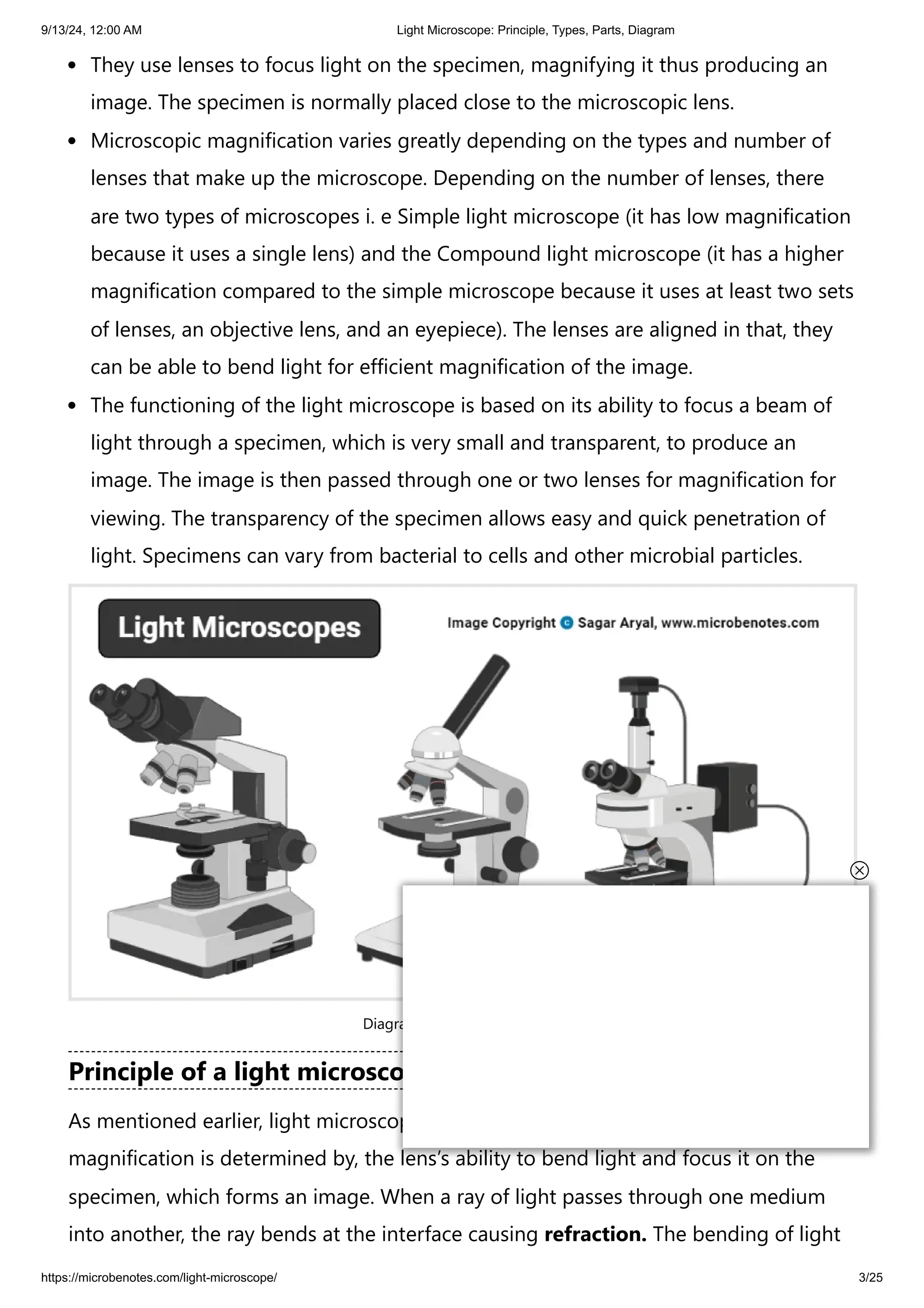
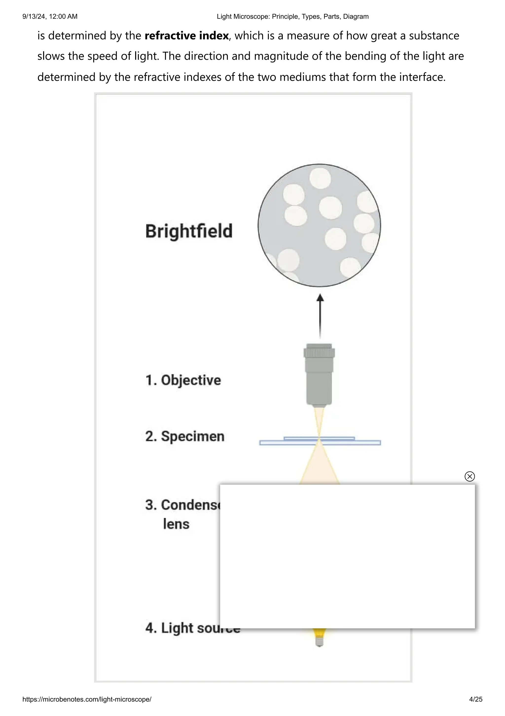
9/13/24, 12:00 AM Light Microscope: Principle, Types, Parts, Diagram
https://microbenotes.com/light-microscope/ 1/25
Home » Microscopy
Light Microscope: Principle, Types, Parts, Diagram
April 13, 2024 by Faith Mokobi
Edited By: Sagar Aryal
The evolution of the Microbiology field put to perspective the need to identify, view,
observe and understand microorganisms, including their structural morphologies
and mechanisms. Microbiology’s scope is to study organisms and minute agents that
can only be examined and observed with a microscope.
Although scientifically, the first simple microscope was discovered by two Dutch
scientists, Zaccharias Janssen and his father, Hans who made spectacles, were the first
to experiment with their lenses by combining lenses in a tube and observed that the
objects that were nearby, appeared closer and larger. Despite not being included as a
scientific discovery, this act paved the way for scientific evolution.
From the History of Microbiology, Antony Van Lewnehoueek an amateur
Microbiologist made the first simple microscope, that enabled him to observe the
presence of tiny living organisms in pond water that appeared like dots. His simple
microscope was made up of a double convex glass lens that was held between two
silver plates.
The application of microscopy in Microbiology enhanced the visualization of cells
and microorganisms by magnifying their images to make them larger.
Microbe Notes
Menu
The light microscope is also known as an optical microscope.
Table of Contents
What is a light microscope?
Principle of a light microscope (optical microscope)
Search Notes
9/13/24, 12:00 AM Light Microscope: Principle, Types, Parts, Diagram
https://microbenotes.com/light-microscope/ 2/25
Interesting Science Videos
What is a light microscope?
A light microscope is a biology laboratory instrument or tool, that uses visible
light to detect and magnify very small objects and enlarge them.
Types of light microscopes (optical microscope)
Brightfield Light Microscope (Compound light microscope)
Parts of a bright-field microscope (Compound light microscope)
Magnification by Bright field Microscope (Compound light microscope)
Applications of the Bright Field Light Microscope (Compound light microscope)
Light Microscope Free Worksheet
Phase Contrast Microscope
Parts of the Phase Contrast Microscope
The functioning of the Phase Contrast microscope
Applications of Phase-Contrast Microscope
Dark-Field Light Microscope
Applications of the Dark Field Microscope
The Fluorescent Microscope
Applications of the Fluorescent Microscope
References
9/13/24, 12:00 AM Light Microscope: Principle, Types, Parts, Diagram
https://microbenotes.com/light-microscope/ 3/25
They use lenses to focus light on the specimen, magnifying it thus producing an
image. The specimen is normally placed close to the microscopic lens.
Microscopic magnification varies greatly depending on the types and number of
lenses that make up the microscope. Depending on the number of lenses, there
are two types of microscopes i. e Simple light microscope (it has low magnification
because it uses a single lens) and the Compound light microscope (it has a higher
magnification compared to the simple microscope because it uses at least two sets
of lenses, an objective lens, and an eyepiece). The lenses are aligned in that, they
can be able to bend light for efficient magnification of the image.
The functioning of the light microscope is based on its ability to focus a beam of
light through a specimen, which is very small and transparent, to produce an
image. The image is then passed through one or two lenses for magnification for
viewing. The transparency of the specimen allows easy and quick penetration of
light. Specimens can vary from bacterial to cells and other microbial particles.
Diagram of Light Microscopes
Principle of a light microscope (optical microscope)
As mentioned earlier, light microscopes visualize an image by using a glass lens, and
magnification is determined by, the lens’s ability to bend light and focus it on the
specimen, which forms an image. When a ray of light passes through one medium
into another, the ray bends at the interface causing refraction. The bending of light
9/13/24, 12:00 AM Light Microscope: Principle, Types, Parts, Diagram
https://microbenotes.com/light-microscope/ 4/25
is determined by the refractive index, which is a measure of how great a substance
slows the speed of light. The direction and magnitude of the bending of the light are
determined by the refractive indexes of the two mediums that form the interface.
9/13/24, 12:00 AM Light Microscope: Principle, Types, Parts, Diagram
https://microbenotes.com/light-microscope/ 5/25
Principle of a light microscope
A medium with a lower refractive index such as glass to air normally speeds up the
light penetration and makes light bend away from the normal and when light is
passed through a medium with a greater refractive index such as air to glass, it
normally slows down and bends towards the normal, perpendicularly to the surface.
If an object is put between these two mediums i.e between water and air, in this case,
a prism, the prism will bend the light at an angle. This is how the microscopic lenses
work, they bend the light at an angle. The lens (convex) on receiving the light rays,
focuses the rays at a specific point known as the focal point (F-point). The measure
of distance from the center of the lens and the focal point is known as the focal
length.
A microscope uses lenses whose strength is predetermined, in that, the strength of a
lens is directly related to the focal length i.e short focal length magnifies objects
more than lenses with a long focal length.
Microscopy works strictly with a factor of resolution whereby resolution is the ability
of a lens to be able to differentiate small objects that are closely packed together.
The resolution of a light microscope is determined by a numerical aperture of its
lens system and by the wavelength of the light it employs; a numerical aperture is a
definition of the light wavelengths produced when the specimen is illuminated.
A minimum distance (d) between two objects that distinguishes them to be two
separate entities, determined by the wavelengths of the light can be calculated by an
Abbe equation using the wavelength of the light that illuminated the specimen
(Lambda, λ) and the numerical aperture (NA, n sin Ɵ) i.e. d=0.5 λ/n sin Ɵ
9/13/24, 12:00 AM Light Microscope: Principle, Types, Parts, Diagram
https://microbenotes.com/light-microscope/ 6/25
Figure: Labeled Diagram of a Light Microscope.
Types of light microscopes (optical microscope)
With the evolved field of Microbiology, the microscopes
used to view specimens are both simple and compound light microscopes, all using
lenses. The difference is simple light microscopes use a single lens for magnification
while compound lenses use two or more lenses for magnifications. This means, that
a series of lenses are placed in an order such that, one lens magnifies the image
further than the initial lens.
9/13/24, 12:00 AM Light Microscope: Principle, Types, Parts, Diagram
https://microbenotes.com/light-microscope/ 7/25
The modern types of Light Microscopes include:
1. Bright field Light Microscope
2. Phase Contrast Light Microscope
3. Dark-Field Light Microscope
4. Fluorescence Light Microscope
Brightfield Light Microscope (Compound light microscope)
This is the most basic optical Microscope used in microbiology laboratories which
produces a dark image against a bright background. Made up of two lenses, it is
widely used to view plant and animal cell organelles including some parasites such as
Paramecium after staining with basic stains. Its functionality is based on being able to
provide a high-resolution image, which highly depends on the proper use of the
microscope. This means that an adequate amount of light will enable sufficient
focusing of the image, to produce a quality image. It is also known as a compound
light microscope.
Parts of a bright-field microscope (Compound light microscope)
9/13/24, 12:00 AM Light Microscope: Principle, Types, Parts, Diagram
https://microbenotes.com/light-microscope/ 8/25
It is composed of:
Two lenses which include the objective lens and the eyepiece or ocular lens.
Objective lens is made up of six or more glasses, which make the image clear from
the object
The condenser is mounted below the stage which focuses a beam of light onto the
specimen. It can be fixed or movable, to adjust the quality of light, but this entirely
depends on the microscope.
They are held together by a sturdy metallic curved back used as an arm and a
stand at the bottom, known as the base, of the microscope. The arm and the base
hold all the parts of the microscope.
The stage where the specimen is placed, allowing movement of the specimen
around for better viewing with the flexible knobs and it is where the light is
focused on.
9/13/24, 12:00 AM Light Microscope: Principle, Types, Parts, Diagram
https://microbenotes.com/light-microscope/ 9/25
Two focusing knobs i.e the fine adjustment knob and the coarse adjustment knob,
found on the microscopes’ arm, which can move the stage or the nosepiece to
focus on the image. the sharpen the image clarity.
It has a light illuminator or a mirror found at the base or on the microbes of the
nosepiece.
The nosepiece has about three to five objective lenses with different magnifying
power. It can move round to any position depending on the objective lens to focus
on the image.
An aperture diaphragm also is known as the contrast, which controls the diameter
of the beam of light that passes through the condenser, in that, when the
condenser is almost closed, the light comes through to the center of the
condenser creating high contrast. But when the condenser is widely open, the
image is very bright with very low contrast.
Magnification by Bright field Microscope (Compound light
microscope)
During visualization, the objective lens remains parfocal which means, when the
objective lens is changed, the image still remains in focus. The objective lens plays a
major role in focusing the image on the condenser forming an enlarged clear image
within the microscope, which is then further magnified by the eyepiece to a primary
image.
What is seen in the microscope as an enlarged clear image of the specimen is known
as the virtual image. To calculate the magnification, multiply the objective and
eyepiece objective magnification together. The magnification is standard, i.e not too
high nor too low, and therefore depending on the magnification power of the lenses,
it will range between 40X and 100oX.
Calculation of magnification = Magnification of objective lens/magnification of the
eyepiece lens
The objective lens plays a vital role in not only enlarging the image but also making it
clear for viewing, a feature known as resolution. Resolution according to Prescott, is
the ability of a lens to separate or distinguish between small objects closely linked
together.
9/13/24, 12:00 AM Light Microscope: Principle, Types, Parts, Diagram
https://microbenotes.com/light-microscope/ 10/25
Whereas the eyepiece magnifies the image at the end of the viewing, its
magnification range is lower than that of the objective lens at 8X-12X (10X standard)
and that of the objective lens at 40X-100X, magnification, and resolution of the
microscope is highly dependant on the objective lens.
Applications of the Bright Field Light Microscope (Compound light
microscope)
Vastly used in Microbiology, this microscope is used to view fixed and live
specimens, that have been stained with basic stains. This gives contrast for easy
visibility under the microscope. Therefore it can be used to identify basic bacteria
cells and parasitic protozoans such as Paramecium.
Light Microscope Free Worksheet
Answer Key
9/13/24, 12:00 AM Light Microscope: Principle, Types, Parts, Diagram
https://microbenotes.com/light-microscope/ 11/25
Phase Contrast Microscope
This is a type of optical microscope whereby small light deviations known as phase
shifts occur during light penetration into the unstained specimen. These phase
shifts are converted into the image to mean, when light passes through the
opaque specimen, the phase shifts brighten the specimen forming an illuminated
(bright) image in the background.
The phase-contrast microscope produces high contrast images when using a
transparent specimen more so those of microbial cultures, thin tissue fragments,
cell tissues, and subcellular particles.
The principle behind the working of the phase-contrast microscope is the use of an
optical method to transform a specimen into an amplitude image, that’s viewed by
the eyepiece of the microscope.
9/13/24, 12:00 AM Light Microscope: Principle, Types, Parts, Diagram
https://microbenotes.com/light-microscope/ 12/25
The PCM can be used to view unstained cells also known as the phase objects,
which means that the morphology of the cell is maintained and the cells can be
observed in their natural state, in high contrast and efficient clarity. This is because
if the specimens are stained and fixed, they kill most cells, a characteristic that is
uniquely undone by the brightfield light microscope.
The shifts that occur during light penetration, become converted to changes in
amplitude which causes the image contrast.
Coupled with contrast-enhancing elements such as fluorescence, they produce
better visuals of the specimens’ image.
Parts of the Phase Contrast Microscope
The instrumentation of the Phase Contrast Microscope is based on its light pathways
from receiving the source of light to the visualization of the image.
Therefore its sequentially made up of:
Light source (Mercury arc lamp)
Collective lens
Aperture
Condenser
Condenser annular
Specimen
Objective
Phase plate
Deflected light
Phase ring
9/13/24, 12:00 AM Light Microscope: Principle, Types, Parts, Diagram
https://microbenotes.com/light-microscope/ 13/25
Phase Contrast Microscopy
The functioning of the Phase Contrast microscope
The change is caused by the deviated scattered (Deflected) light and the
undeviated light that reaches the specimen which is absorbed, create at a certain
wavelength, producing color. The difference created by the scattered light and that
of the absorbed light is known as amplitude variations. These amplitude
variations are sensitive to allowing visualization by photographic equipment like
the Phase Contrast Microscope, hence seen by the human eye.
The Condenser of the phase-contrast microscope has an opaque disk that is
known as an annular ring, with a transparent ring that produces a cone of light,
that passes through a specimen. Due to light variations some light bends at the
specimen, caused by variations in light density, forming an image at the objective
lens. The undeviated light will strike the phase ring on the phase plate and the
deviated light will miss the phase ring passing through the phase plate directly,
this forms an image.
The Phase-Contrast Microscope is designed with objective lenses that have the ability
to perform multiple functions when combined with contrast-enhancing techniques,
for example, fluorescence. The objective lenses are located in the internal phase plate
with variation in the light absorption and phase displacement i.e undiffraction,
9/13/24, 12:00 AM Light Microscope: Principle, Types, Parts, Diagram
https://microbenotes.com/light-microscope/ 14/25
creating a wide spectrum for contrasting the specimen and forming a strong contrast
in the background.
Applications of Phase-Contrast Microscope
Determine morphologies of living cells such as plant and animal cells
Studying microbial motility and structures of locomotion
To detect certain microbial elements such as the bacterial endospores
Dark-Field Light Microscope
This is a specialized type of bright field light microscope that has several similarities
to the Phase-Contrast Microscope. To make a dark field Microscope, place a
darkfield stop underneath and a condenser lens which produces a hollow cone beam
of light that enters the objective only, from the specimen (Prescott, pg 22).
Darkfield Microscope
This technique is used to visualize living unstained cells. This is affected by the way
illumination is done on the specimen in that, when a hollow cone beam of light is
transmitted to the specimen, deviated light (unreflected/unrefracted) rays do not
pass through the objectives but the undeviated (reflected/refracted) light passes
through the objectives to the specimen forming an image.
This makes the surrounding field of the specimen appear black while the specimen
will appear illuminated. This is enabled by the dark background this the name, dark-
9/13/24, 12:00 AM Light Microscope: Principle, Types, Parts, Diagram
https://microbenotes.com/light-microscope/ 15/25
field Microscopy.
Applications of the Dark Field Microscope
It is used to visualize the internal organs of larger cells such as the eukaryotic cells
Identification of bacterial cells with distinctive shapes such as Treponema pallidum,
a causative agent of syphilis.
The Fluorescent Microscope
The above-discussed microscopes will normally produce images after a light has
been transmitted and passed through the specimen.
In the case of the fluorescent Microscope, the specimen emits light. How? By adding
a dye molecule to the specimen. This dye molecule will normally become excited
when it absorbs light energy, hence it releases any trapped energy as light. The light
energy that is released by the excited molecule has a long wavelength compared to
its radiating light. The dye molecule is normally a fluorochrome, that fluoresces when
exposed to the light of a certain specific wavelength. The image formed is a
fluorochrome-labeled image from the emitted light
The principle behind this working mechanism is that the fluorescent microscope will
expose the specimen to ultra or violet or blue light, which forms an image of the
specimen that is emanated by the fluorescent light. They have a mercury vapor arc
lamp that produces an intense beam of light that passes through an exciter filter. The
exciter filter functions to transmit a specific wavelength to the fluorochrome stained
specimen, producing the fluorochrome-labeled image, at the objective.
After the objective, there is a barrier filter that functions primarily to remove any
ultraviolet radiation that may be harmful to the viewer’s light, thus reducing the
contrast of the image.
9/13/24, 12:00 AM Light Microscope: Principle, Types, Parts, Diagram
https://microbenotes.com/light-microscope/ 16/25
Fluorescence Microscopy
Applications of the Fluorescent Microscope
Used in the visualization of bacterial agents such as Mycobacterium tuberculosis.
Used to identify specific antibodies produced against bacterial antigens/pathogens
in immunofluorescence techniques by labeling the antibodies with fluorochromes.
Used in ecological studies to identify and observe microorganisms labeled by the
fluorochromes
It can also be used to differentiate between dead and live bacteria by the color
they emit when treated with special stains
Besides the above-discussed microscopes, there is one not commonly used
microscope known as the Differential Interference Contrast Microscopy. It is very
similar to the phase-contrast microscope whereby the images are formed from the
variations in the light either deviated and or undeviated. The difference is, here two
beams of light are emitted to the specimen and focused by a prism. One beam
passes through the prism to the specimen while another passes through the glass
slide clear area without the specimen. The two beams then combine and interfere
with each other to form an image. It can be used to view cell structures such as
endospores, bacterial cell walls, nuclei, and granules for unstained specimens.
References
9/13/24, 12:00 AM Light Microscope: Principle, Types, Parts, Diagram
https://microbenotes.com/light-microscope/ 17/25
1. Microbiology by Laning M. Prescott, 5th Edition
2. https://science.umd.edu/CBMG/faculty/wolniak/wolniakmicro.html
3. https://en.wikipedia.org/wiki/Bright-field_microscopy
4. https://sciencing.com/calculate-magnification-light-microscope-7558311.html
5. https://www.microscopemaster.com/brightfield-microscopy.html
6. https://en.wikipedia.org/wiki/Phase-contrast_microscopy
7. https://www.microscopyu.com/techniques/phase-contrast/introduction-to-phase-
contrast-microscopy
8. https://www.olympus-lifescience.com/en/microscope-
resource/primer/techniques/phasecontrast/phase/
9. https://laboratoryinfo.com/types-of-microscopes/
About Author
Faith Mokobi
Faith Mokobi is a passionate scientist and graduate student currently pursuing her
Ph.D. in Nanoengineering (Synthetic Biology specialization) from Joint School of
Nanoscience and Nanoengineering, North Carolina A and T State University, North
Carolina, USA. She has a background in Immunology and Microbiology
(MSc./BSc.). With extensive higher education teaching and research experience in
Biomedical studies, metagenomic studies, and drug resistance, Faith is currently
integrating her Biomedical experience in nanotechnology and cancer theranostics.
15 thoughts on “Light Microscope: Principle, Types, Parts, Diagram”
G.Ramya
August 18, 2024 at 1:01 PM
9/13/24, 12:00 AM Light Microscope: Principle, Types, Parts, Diagram
https://microbenotes.com/light-microscope/ 18/25
Thanks to your Information
Reply
John Sini Hanti
August 17, 2024 at 6:49 AM
This is very informative, thanks. But how can I cite this article
Reply
Sagar Aryal
August 17, 2024 at 1:04 PM
You can cite as: “Mokobi, F. (2024, April 15). Light Microscope: Principle,
types, parts, diagram. Retrieved from https://microbenotes.com/light-
microscope/“.
Reply
James
March 7, 2024 at 4:00 PM
This is helpful
Reply
9/13/24, 12:00 AM Light Microscope: Principle, Types, Parts, Diagram
https://microbenotes.com/light-microscope/ 19/25
angel
January 29, 2024 at 8:08 PM
Hi Thank you so much for this article really helpfull.
Reply
Thelma Nachande
May 4, 2023 at 1:13 AM
Thanks for this article it was helpful,continue the good work
Reply
roby
November 15, 2022 at 8:48 AM
thanks that was helpful
Reply
JANAN
November 5, 2022 at 9:30 PM
Thank for the article. It’s has been of great benefits
9/13/24, 12:00 AM Light Microscope: Principle, Types, Parts, Diagram
https://microbenotes.com/light-microscope/ 20/25
Reply
Benson kasenga
July 25, 2022 at 12:27 AM
That’s most informative article who wrote it? He did a great job to us he
helped us in our education
Reply
Moses
October 3, 2021 at 2:03 AM
This is a great article, who wrote it?
Reply
Salahudeen
July 8, 2021 at 2:17 AM
Thoroughly researched article. Helpful.
Reply
9/13/24, 12:00 AM Light Microscope: Principle, Types, Parts, Diagram
https://microbenotes.com/light-microscope/ 21/25
Jerrrline Teah
September 6, 2020 at 5:24 AM
Thanks for this article. it was of great benefit to me.
Reply
sadaf
May 7, 2021 at 12:53 PM
this is one of the best article i found…this is really informative..thnks
Reply
Rehmat Ali Barki
June 30, 2021 at 11:44 PM
Thanks for this article, this is very informative
Wow amazing
Reply
Student microscope
August 21, 2020 at 4:53 PM
Thanks for sharing such an amazing article, really informative
9/13/24, 12:00 AM Light Microscope: Principle, Types, Parts, Diagram
https://microbenotes.com/light-microscope/ 22/25
Reply
Leave a Comment
Name *
Email *
Save my name, email, and website in this browser for the next time I comment.
Post Comment
Topics / Categories
Agricultural Microbiology (13)
Bacteriology (124)
Basic Microbiology (61)
Biochemical Test (114)
Biochemistry (165)
Bioinformatics (23)
Biology (209)
Biotechnology (31)
Cell Biology (107)
9/13/24, 12:00 AM Light Microscope: Principle, Types, Parts, Diagram
https://microbenotes.com/light-microscope/ 23/25
Culture Media (67)
Difference Between (89)
Diseases (32)
Environmental Microbiology (7)
Epidemiology (23)
Food Microbiology (51)
Genetics (79)
Human Anatomy and Physiology (75)
Immunology (115)
Instrumentation (64)
Microscopy (27)
Molecular Biology (78)
Mycology (33)
Parasitology (26)
Pharmacology (14)
Phycology (2)
Protocols (9)
Research Methodology (20)
Staining (29)
Syllabus (18)
Virology (50)
9/13/24, 12:00 AM Light Microscope: Principle, Types, Parts, Diagram
https://microbenotes.com/light-microscope/ 24/25
LEAM Solution Fluorescence Microscope
Mycorrhiza: Definition, Types and Significances
Marine Ecosystem and its Abiotic and Biotic Components
Homeostasis: Definition, Types, Examples, Applications
Neutrophils: Definition, Structure, Count, Range, Functions
Whole Transcriptome Sequencing (WTS) / Total RNA-Seq
Our Team
Advisory Board
About Us
Contact Us
Privacy Policy
9/13/24, 12:00 AM Light Microscope: Principle, Types, Parts, Diagram
https://microbenotes.com/light-microscope/ 25/25
© 2024 Microbe Notes. Made with ♡ by Sagar Aryal.
Cookie Policy
Disclaimer (Terms and Conditions)

Light Microscope_ Principle, Types, Parts, Diagram.pdf

  • 1.
    9/13/24, 12:00 AMLight Microscope: Principle, Types, Parts, Diagram https://microbenotes.com/light-microscope/ 1/25 Home » Microscopy Light Microscope: Principle, Types, Parts, Diagram April 13, 2024 by Faith Mokobi Edited By: Sagar Aryal The evolution of the Microbiology field put to perspective the need to identify, view, observe and understand microorganisms, including their structural morphologies and mechanisms. Microbiology’s scope is to study organisms and minute agents that can only be examined and observed with a microscope. Although scientifically, the first simple microscope was discovered by two Dutch scientists, Zaccharias Janssen and his father, Hans who made spectacles, were the first to experiment with their lenses by combining lenses in a tube and observed that the objects that were nearby, appeared closer and larger. Despite not being included as a scientific discovery, this act paved the way for scientific evolution. From the History of Microbiology, Antony Van Lewnehoueek an amateur Microbiologist made the first simple microscope, that enabled him to observe the presence of tiny living organisms in pond water that appeared like dots. His simple microscope was made up of a double convex glass lens that was held between two silver plates. The application of microscopy in Microbiology enhanced the visualization of cells and microorganisms by magnifying their images to make them larger. Microbe Notes Menu The light microscope is also known as an optical microscope. Table of Contents What is a light microscope? Principle of a light microscope (optical microscope) Search Notes
  • 2.
    9/13/24, 12:00 AMLight Microscope: Principle, Types, Parts, Diagram https://microbenotes.com/light-microscope/ 2/25 Interesting Science Videos What is a light microscope? A light microscope is a biology laboratory instrument or tool, that uses visible light to detect and magnify very small objects and enlarge them. Types of light microscopes (optical microscope) Brightfield Light Microscope (Compound light microscope) Parts of a bright-field microscope (Compound light microscope) Magnification by Bright field Microscope (Compound light microscope) Applications of the Bright Field Light Microscope (Compound light microscope) Light Microscope Free Worksheet Phase Contrast Microscope Parts of the Phase Contrast Microscope The functioning of the Phase Contrast microscope Applications of Phase-Contrast Microscope Dark-Field Light Microscope Applications of the Dark Field Microscope The Fluorescent Microscope Applications of the Fluorescent Microscope References
  • 3.
    9/13/24, 12:00 AMLight Microscope: Principle, Types, Parts, Diagram https://microbenotes.com/light-microscope/ 3/25 They use lenses to focus light on the specimen, magnifying it thus producing an image. The specimen is normally placed close to the microscopic lens. Microscopic magnification varies greatly depending on the types and number of lenses that make up the microscope. Depending on the number of lenses, there are two types of microscopes i. e Simple light microscope (it has low magnification because it uses a single lens) and the Compound light microscope (it has a higher magnification compared to the simple microscope because it uses at least two sets of lenses, an objective lens, and an eyepiece). The lenses are aligned in that, they can be able to bend light for efficient magnification of the image. The functioning of the light microscope is based on its ability to focus a beam of light through a specimen, which is very small and transparent, to produce an image. The image is then passed through one or two lenses for magnification for viewing. The transparency of the specimen allows easy and quick penetration of light. Specimens can vary from bacterial to cells and other microbial particles. Diagram of Light Microscopes Principle of a light microscope (optical microscope) As mentioned earlier, light microscopes visualize an image by using a glass lens, and magnification is determined by, the lens’s ability to bend light and focus it on the specimen, which forms an image. When a ray of light passes through one medium into another, the ray bends at the interface causing refraction. The bending of light
  • 4.
    9/13/24, 12:00 AMLight Microscope: Principle, Types, Parts, Diagram https://microbenotes.com/light-microscope/ 4/25 is determined by the refractive index, which is a measure of how great a substance slows the speed of light. The direction and magnitude of the bending of the light are determined by the refractive indexes of the two mediums that form the interface.
  • 5.
    9/13/24, 12:00 AMLight Microscope: Principle, Types, Parts, Diagram https://microbenotes.com/light-microscope/ 5/25 Principle of a light microscope A medium with a lower refractive index such as glass to air normally speeds up the light penetration and makes light bend away from the normal and when light is passed through a medium with a greater refractive index such as air to glass, it normally slows down and bends towards the normal, perpendicularly to the surface. If an object is put between these two mediums i.e between water and air, in this case, a prism, the prism will bend the light at an angle. This is how the microscopic lenses work, they bend the light at an angle. The lens (convex) on receiving the light rays, focuses the rays at a specific point known as the focal point (F-point). The measure of distance from the center of the lens and the focal point is known as the focal length. A microscope uses lenses whose strength is predetermined, in that, the strength of a lens is directly related to the focal length i.e short focal length magnifies objects more than lenses with a long focal length. Microscopy works strictly with a factor of resolution whereby resolution is the ability of a lens to be able to differentiate small objects that are closely packed together. The resolution of a light microscope is determined by a numerical aperture of its lens system and by the wavelength of the light it employs; a numerical aperture is a definition of the light wavelengths produced when the specimen is illuminated. A minimum distance (d) between two objects that distinguishes them to be two separate entities, determined by the wavelengths of the light can be calculated by an Abbe equation using the wavelength of the light that illuminated the specimen (Lambda, λ) and the numerical aperture (NA, n sin Ɵ) i.e. d=0.5 λ/n sin Ɵ
  • 6.
    9/13/24, 12:00 AMLight Microscope: Principle, Types, Parts, Diagram https://microbenotes.com/light-microscope/ 6/25 Figure: Labeled Diagram of a Light Microscope. Types of light microscopes (optical microscope) With the evolved field of Microbiology, the microscopes used to view specimens are both simple and compound light microscopes, all using lenses. The difference is simple light microscopes use a single lens for magnification while compound lenses use two or more lenses for magnifications. This means, that a series of lenses are placed in an order such that, one lens magnifies the image further than the initial lens.
  • 7.
    9/13/24, 12:00 AMLight Microscope: Principle, Types, Parts, Diagram https://microbenotes.com/light-microscope/ 7/25 The modern types of Light Microscopes include: 1. Bright field Light Microscope 2. Phase Contrast Light Microscope 3. Dark-Field Light Microscope 4. Fluorescence Light Microscope Brightfield Light Microscope (Compound light microscope) This is the most basic optical Microscope used in microbiology laboratories which produces a dark image against a bright background. Made up of two lenses, it is widely used to view plant and animal cell organelles including some parasites such as Paramecium after staining with basic stains. Its functionality is based on being able to provide a high-resolution image, which highly depends on the proper use of the microscope. This means that an adequate amount of light will enable sufficient focusing of the image, to produce a quality image. It is also known as a compound light microscope. Parts of a bright-field microscope (Compound light microscope)
  • 8.
    9/13/24, 12:00 AMLight Microscope: Principle, Types, Parts, Diagram https://microbenotes.com/light-microscope/ 8/25 It is composed of: Two lenses which include the objective lens and the eyepiece or ocular lens. Objective lens is made up of six or more glasses, which make the image clear from the object The condenser is mounted below the stage which focuses a beam of light onto the specimen. It can be fixed or movable, to adjust the quality of light, but this entirely depends on the microscope. They are held together by a sturdy metallic curved back used as an arm and a stand at the bottom, known as the base, of the microscope. The arm and the base hold all the parts of the microscope. The stage where the specimen is placed, allowing movement of the specimen around for better viewing with the flexible knobs and it is where the light is focused on.
  • 9.
    9/13/24, 12:00 AMLight Microscope: Principle, Types, Parts, Diagram https://microbenotes.com/light-microscope/ 9/25 Two focusing knobs i.e the fine adjustment knob and the coarse adjustment knob, found on the microscopes’ arm, which can move the stage or the nosepiece to focus on the image. the sharpen the image clarity. It has a light illuminator or a mirror found at the base or on the microbes of the nosepiece. The nosepiece has about three to five objective lenses with different magnifying power. It can move round to any position depending on the objective lens to focus on the image. An aperture diaphragm also is known as the contrast, which controls the diameter of the beam of light that passes through the condenser, in that, when the condenser is almost closed, the light comes through to the center of the condenser creating high contrast. But when the condenser is widely open, the image is very bright with very low contrast. Magnification by Bright field Microscope (Compound light microscope) During visualization, the objective lens remains parfocal which means, when the objective lens is changed, the image still remains in focus. The objective lens plays a major role in focusing the image on the condenser forming an enlarged clear image within the microscope, which is then further magnified by the eyepiece to a primary image. What is seen in the microscope as an enlarged clear image of the specimen is known as the virtual image. To calculate the magnification, multiply the objective and eyepiece objective magnification together. The magnification is standard, i.e not too high nor too low, and therefore depending on the magnification power of the lenses, it will range between 40X and 100oX. Calculation of magnification = Magnification of objective lens/magnification of the eyepiece lens The objective lens plays a vital role in not only enlarging the image but also making it clear for viewing, a feature known as resolution. Resolution according to Prescott, is the ability of a lens to separate or distinguish between small objects closely linked together.
  • 10.
    9/13/24, 12:00 AMLight Microscope: Principle, Types, Parts, Diagram https://microbenotes.com/light-microscope/ 10/25 Whereas the eyepiece magnifies the image at the end of the viewing, its magnification range is lower than that of the objective lens at 8X-12X (10X standard) and that of the objective lens at 40X-100X, magnification, and resolution of the microscope is highly dependant on the objective lens. Applications of the Bright Field Light Microscope (Compound light microscope) Vastly used in Microbiology, this microscope is used to view fixed and live specimens, that have been stained with basic stains. This gives contrast for easy visibility under the microscope. Therefore it can be used to identify basic bacteria cells and parasitic protozoans such as Paramecium. Light Microscope Free Worksheet Answer Key
  • 11.
    9/13/24, 12:00 AMLight Microscope: Principle, Types, Parts, Diagram https://microbenotes.com/light-microscope/ 11/25 Phase Contrast Microscope This is a type of optical microscope whereby small light deviations known as phase shifts occur during light penetration into the unstained specimen. These phase shifts are converted into the image to mean, when light passes through the opaque specimen, the phase shifts brighten the specimen forming an illuminated (bright) image in the background. The phase-contrast microscope produces high contrast images when using a transparent specimen more so those of microbial cultures, thin tissue fragments, cell tissues, and subcellular particles. The principle behind the working of the phase-contrast microscope is the use of an optical method to transform a specimen into an amplitude image, that’s viewed by the eyepiece of the microscope.
  • 12.
    9/13/24, 12:00 AMLight Microscope: Principle, Types, Parts, Diagram https://microbenotes.com/light-microscope/ 12/25 The PCM can be used to view unstained cells also known as the phase objects, which means that the morphology of the cell is maintained and the cells can be observed in their natural state, in high contrast and efficient clarity. This is because if the specimens are stained and fixed, they kill most cells, a characteristic that is uniquely undone by the brightfield light microscope. The shifts that occur during light penetration, become converted to changes in amplitude which causes the image contrast. Coupled with contrast-enhancing elements such as fluorescence, they produce better visuals of the specimens’ image. Parts of the Phase Contrast Microscope The instrumentation of the Phase Contrast Microscope is based on its light pathways from receiving the source of light to the visualization of the image. Therefore its sequentially made up of: Light source (Mercury arc lamp) Collective lens Aperture Condenser Condenser annular Specimen Objective Phase plate Deflected light Phase ring
  • 13.
    9/13/24, 12:00 AMLight Microscope: Principle, Types, Parts, Diagram https://microbenotes.com/light-microscope/ 13/25 Phase Contrast Microscopy The functioning of the Phase Contrast microscope The change is caused by the deviated scattered (Deflected) light and the undeviated light that reaches the specimen which is absorbed, create at a certain wavelength, producing color. The difference created by the scattered light and that of the absorbed light is known as amplitude variations. These amplitude variations are sensitive to allowing visualization by photographic equipment like the Phase Contrast Microscope, hence seen by the human eye. The Condenser of the phase-contrast microscope has an opaque disk that is known as an annular ring, with a transparent ring that produces a cone of light, that passes through a specimen. Due to light variations some light bends at the specimen, caused by variations in light density, forming an image at the objective lens. The undeviated light will strike the phase ring on the phase plate and the deviated light will miss the phase ring passing through the phase plate directly, this forms an image. The Phase-Contrast Microscope is designed with objective lenses that have the ability to perform multiple functions when combined with contrast-enhancing techniques, for example, fluorescence. The objective lenses are located in the internal phase plate with variation in the light absorption and phase displacement i.e undiffraction,
  • 14.
    9/13/24, 12:00 AMLight Microscope: Principle, Types, Parts, Diagram https://microbenotes.com/light-microscope/ 14/25 creating a wide spectrum for contrasting the specimen and forming a strong contrast in the background. Applications of Phase-Contrast Microscope Determine morphologies of living cells such as plant and animal cells Studying microbial motility and structures of locomotion To detect certain microbial elements such as the bacterial endospores Dark-Field Light Microscope This is a specialized type of bright field light microscope that has several similarities to the Phase-Contrast Microscope. To make a dark field Microscope, place a darkfield stop underneath and a condenser lens which produces a hollow cone beam of light that enters the objective only, from the specimen (Prescott, pg 22). Darkfield Microscope This technique is used to visualize living unstained cells. This is affected by the way illumination is done on the specimen in that, when a hollow cone beam of light is transmitted to the specimen, deviated light (unreflected/unrefracted) rays do not pass through the objectives but the undeviated (reflected/refracted) light passes through the objectives to the specimen forming an image. This makes the surrounding field of the specimen appear black while the specimen will appear illuminated. This is enabled by the dark background this the name, dark-
  • 15.
    9/13/24, 12:00 AMLight Microscope: Principle, Types, Parts, Diagram https://microbenotes.com/light-microscope/ 15/25 field Microscopy. Applications of the Dark Field Microscope It is used to visualize the internal organs of larger cells such as the eukaryotic cells Identification of bacterial cells with distinctive shapes such as Treponema pallidum, a causative agent of syphilis. The Fluorescent Microscope The above-discussed microscopes will normally produce images after a light has been transmitted and passed through the specimen. In the case of the fluorescent Microscope, the specimen emits light. How? By adding a dye molecule to the specimen. This dye molecule will normally become excited when it absorbs light energy, hence it releases any trapped energy as light. The light energy that is released by the excited molecule has a long wavelength compared to its radiating light. The dye molecule is normally a fluorochrome, that fluoresces when exposed to the light of a certain specific wavelength. The image formed is a fluorochrome-labeled image from the emitted light The principle behind this working mechanism is that the fluorescent microscope will expose the specimen to ultra or violet or blue light, which forms an image of the specimen that is emanated by the fluorescent light. They have a mercury vapor arc lamp that produces an intense beam of light that passes through an exciter filter. The exciter filter functions to transmit a specific wavelength to the fluorochrome stained specimen, producing the fluorochrome-labeled image, at the objective. After the objective, there is a barrier filter that functions primarily to remove any ultraviolet radiation that may be harmful to the viewer’s light, thus reducing the contrast of the image.
  • 16.
    9/13/24, 12:00 AMLight Microscope: Principle, Types, Parts, Diagram https://microbenotes.com/light-microscope/ 16/25 Fluorescence Microscopy Applications of the Fluorescent Microscope Used in the visualization of bacterial agents such as Mycobacterium tuberculosis. Used to identify specific antibodies produced against bacterial antigens/pathogens in immunofluorescence techniques by labeling the antibodies with fluorochromes. Used in ecological studies to identify and observe microorganisms labeled by the fluorochromes It can also be used to differentiate between dead and live bacteria by the color they emit when treated with special stains Besides the above-discussed microscopes, there is one not commonly used microscope known as the Differential Interference Contrast Microscopy. It is very similar to the phase-contrast microscope whereby the images are formed from the variations in the light either deviated and or undeviated. The difference is, here two beams of light are emitted to the specimen and focused by a prism. One beam passes through the prism to the specimen while another passes through the glass slide clear area without the specimen. The two beams then combine and interfere with each other to form an image. It can be used to view cell structures such as endospores, bacterial cell walls, nuclei, and granules for unstained specimens. References
  • 17.
    9/13/24, 12:00 AMLight Microscope: Principle, Types, Parts, Diagram https://microbenotes.com/light-microscope/ 17/25 1. Microbiology by Laning M. Prescott, 5th Edition 2. https://science.umd.edu/CBMG/faculty/wolniak/wolniakmicro.html 3. https://en.wikipedia.org/wiki/Bright-field_microscopy 4. https://sciencing.com/calculate-magnification-light-microscope-7558311.html 5. https://www.microscopemaster.com/brightfield-microscopy.html 6. https://en.wikipedia.org/wiki/Phase-contrast_microscopy 7. https://www.microscopyu.com/techniques/phase-contrast/introduction-to-phase- contrast-microscopy 8. https://www.olympus-lifescience.com/en/microscope- resource/primer/techniques/phasecontrast/phase/ 9. https://laboratoryinfo.com/types-of-microscopes/ About Author Faith Mokobi Faith Mokobi is a passionate scientist and graduate student currently pursuing her Ph.D. in Nanoengineering (Synthetic Biology specialization) from Joint School of Nanoscience and Nanoengineering, North Carolina A and T State University, North Carolina, USA. She has a background in Immunology and Microbiology (MSc./BSc.). With extensive higher education teaching and research experience in Biomedical studies, metagenomic studies, and drug resistance, Faith is currently integrating her Biomedical experience in nanotechnology and cancer theranostics. 15 thoughts on “Light Microscope: Principle, Types, Parts, Diagram” G.Ramya August 18, 2024 at 1:01 PM
  • 18.
    9/13/24, 12:00 AMLight Microscope: Principle, Types, Parts, Diagram https://microbenotes.com/light-microscope/ 18/25 Thanks to your Information Reply John Sini Hanti August 17, 2024 at 6:49 AM This is very informative, thanks. But how can I cite this article Reply Sagar Aryal August 17, 2024 at 1:04 PM You can cite as: “Mokobi, F. (2024, April 15). Light Microscope: Principle, types, parts, diagram. Retrieved from https://microbenotes.com/light- microscope/“. Reply James March 7, 2024 at 4:00 PM This is helpful Reply
  • 19.
    9/13/24, 12:00 AMLight Microscope: Principle, Types, Parts, Diagram https://microbenotes.com/light-microscope/ 19/25 angel January 29, 2024 at 8:08 PM Hi Thank you so much for this article really helpfull. Reply Thelma Nachande May 4, 2023 at 1:13 AM Thanks for this article it was helpful,continue the good work Reply roby November 15, 2022 at 8:48 AM thanks that was helpful Reply JANAN November 5, 2022 at 9:30 PM Thank for the article. It’s has been of great benefits
  • 20.
    9/13/24, 12:00 AMLight Microscope: Principle, Types, Parts, Diagram https://microbenotes.com/light-microscope/ 20/25 Reply Benson kasenga July 25, 2022 at 12:27 AM That’s most informative article who wrote it? He did a great job to us he helped us in our education Reply Moses October 3, 2021 at 2:03 AM This is a great article, who wrote it? Reply Salahudeen July 8, 2021 at 2:17 AM Thoroughly researched article. Helpful. Reply
  • 21.
    9/13/24, 12:00 AMLight Microscope: Principle, Types, Parts, Diagram https://microbenotes.com/light-microscope/ 21/25 Jerrrline Teah September 6, 2020 at 5:24 AM Thanks for this article. it was of great benefit to me. Reply sadaf May 7, 2021 at 12:53 PM this is one of the best article i found…this is really informative..thnks Reply Rehmat Ali Barki June 30, 2021 at 11:44 PM Thanks for this article, this is very informative Wow amazing Reply Student microscope August 21, 2020 at 4:53 PM Thanks for sharing such an amazing article, really informative
  • 22.
    9/13/24, 12:00 AMLight Microscope: Principle, Types, Parts, Diagram https://microbenotes.com/light-microscope/ 22/25 Reply Leave a Comment Name * Email * Save my name, email, and website in this browser for the next time I comment. Post Comment Topics / Categories Agricultural Microbiology (13) Bacteriology (124) Basic Microbiology (61) Biochemical Test (114) Biochemistry (165) Bioinformatics (23) Biology (209) Biotechnology (31) Cell Biology (107)
  • 23.
    9/13/24, 12:00 AMLight Microscope: Principle, Types, Parts, Diagram https://microbenotes.com/light-microscope/ 23/25 Culture Media (67) Difference Between (89) Diseases (32) Environmental Microbiology (7) Epidemiology (23) Food Microbiology (51) Genetics (79) Human Anatomy and Physiology (75) Immunology (115) Instrumentation (64) Microscopy (27) Molecular Biology (78) Mycology (33) Parasitology (26) Pharmacology (14) Phycology (2) Protocols (9) Research Methodology (20) Staining (29) Syllabus (18) Virology (50)
  • 24.
    9/13/24, 12:00 AMLight Microscope: Principle, Types, Parts, Diagram https://microbenotes.com/light-microscope/ 24/25 LEAM Solution Fluorescence Microscope Mycorrhiza: Definition, Types and Significances Marine Ecosystem and its Abiotic and Biotic Components Homeostasis: Definition, Types, Examples, Applications Neutrophils: Definition, Structure, Count, Range, Functions Whole Transcriptome Sequencing (WTS) / Total RNA-Seq Our Team Advisory Board About Us Contact Us Privacy Policy
  • 25.
    9/13/24, 12:00 AMLight Microscope: Principle, Types, Parts, Diagram https://microbenotes.com/light-microscope/ 25/25 © 2024 Microbe Notes. Made with ♡ by Sagar Aryal. Cookie Policy Disclaimer (Terms and Conditions)