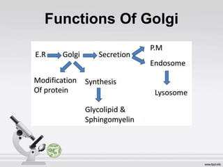
The Golgi
Apparatus
By: Rohit Satyam
Department Of Biotechnology, II year
NIET, Greater Noida
Group Members
1.Nishtha Singh
2.Rohit Panchal
3.Rohit Satyam
4.Shristi Singh
5.Naresh Sharma
Discovery
• The Golgi apparatus is noticeable with both
light and electron microscope. It is also
called Golgi complex.
• The Golgi complex was discovered by an
Italian physician and Noble Laureate
Camillo Golgi in 1898 during an
investigation of the nervous system.
• Its electron microscopic structure was
described by Dalton and Felix in 1954.
Camilo Golgi
Nobel Prize For the “ Black Stain”
Structure of Golgi Complex
• It varies in size and form in different cell types but usually has
similar organization for any one kind of cells.
• Electron microscope shows it as a central stack (pile) of parallel,
flattened, intercommunicating sacs or cisternae and many
peripheral tubules and vesicles.
• Consists of 4 functionally distinct region- cis face (entry face),
middle cisterna, trans cisterna, trans face(exit face).
• Each region contain different enzymes which selectively modify
the contents depending on where they reside.
Cisternae:
• The cisternae vary in number from 3-7 in most animal cells
and from 10-20 in plant cells.
• Usually equally spaced in the stack, separated from each
other by thin layers of intercisternal cytoplasm.
• Cisternae may be flat but are often curved.
• Golgi complex has a distinct polarity, the two poles are called
cis face and trans face, which act respectively as the
receiving and shipping departments.
• Convex side of stack -> forming (cis) face.
• Concave side of stack -> maturing (trans) face.
Location
• The Golgi apparatus is present in all Eukaryotic cells and absent in
Prokaryotes.
• The Golgi apparatus is specially extensive in the secretory cells.
• It is absent in few cell types, such as the mammalian RBCs, sperm
cells of Bryophytes and Pteridophytes and sieve tubes of plants.
• A cell may have one large Golgi complex or several very small
ones. It occupies different positions in different kind of cells.
• In secretory and absorptive cells, it usually lies between the
nucleus and Plasma Membrane.
• The invertebrate and plant cells usually have several small Golgi
complexes, called Dictyosomes, scattered throughout the
cytoplasm.
Functions
• Secretion
• Synthesis
• Sulfation
• Apoptosis
• Phosphorylation
• Cell-specific functions
Functions Of Golgi
SECRETION
• Although the Golgi apparatus is involved in many different
cellular processes ,its principle role in many cells is in
secretion. Golgi plays an important role in the synthesis of
proteoglycans, which are molecules present in the
extracellular matrix of animals.
• Secretory materials reach the Golgi complex from Smooth
Endoplasmic Reticulum (SER) by way of transport vesicles
which bud off from SER and fuse with Golgi cisternae on
the cis face.
• From the trans face Secretory vesicles arises that carry the
processed material to their destination.
• Tubules: Small, round tubules arise from the periphery of the
cisternae. Some of these enlarge at their ends to form vesicles.
• Vesicles: the vesicles lie near the ends and concave surface of
the Golgi complex.
• They are of two types: smooth or secretory vesicles and
coated vesicles.
• All the Golgi elements are filled with a fluid, the Golgi matrix.
Path of secretion
Diagram of secretory process from
endoplasmic reticulum (orange) to
Golgi apparatus (pink).
1. Nuclear membrane
2. Nuclear pore
3. Rough endoplasmic reticulum
(RER)
4. Smooth endoplasmic reticulum
(SER)
5. Ribosome attached to RER
6. Macromolecules
7. Transport vesicles
8. Golgi apparatus
9.Cis face of Golgi apparatus
10. Trans face of Golgi apparatus
11. Cisternae of the Golgi Apparatus
SYNTHESIS
• It is also a major site of carbohydrate synthesis.
• This includes the production of glycosaminoglycan (GAGs),
long unbranched polysaccharides which the Golgi then attaches
to a protein synthesised in the endoplasmic reticulum to
form proteoglycans.
• Enzymes in the Golgi polymerize several of these GAGs.
• The Golgi apparatus is also involved in lipid Transport
and lysosome formation.
SULFATION
• Another task of the Golgi involves the sulfation (Adding of
sulpho group, HSO3
- ) certain molecules passing through its
lumen via sulfotranferases that gain their sulphur molecule from
a donor.
• This process occurs on the GAGs of proteoglycans as well as
on the core protein.
• Sulfation is generally performed in the trans-Golgi network.
• The level of sulfation is very important to the proteoglycans'
signalling abilities as well as giving the proteoglycan its overall
negative charge.
Protein modification
• Start in the E.R after translation - addition of 2
N-acetylglucosamine(NAG) & mannose.
• In Golgi, glycosyltransferase & glycosidase is
present – help in addition & removal of sugar
residues in N-linked oligosaccharides.
APOPTOSIS
• The Golgi has a putative role in apoptosis.
• A newly characterized protein, GAAP (Golgi anti-
apoptotic protein), almost exclusively resides in the
Golgi and protects cells from apoptosis by an as-yet
undefined mechanism.
PHOSPHORYLATION
• The phosphorylation of molecules requires energy in
the form of ATP .
• That ATP is imported into the lumen of the Golgi utilised
by resident kinases such as casein kinase 1and casein
kinase 2.
• One molecule that is phosphorylated in the Golgi is
Apolipoprotein, which forms a molecule known as VLDL
that is a constituent of blood serum.
• It is thought that the phosphorylation of these
molecules is important to help aid in their sorting for
secretion into the blood serum.
Vesicular Transport
• Vesicles leaving RER transported to the cis face of GA,
fuse with the membrane and empty the contents into the
lumen.
• Molecules inside the lumen are modified and sorted for
transport to the next destination.
• Proteins destined for places other than ER and GA, moves
to trans face.
• Gets placed on either of the 3 vesicles, i.e.
Exocytotic , Secretory and Lysosomal vesicles.
Cell Specific Functions
• Formation of cell wall and cell plate in Plant Tissues.
• Acrosome development in sperm cells.
• Secretion of Zymogen in the Exocrine cells of Pancreas.
• Secretion and transformation of Lipid in the liver cells.
• Similar secretory functions are carried out in the Brunner’s
gland cells, alveolar epithelium, Paneth cells, connective
tissues as well.
Golgi Bodies And Alzheimer
Disease
• Golgi apparatus is found to play an important role in
Alzheimer’s disease. Alzheimer’s disease is a brain
disorder where brain cells are being destroyed. This is
what gives a person memory loss and this disease can
lead to death. As the death of neurons increases the
affected brain region begins to shrink. The cause of this
disease is because there is too little removal of a specific
type of protein.
• Therefore, the Golgi apparatus isn’t functioning as it is
suppose to in collecting, transporting, and distributing the
protein molecules.
Summary
• GA is made of a stack of membrane bound sacs.
• It receives most of the substances from Endoplasmic
Reticulum.
• It produces vesicles which carry secretions on to the
cell surface.
• Basically, it functions as a “post office”- where the
proteins and lipids are taken, labelled and transported
to other locations within the body.
Thank
You

Golgi bodies

  • 1.
    The Golgi Apparatus By: RohitSatyam Department Of Biotechnology, II year NIET, Greater Noida
  • 2.
    Group Members 1.Nishtha Singh 2.RohitPanchal 3.Rohit Satyam 4.Shristi Singh 5.Naresh Sharma
  • 3.
    Discovery • The Golgiapparatus is noticeable with both light and electron microscope. It is also called Golgi complex. • The Golgi complex was discovered by an Italian physician and Noble Laureate Camillo Golgi in 1898 during an investigation of the nervous system. • Its electron microscopic structure was described by Dalton and Felix in 1954. Camilo Golgi Nobel Prize For the “ Black Stain”
  • 4.
    Structure of GolgiComplex • It varies in size and form in different cell types but usually has similar organization for any one kind of cells. • Electron microscope shows it as a central stack (pile) of parallel, flattened, intercommunicating sacs or cisternae and many peripheral tubules and vesicles. • Consists of 4 functionally distinct region- cis face (entry face), middle cisterna, trans cisterna, trans face(exit face). • Each region contain different enzymes which selectively modify the contents depending on where they reside.
  • 5.
    Cisternae: • The cisternaevary in number from 3-7 in most animal cells and from 10-20 in plant cells. • Usually equally spaced in the stack, separated from each other by thin layers of intercisternal cytoplasm. • Cisternae may be flat but are often curved. • Golgi complex has a distinct polarity, the two poles are called cis face and trans face, which act respectively as the receiving and shipping departments. • Convex side of stack -> forming (cis) face. • Concave side of stack -> maturing (trans) face.
  • 7.
    Location • The Golgiapparatus is present in all Eukaryotic cells and absent in Prokaryotes. • The Golgi apparatus is specially extensive in the secretory cells. • It is absent in few cell types, such as the mammalian RBCs, sperm cells of Bryophytes and Pteridophytes and sieve tubes of plants. • A cell may have one large Golgi complex or several very small ones. It occupies different positions in different kind of cells. • In secretory and absorptive cells, it usually lies between the nucleus and Plasma Membrane. • The invertebrate and plant cells usually have several small Golgi complexes, called Dictyosomes, scattered throughout the cytoplasm.
  • 9.
    Functions • Secretion • Synthesis •Sulfation • Apoptosis • Phosphorylation • Cell-specific functions
  • 10.
  • 11.
    SECRETION • Although theGolgi apparatus is involved in many different cellular processes ,its principle role in many cells is in secretion. Golgi plays an important role in the synthesis of proteoglycans, which are molecules present in the extracellular matrix of animals. • Secretory materials reach the Golgi complex from Smooth Endoplasmic Reticulum (SER) by way of transport vesicles which bud off from SER and fuse with Golgi cisternae on the cis face. • From the trans face Secretory vesicles arises that carry the processed material to their destination.
  • 12.
    • Tubules: Small,round tubules arise from the periphery of the cisternae. Some of these enlarge at their ends to form vesicles. • Vesicles: the vesicles lie near the ends and concave surface of the Golgi complex. • They are of two types: smooth or secretory vesicles and coated vesicles. • All the Golgi elements are filled with a fluid, the Golgi matrix.
  • 14.
    Path of secretion Diagramof secretory process from endoplasmic reticulum (orange) to Golgi apparatus (pink). 1. Nuclear membrane 2. Nuclear pore 3. Rough endoplasmic reticulum (RER) 4. Smooth endoplasmic reticulum (SER) 5. Ribosome attached to RER 6. Macromolecules 7. Transport vesicles 8. Golgi apparatus 9.Cis face of Golgi apparatus 10. Trans face of Golgi apparatus 11. Cisternae of the Golgi Apparatus
  • 15.
    SYNTHESIS • It isalso a major site of carbohydrate synthesis. • This includes the production of glycosaminoglycan (GAGs), long unbranched polysaccharides which the Golgi then attaches to a protein synthesised in the endoplasmic reticulum to form proteoglycans. • Enzymes in the Golgi polymerize several of these GAGs. • The Golgi apparatus is also involved in lipid Transport and lysosome formation.
  • 16.
    SULFATION • Another taskof the Golgi involves the sulfation (Adding of sulpho group, HSO3 - ) certain molecules passing through its lumen via sulfotranferases that gain their sulphur molecule from a donor. • This process occurs on the GAGs of proteoglycans as well as on the core protein. • Sulfation is generally performed in the trans-Golgi network. • The level of sulfation is very important to the proteoglycans' signalling abilities as well as giving the proteoglycan its overall negative charge.
  • 17.
    Protein modification • Startin the E.R after translation - addition of 2 N-acetylglucosamine(NAG) & mannose. • In Golgi, glycosyltransferase & glycosidase is present – help in addition & removal of sugar residues in N-linked oligosaccharides.
  • 18.
    APOPTOSIS • The Golgihas a putative role in apoptosis. • A newly characterized protein, GAAP (Golgi anti- apoptotic protein), almost exclusively resides in the Golgi and protects cells from apoptosis by an as-yet undefined mechanism.
  • 19.
    PHOSPHORYLATION • The phosphorylationof molecules requires energy in the form of ATP . • That ATP is imported into the lumen of the Golgi utilised by resident kinases such as casein kinase 1and casein kinase 2. • One molecule that is phosphorylated in the Golgi is Apolipoprotein, which forms a molecule known as VLDL that is a constituent of blood serum. • It is thought that the phosphorylation of these molecules is important to help aid in their sorting for secretion into the blood serum.
  • 20.
    Vesicular Transport • Vesiclesleaving RER transported to the cis face of GA, fuse with the membrane and empty the contents into the lumen. • Molecules inside the lumen are modified and sorted for transport to the next destination. • Proteins destined for places other than ER and GA, moves to trans face. • Gets placed on either of the 3 vesicles, i.e. Exocytotic , Secretory and Lysosomal vesicles.
  • 21.
    Cell Specific Functions •Formation of cell wall and cell plate in Plant Tissues. • Acrosome development in sperm cells. • Secretion of Zymogen in the Exocrine cells of Pancreas. • Secretion and transformation of Lipid in the liver cells. • Similar secretory functions are carried out in the Brunner’s gland cells, alveolar epithelium, Paneth cells, connective tissues as well.
  • 22.
    Golgi Bodies AndAlzheimer Disease
  • 23.
    • Golgi apparatusis found to play an important role in Alzheimer’s disease. Alzheimer’s disease is a brain disorder where brain cells are being destroyed. This is what gives a person memory loss and this disease can lead to death. As the death of neurons increases the affected brain region begins to shrink. The cause of this disease is because there is too little removal of a specific type of protein. • Therefore, the Golgi apparatus isn’t functioning as it is suppose to in collecting, transporting, and distributing the protein molecules.
  • 24.
    Summary • GA ismade of a stack of membrane bound sacs. • It receives most of the substances from Endoplasmic Reticulum. • It produces vesicles which carry secretions on to the cell surface. • Basically, it functions as a “post office”- where the proteins and lipids are taken, labelled and transported to other locations within the body.
  • 25.

Editor's Notes

  • #11 Sphingosine (2-amino-4-octadecene-1,3-diol) is an 18-carbon amino alcohol with an unsaturated hydrocarbon chain, which forms a primary part of sphingolipids. Sphingolipids are a class of lipids containing a backbone of sphingoid bases, a set of aliphatic amino alcohols that includes sphingosine. These compounds play important roles in signal transmission and cell recognition. Sphingomyelin (SPH, ˌsfɪŋɡoˈmaɪəlɪn) is a type of sphingolipid found in animal cell membranes, especially in the membranous myelin sheath that surrounds some nerve cell axons. The membranous myelin sheath that surrounds and electrically insulates many nerve cell axons is particularly rich in sphingomyelin, suggesting its role as an insulator of nerve fibers. They also take part in signal transduction and Apoptosis.
  • #16 Ever touched a slug, or a frog? Slimy! That sliminess is also found protecting different organs of your body, like in your gut and your throat. Some of the components of that slime include proteoglycans or PGs. PGs are protein molecules found in the space between our cells, a physiological component known as theextracellular matrix (ECM). ECM is like bubblegum in that it keeps our cells together and mobile as a group. It also helps to protect them from outside forces and chemicals that would tear them apart.
  • #22 A zymogen, also called a proenzyme, is an inactive precursor of an enzyme. A zymogen requires a biochemical change (such as a hydrolysis reaction revealing the active site, or changing the configuration to reveal the active site) for it to become an active enzyme. The biochemical change usually occurs in Golgi bodies, where a specific part of the precursor enzyme is cleaved in order to activate it. The pancreas secretes zymogens partly to prevent the enzymes from digesting proteins in the cells in which they are synthesised. Enzymes like pepsin are created in the form of pepsinogen, an inactive zymogen. Pepsinogen is activated when chief cells release it into the gastric acid, whose hydrochloric acid partially activates it. Another partially activated pepsinogen completes the activation by removing the peptide, turning the pepsinogen into pepsin. Accidental activation of zymogens can happen when the secretion duct in the pancreas is blocked by a gallstone resulting in acute pancreatitis.