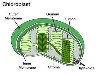
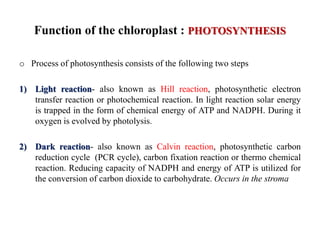
Chloroplasts are organelles found in plant cells and algae that conduct photosynthesis. They have their own DNA and can synthesize some of their own proteins, making them semi-autonomous. Chloroplasts contain chlorophyll and carotenoids which capture light energy. Their internal structure includes an envelope, stroma, and thylakoids where the light reactions take place. It is believed that chloroplasts originated through endosymbiosis between cyanobacteria and eukaryotic cells. The two main stages of photosynthesis are the light reactions on the thylakoid membranes which produce ATP and NADPH, and the dark reactions in the stroma that use these products to fix carbon into sugars.
This power point presentation consists of 64 slides including information about plant and other type of cell wall. Chemical composition, structure, function and properties of cell wall have been explained. Ultra structure of plant cell wall has also been high lighted. Algal,Fungal,Bacterial and Archaeal cell walls have also been explained.
DNA is tightly packed in the nucleus of every cell. DNA wraps around special proteins called histones, which form loops of DNA called nucleosomes. These nucleosomes coil and stack together to form fibers called chromatin. Chromatin in turn forms larger loops and coils to form chromosomes.
DNA packaging is crucial because it makes sure that those excessive DNA are able to fit nicely in a cell that is many times smaller.
The DNA in bacterial cells are either circular or linear. To accommodate the size of bacterial cell, supercoiled DNA are folded into loops with each loop resembles shape of bead-like packets containing small basic proteins that is analogous to histone found in Eukaryotes.
Basics of Undergraduate/university fellows
Nucleosome model of chromosome is proposed by ROGER KORNBERG (son of Arthur
Kornberg) in 1974.
It was confirmed and crystalised by P. Oudet et al., (1975).
Nucleosome is the lowest level of Chromosome organization in eukaryotic cells.
Nucleosome model is a scientific model which explains the organization of DNA and
associated proteins in the chromosomes.
Nucleosome model also explains the exact mechanism of the folding of DNA in
thenucleus.
It is the most accepted model of chromatin organization.
Details of cytoskeleton element-microtubule. The Microtubule associated protein-type and function, Treadmilling and dynamic instability, Structure of cilia and flagella
The plastid (Greek: πλαστός; plastós: formed, molded – plural plastids) is a major organelle found in the cells of plants and algae. Plastids are the site of manufacture and storage of important chemical compounds used by the cell. They often contain pigments used in photosynthesis, and the types of pigments present can change or determine the cell's colour. They possess a double-stranded DNA molecule, which is circular, like that of prokaryotes.
The presentation describes the advantages of plastid transformation over 'conventional' nuclear transformation, hurdles to plastid transformation, its advantages. The presentation also covers some successful plastid engineering and its potential.
For this assignment, we were instructed to create a powerpoint presentation of at least 12 slides that adequately covered an academic subject of our choice. All sources for media is cited in the work cited at the end of the presentation.
It is a process used by plants & other organisms to convert light energy into chemical energy that can be later used by organisms as a fuel. i.e; energy transformation
The term Chloroplast was first described by Nehemiah Grew and Antonie Van Leeuwenhoek.
“Chloro” means green while“ Plast” means living.
Chlorophyll pigments present in the chloroplast imparts the green colour to plants.
Chloroplasts are present in plants and other eukaryotic organisms that conducts photosynthesis
Responsible for photosynthesis, are in many respects similar to mitochondria.
Chloroplasts are larger and more complex than mitochondria, and they perform several critical tasks in addition to the generation of ATP.
Chloroplasts synthesize amino acids, fatty acids, and the lipid components of their own membranes.
The reduction of nitrite (NO2-) to ammonia (NH3), an essential step in the incorporation of nitrogen into organic compounds, also occurs in chloroplasts.
The increased availability of biomedical data, particularly in the public domain, offers the opportunity to better understand human health and to develop effective therapeutics for a wide range of unmet medical needs. However, data scientists remain stymied by the fact that data remain hard to find and to productively reuse because data and their metadata i) are wholly inaccessible, ii) are in non-standard or incompatible representations, iii) do not conform to community standards, and iv) have unclear or highly restricted terms and conditions that preclude legitimate reuse. These limitations require a rethink on data can be made machine and AI-ready - the key motivation behind the FAIR Guiding Principles. Concurrently, while recent efforts have explored the use of deep learning to fuse disparate data into predictive models for a wide range of biomedical applications, these models often fail even when the correct answer is already known, and fail to explain individual predictions in terms that data scientists can appreciate. These limitations suggest that new methods to produce practical artificial intelligence are still needed.
In this talk, I will discuss our work in (1) building an integrative knowledge infrastructure to prepare FAIR and "AI-ready" data and services along with (2) neurosymbolic AI methods to improve the quality of predictions and to generate plausible explanations. Attention is given to standards, platforms, and methods to wrangle knowledge into simple, but effective semantic and latent representations, and to make these available into standards-compliant and discoverable interfaces that can be used in model building, validation, and explanation. Our work, and those of others in the field, creates a baseline for building trustworthy and easy to deploy AI models in biomedicine.
Bio
Dr. Michel Dumontier is the Distinguished Professor of Data Science at Maastricht University, founder and executive director of the Institute of Data Science, and co-founder of the FAIR (Findable, Accessible, Interoperable and Reusable) data principles. His research explores socio-technological approaches for responsible discovery science, which includes collaborative multi-modal knowledge graphs, privacy-preserving distributed data mining, and AI methods for drug discovery and personalized medicine. His work is supported through the Dutch National Research Agenda, the Netherlands Organisation for Scientific Research, Horizon Europe, the European Open Science Cloud, the US National Institutes of Health, and a Marie-Curie Innovative Training Network. He is the editor-in-chief for the journal Data Science and is internationally recognized for his contributions in bioinformatics, biomedical informatics, and semantic technologies including ontologies and linked data.
Earliest Galaxies in the JADES Origins Field: Luminosity Function and Cosmic ...Sérgio Sacani
We characterize the earliest galaxy population in the JADES Origins Field (JOF), the deepest
imaging field observed with JWST. We make use of the ancillary Hubble optical images (5 filters
spanning 0.4−0.9µm) and novel JWST images with 14 filters spanning 0.8−5µm, including 7 mediumband filters, and reaching total exposure times of up to 46 hours per filter. We combine all our data
at > 2.3µm to construct an ultradeep image, reaching as deep as ≈ 31.4 AB mag in the stack and
30.3-31.0 AB mag (5σ, r = 0.1” circular aperture) in individual filters. We measure photometric
redshifts and use robust selection criteria to identify a sample of eight galaxy candidates at redshifts
z = 11.5 − 15. These objects show compact half-light radii of R1/2 ∼ 50 − 200pc, stellar masses of
M⋆ ∼ 107−108M⊙, and star-formation rates of SFR ∼ 0.1−1 M⊙ yr−1
. Our search finds no candidates
at 15 < z < 20, placing upper limits at these redshifts. We develop a forward modeling approach to
infer the properties of the evolving luminosity function without binning in redshift or luminosity that
marginalizes over the photometric redshift uncertainty of our candidate galaxies and incorporates the
impact of non-detections. We find a z = 12 luminosity function in good agreement with prior results,
and that the luminosity function normalization and UV luminosity density decline by a factor of ∼ 2.5
from z = 12 to z = 14. We discuss the possible implications of our results in the context of theoretical
models for evolution of the dark matter halo mass function.
Nutraceutical market, scope and growth: Herbal drug technologyLokesh Patil
As consumer awareness of health and wellness rises, the nutraceutical market—which includes goods like functional meals, drinks, and dietary supplements that provide health advantages beyond basic nutrition—is growing significantly. As healthcare expenses rise, the population ages, and people want natural and preventative health solutions more and more, this industry is increasing quickly. Further driving market expansion are product formulation innovations and the use of cutting-edge technology for customized nutrition. With its worldwide reach, the nutraceutical industry is expected to keep growing and provide significant chances for research and investment in a number of categories, including vitamins, minerals, probiotics, and herbal supplements.
Cancer cell metabolism: special Reference to Lactate PathwayAADYARAJPANDEY1
Normal Cell Metabolism:
Cellular respiration describes the series of steps that cells use to break down sugar and other chemicals to get the energy we need to function.
Energy is stored in the bonds of glucose and when glucose is broken down, much of that energy is released.
Cell utilize energy in the form of ATP.
The first step of respiration is called glycolysis. In a series of steps, glycolysis breaks glucose into two smaller molecules - a chemical called pyruvate. A small amount of ATP is formed during this process.
Most healthy cells continue the breakdown in a second process, called the Kreb's cycle. The Kreb's cycle allows cells to “burn” the pyruvates made in glycolysis to get more ATP.
The last step in the breakdown of glucose is called oxidative phosphorylation (Ox-Phos).
It takes place in specialized cell structures called mitochondria. This process produces a large amount of ATP. Importantly, cells need oxygen to complete oxidative phosphorylation.
If a cell completes only glycolysis, only 2 molecules of ATP are made per glucose. However, if the cell completes the entire respiration process (glycolysis - Kreb's - oxidative phosphorylation), about 36 molecules of ATP are created, giving it much more energy to use.
IN CANCER CELL:
Unlike healthy cells that "burn" the entire molecule of sugar to capture a large amount of energy as ATP, cancer cells are wasteful.
Cancer cells only partially break down sugar molecules. They overuse the first step of respiration, glycolysis. They frequently do not complete the second step, oxidative phosphorylation.
This results in only 2 molecules of ATP per each glucose molecule instead of the 36 or so ATPs healthy cells gain. As a result, cancer cells need to use a lot more sugar molecules to get enough energy to survive.
introduction to WARBERG PHENOMENA:
WARBURG EFFECT Usually, cancer cells are highly glycolytic (glucose addiction) and take up more glucose than do normal cells from outside.
Otto Heinrich Warburg (; 8 October 1883 – 1 August 1970) In 1931 was awarded the Nobel Prize in Physiology for his "discovery of the nature and mode of action of the respiratory enzyme.
WARNBURG EFFECT : cancer cells under aerobic (well-oxygenated) conditions to metabolize glucose to lactate (aerobic glycolysis) is known as the Warburg effect. Warburg made the observation that tumor slices consume glucose and secrete lactate at a higher rate than normal tissues.
Slide 1: Title Slide
Extrachromosomal Inheritance
Slide 2: Introduction to Extrachromosomal Inheritance
Definition: Extrachromosomal inheritance refers to the transmission of genetic material that is not found within the nucleus.
Key Components: Involves genes located in mitochondria, chloroplasts, and plasmids.
Slide 3: Mitochondrial Inheritance
Mitochondria: Organelles responsible for energy production.
Mitochondrial DNA (mtDNA): Circular DNA molecule found in mitochondria.
Inheritance Pattern: Maternally inherited, meaning it is passed from mothers to all their offspring.
Diseases: Examples include Leber’s hereditary optic neuropathy (LHON) and mitochondrial myopathy.
Slide 4: Chloroplast Inheritance
Chloroplasts: Organelles responsible for photosynthesis in plants.
Chloroplast DNA (cpDNA): Circular DNA molecule found in chloroplasts.
Inheritance Pattern: Often maternally inherited in most plants, but can vary in some species.
Examples: Variegation in plants, where leaf color patterns are determined by chloroplast DNA.
Slide 5: Plasmid Inheritance
Plasmids: Small, circular DNA molecules found in bacteria and some eukaryotes.
Features: Can carry antibiotic resistance genes and can be transferred between cells through processes like conjugation.
Significance: Important in biotechnology for gene cloning and genetic engineering.
Slide 6: Mechanisms of Extrachromosomal Inheritance
Non-Mendelian Patterns: Do not follow Mendel’s laws of inheritance.
Cytoplasmic Segregation: During cell division, organelles like mitochondria and chloroplasts are randomly distributed to daughter cells.
Heteroplasmy: Presence of more than one type of organellar genome within a cell, leading to variation in expression.
Slide 7: Examples of Extrachromosomal Inheritance
Four O’clock Plant (Mirabilis jalapa): Shows variegated leaves due to different cpDNA in leaf cells.
Petite Mutants in Yeast: Result from mutations in mitochondrial DNA affecting respiration.
Slide 8: Importance of Extrachromosomal Inheritance
Evolution: Provides insight into the evolution of eukaryotic cells.
Medicine: Understanding mitochondrial inheritance helps in diagnosing and treating mitochondrial diseases.
Agriculture: Chloroplast inheritance can be used in plant breeding and genetic modification.
Slide 9: Recent Research and Advances
Gene Editing: Techniques like CRISPR-Cas9 are being used to edit mitochondrial and chloroplast DNA.
Therapies: Development of mitochondrial replacement therapy (MRT) for preventing mitochondrial diseases.
Slide 10: Conclusion
Summary: Extrachromosomal inheritance involves the transmission of genetic material outside the nucleus and plays a crucial role in genetics, medicine, and biotechnology.
Future Directions: Continued research and technological advancements hold promise for new treatments and applications.
Slide 11: Questions and Discussion
Invite Audience: Open the floor for any questions or further discussion on the topic.
2. • Type of the plastids
• Historical
• Distribution
• Chloroplast as semiautonomous organelle
• Chemical composition
• Ultrastructure
• Biogenesis
• The symbiotic origin of chloroplast
• Function of the chloroplast-Photosynthesis
Outlines
3. Types of the plastids
• The term plastid was used by Schimper in 1885 and he classified plastids as
following
1. Leucoplasts-(Gr., leuco=white; plast=living) are the colourless plastids
are found in embryonic and germ cells. Found in those regions of the plant
which are not receiving light. They store carbohydrates, lipids and protein
and accordingly are of following types i) Amyloplast ii) Elaioplast iii)
Proteinoplast
2. Chromoplast-(Gr., chroma=colour; plast=living) are the coloured plastids
containing carotenoids and other pigments. They impart colour (yellow,
orange and red) to certain portion of plants such as flower petals (rose),
fruits (tomato), roots (carrot). Two types i) Phaeoplast ii) Rhodoplast
3. Chloroplast- (Gr., chloro=green plast=living)- occurs mostly in the green
algae and plants and contain pigments like chlorophyll-a and chlorophyll-
b and DNA and RNA
4. o According to Schimper different kinds of plastids can transform into one
another
Leucoplasts
Chloroplasts
Chromoplasts
5. Historical
o Chloroplast were described as early as seventeenth century by Nehemiah
Grew and Antonie van Leeuwenhoek
o The term Plastid was used by Schimper in 1885 he also classified the
plastids of plant
o A Meyer, F. Schmitz and A.F.W. Schimper showed that chloroplasts always
arise from pre-existing chloroplast.
o Wilstatter and Stoll isolated and characterized green pigments- Chlorophyll
a and b
o Julius Sachs showed that chlorophyll is confined to chloroplast not
distributed throughout the plant cell
6. Distribution
o The chloroplasts remain distributed homogeneously in the cytoplasm of
plant cell.
o The algae usually have a single huge chloroplast the cells of higher plants
have 20-40 chloroplast.
o When the number of chloroplast is inadequate, it is increased by division
when excessive, it is reduced by degeneration.
7. Chloroplast as semiautonomous organelle
o Like the mitochondria the chloroplast have their own DNA, RNA and
protein synthetic machinery.
a. DNA of chloroplast- Ris and plant (1962) reported DNA in chloroplast.
Chloroplast DNA is double helical circle with an average length of 45µm
(about 135,000bp)
b. Ribosomes- ribosomes of chloroplast are smaller than cytoplasmic
ribosome and are of 70S type and resemble with the bacterial ribosomes.
Contain t-RNA
c. Protein synthesis-DNA of chloroplast codes for chloroplast mRNA,
rRNA, tRNA and ribosomal proteins.
10. Ultrastructure
o A chloroplast comprises the following three main components
1. Envelope- The entire chloroplast is bounded by an envelope which is
made of a double unit membranes. Across this double membrane envelope
exchange of molecules between chloroplast and cytosol occurs.
2. Stroma- The matrix or stroma fills most of the volume of the chloroplast
and is a kind of gel-fluid phase that surrounds the thylakoids (grana). It
contains proteins, ribosomes and DNA. The stroma is the site of CO2
fixation and where the synthesis of sugar, starch, fatty acids and some
proteins occurs
3. Thylakoids- The thylakoids consists of flattened and closed vesicles
arranged as a membrane network, Thylakoids may be stacked like a neat
pile of coins forming grana. There may be 40-60 grana in the matrix of a
chloroplast. Light reactions occurs in thylakoids membrane
11.
12. Biogenesis of chloroplast
o The chloroplasts never originates de novo.
o Chloroplast multiply by fission a process that implies growth of daughter
organelles
o During the development of the chloroplast the first structure to appear is the
so-called proplastid.
o Proplastid is then develops into chloroplast
13. The symbiotic origin of chloroplast
o Chloroplast divide, grow and differentiate; they contain circular DNA,
ribosomal RNA, messenger RNA and are able to conduct protein synthesis
o By visualizing these similarities between micro-organisms and chloroplast
it has been suggested that chloroplast might have relationship between
autotrophic micro-organism
o But some of the enzymes of chloroplast are coded by nuclear genes so there
still exist certain doubt about the symbiotic origin of chloroplast
(kirk ,1966)
14. Function of the chloroplast : PHOTOSYNTHESIS
o Process of photosynthesis consists of the following two steps
1) Light reaction- also known as Hill reaction, photosynthetic electron
transfer reaction or photochemical reaction. In light reaction solar energy
is trapped in the form of chemical energy of ATP and NADPH. During it
oxygen is evolved by photolysis.
2) Dark reaction- also known as Calvin reaction, photosynthetic carbon
reduction cycle (PCR cycle), carbon fixation reaction or thermo chemical
reaction. Reducing capacity of NADPH and energy of ATP is utilized for
the conversion of carbon dioxide to carbohydrate. Occurs in the stroma