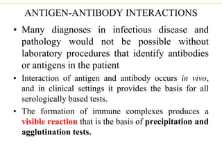
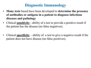
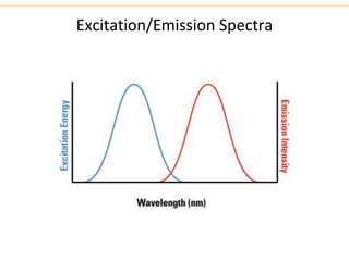
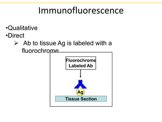
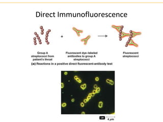
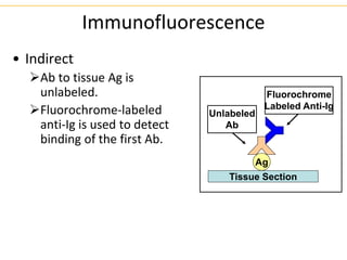
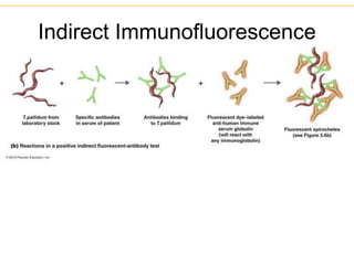
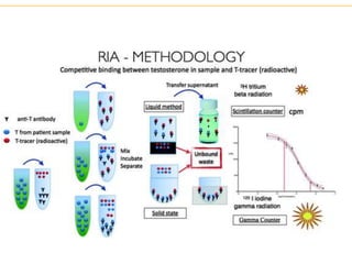
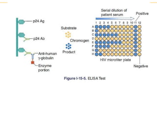
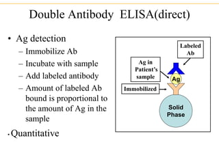
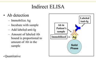
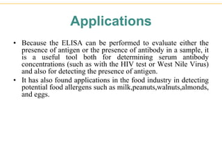
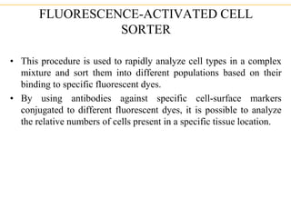
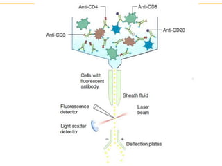
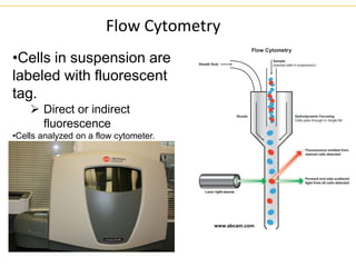
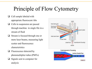
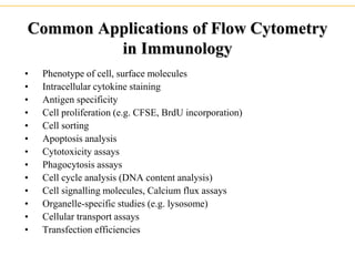
Laboratory Techniques in
Immunology
Practical Applications of
Immunology
ANTIGEN-ANTIBODY INTERACTIONS
• Many diagnoses in infectious disease and
pathology would not be possible without
laboratory procedures that identify antibodies
or antigens in the patient
• Interaction of antigen and antibody occurs in vivo,
and in clinical settings it provides the basis for all
serologically based tests.
• The formation of immune complexes produces a
visible reaction that is the basis of precipitation and
agglutination tests.
Diagnostic Immunology
• Many tests based have been developed to determine the presence
of antibodies or antigens in a patient to diagnose infectious
diseases and pathology
• Clinical sensitivity - ability of a test to provide a positive result if
the patient has the disease (no false negatives).
• Clinical specificity – ability of a test to give a negative result if the
patient does not have disease (no false positives).
Labeled immunoassay
Lattice formation not required
FLUORESCENT ANTIBODY TESTS
• The direct fluorescent antibody test (DFA) is used
to detect and localize antigen in the patient. The
tissue sample to be tested is treated with antibodies
against that particular antigen that have been labeled
with a fluorescent dye. If the antigen is present in the
tissues, the fluorescent-labeled antibodies will bind,
and their binding can be detected with a fluorescence
microscope.
• Variations of this test are used to diagnose respiratory
syncytial virus, herpes simplex- 1 and -2, and
Pneumocystis infections.
Excitation/Emission Spectra
Immunofluorescence
•Qualitative
•Direct
 Ab to tissue Ag is labeled with a
fluorochrome.
Ag
Fluorochrome
Labeled Ab
Tissue Section
Direct Immunofluorescence
• The indirect fluorescent antibody test (IFA) is used to
detect pathogen-specific antibodies in the patient. In this case,
a laboratory-generated sample of infected tissue is mixed with
serum from the patient. A fluorescent dye-labeled anti-
immunoglobulin (raised in animals) is then added. If binding
of antibodies from the patient to the tissue sample occurs, then
the fluorescent antibodies can be bound, and fluorescence can
be detected in the tissue by microscopy.
• This technique is used to detect antinuclear antibodies, anti-
dsDNA antibodies, antithyroid antibodies, antiglomerular
basement-membrane antibodies, and anti-Epstein-Barr virus
viral-capsid antigen antibodies.
Immunofluorescence
• Indirect
Ab to tissue Ag is
unlabeled.
Fluorochrome-labeled
anti-Ig is used to detect
binding of the first Ab.
Ag
Fluorochrome
Labeled Anti-Ig
Tissue Section
Unlabeled
Ab
Indirect Immunofluorescence
•In suspected Graves' disease the
first-line test is antibodies against
TSH receptors (TRAb), which occur
with a prevalence of 90 to 100 %
(ELISA). The detection of
antibodies against TPO can support
the diagnosis (ELISA or IIFT).
•In Hashimoto's thyroiditis
serological antibodies against TPO
are detectable with a prevalence of
up to 90 %, while antibodies against
TG are occur in 60 to 70 % of
cases.
• RADIOIMMUNOASSAY (RIA) AND ENZVME-LINKED
IMMUNOABSORBENT ASSAY (EIA OR ELISA)
• RIA and ELISA are extremely sensitive tests (as little as 10-9 g
of material can be detected) that are common in medical
laboratories.
• They can be used to detect the presence of hormones, drugs,
antibiotics, serum proteins, infectious disease antigens, and
tumor markers.
• Both tests are conducted similarly, but the RIA uses the
detection of a radiolabeled product and the ELISA detects the
presence of enzyme-mediated color changes in a
chromogenic substrate.
ELISA
• In the screening test for HIV infection, the ELISA is
used, with the p24 capsid antigen from the virus
coated on to microtiter plates. The serum from the
patient is then added, followed by addition of an
enzyme-labeled antihuman immunoglobulin.
• Finally, the enzyme substrate is added, and the
production of a color change in the well can be
observed if all reagents bind one another in sequence.
Double Antibody ELISA(direct)
• Ag detection
– Immobilize Ab
– Incubate with sample
– Add labeled antibody
– Amount of labeled Ab
bound is proportional to
the amount of Ag in the
sample
• Quantitative
Solid
Phase
Ag
Immobilized
Ag in
Patient’s
sample
Labeled
Ab
Indirect ELISA
• Ab detection
– Immobilize Ag
– Incubate with sample
– Add labeled anti-Ig
– Amount of labeled Ab
bound is proportional to
amount of Ab in the
sample
• Quantitative
Solid
Phase
Ag
Immobilized
Ab in
Patient’s
sample
Labeled
Anti-Ig
Applications
• Because the ELISA can be performed to evaluate either the
presence of antigen or the presence of antibody in a sample, it
is a useful tool both for determining serum antibody
concentrations (such as with the HIV test or West Nile Virus)
and also for detecting the presence of antigen.
• It has also found applications in the food industry in detecting
potential food allergens such as milk,peanuts,walnuts,almonds,
and eggs.
FLUORESCENCE-ACTIVATED CELL
SORTER
• This procedure is used to rapidly analyze cell types in a complex
mixture and sort them into different populations based on their
binding to specific fluorescent dyes.
• By using antibodies against specific cell-surface markers
conjugated to different fluorescent dyes, it is possible to analyze
the relative numbers of cells present in a specific tissue location.
• As cells pass through the apparatus in a single file, a
computer-generated graph is produced, plotting the intensity
and color of fluorescence of each cell along the axes.
• Each dot on the graph reflects the passage of a cell with a
certain level and color of fluorescence, so the darkly dotted
areas of the graph reflect the presence of many cells of similar
attributes. Cells with high fluorescence from both dyes will
therefore be found in the top right quadrant.
Flow Cytometry
•Cells in suspension are
labeled with fluorescent
tag.
 Direct or indirect
fluorescence
•Cells analyzed on a flow cytometer.
www.abcam.com
Principle of Flow Cytometry
 Cell sample labeled with
appropriate fluorescent Abs
 Cells in suspension are passed
through machine in single file in a
stream of fluid
 Stream is focused through one or
more laser beams, measuring light
scatter and fluorescence
characteristics
 Fluorescence detected by
photomultiplier tubes (PMTs)
 Signals sent to computer for
analysis
Green Fluorescence Intensity
Number
of
Cells
Unstained cells
FITC-labeled cells
One Parameter Histogram
Red Fluorescence Intensity
Green
Fluorescence
Intensity
Two Parameter Histogram
Flow Cytometry Interpretation
Myeloma
Common Applications of Flow Cytometry
in Immunology
• Phenotype of cell, surface molecules
• Intracellular cytokine staining
• Antigen specificity
• Cell proliferation (e.g. CFSE, BrdU incorporation)
• Cell sorting
• Apoptosis analysis
• Cytotoxicity assays
• Phagocytosis assays
• Cell cycle analysis (DNA content analysis)
• Cell signalling molecules, Calcium flux assays
• Organelle-specific studies (e.g. lysosome)
• Cellular transport assays
• Transfection efficiencies

Laboratory Techniques in immunology practice.

  • 1.
  • 2.
    ANTIGEN-ANTIBODY INTERACTIONS • Manydiagnoses in infectious disease and pathology would not be possible without laboratory procedures that identify antibodies or antigens in the patient • Interaction of antigen and antibody occurs in vivo, and in clinical settings it provides the basis for all serologically based tests. • The formation of immune complexes produces a visible reaction that is the basis of precipitation and agglutination tests.
  • 3.
    Diagnostic Immunology • Manytests based have been developed to determine the presence of antibodies or antigens in a patient to diagnose infectious diseases and pathology • Clinical sensitivity - ability of a test to provide a positive result if the patient has the disease (no false negatives). • Clinical specificity – ability of a test to give a negative result if the patient does not have disease (no false positives).
  • 4.
  • 5.
    FLUORESCENT ANTIBODY TESTS •The direct fluorescent antibody test (DFA) is used to detect and localize antigen in the patient. The tissue sample to be tested is treated with antibodies against that particular antigen that have been labeled with a fluorescent dye. If the antigen is present in the tissues, the fluorescent-labeled antibodies will bind, and their binding can be detected with a fluorescence microscope. • Variations of this test are used to diagnose respiratory syncytial virus, herpes simplex- 1 and -2, and Pneumocystis infections.
  • 6.
  • 7.
    Immunofluorescence •Qualitative •Direct  Ab totissue Ag is labeled with a fluorochrome. Ag Fluorochrome Labeled Ab Tissue Section
  • 8.
  • 9.
    • The indirectfluorescent antibody test (IFA) is used to detect pathogen-specific antibodies in the patient. In this case, a laboratory-generated sample of infected tissue is mixed with serum from the patient. A fluorescent dye-labeled anti- immunoglobulin (raised in animals) is then added. If binding of antibodies from the patient to the tissue sample occurs, then the fluorescent antibodies can be bound, and fluorescence can be detected in the tissue by microscopy. • This technique is used to detect antinuclear antibodies, anti- dsDNA antibodies, antithyroid antibodies, antiglomerular basement-membrane antibodies, and anti-Epstein-Barr virus viral-capsid antigen antibodies.
  • 10.
    Immunofluorescence • Indirect Ab totissue Ag is unlabeled. Fluorochrome-labeled anti-Ig is used to detect binding of the first Ab. Ag Fluorochrome Labeled Anti-Ig Tissue Section Unlabeled Ab
  • 11.
  • 12.
    •In suspected Graves'disease the first-line test is antibodies against TSH receptors (TRAb), which occur with a prevalence of 90 to 100 % (ELISA). The detection of antibodies against TPO can support the diagnosis (ELISA or IIFT). •In Hashimoto's thyroiditis serological antibodies against TPO are detectable with a prevalence of up to 90 %, while antibodies against TG are occur in 60 to 70 % of cases.
  • 13.
    • RADIOIMMUNOASSAY (RIA)AND ENZVME-LINKED IMMUNOABSORBENT ASSAY (EIA OR ELISA) • RIA and ELISA are extremely sensitive tests (as little as 10-9 g of material can be detected) that are common in medical laboratories. • They can be used to detect the presence of hormones, drugs, antibiotics, serum proteins, infectious disease antigens, and tumor markers. • Both tests are conducted similarly, but the RIA uses the detection of a radiolabeled product and the ELISA detects the presence of enzyme-mediated color changes in a chromogenic substrate.
  • 15.
    ELISA • In thescreening test for HIV infection, the ELISA is used, with the p24 capsid antigen from the virus coated on to microtiter plates. The serum from the patient is then added, followed by addition of an enzyme-labeled antihuman immunoglobulin. • Finally, the enzyme substrate is added, and the production of a color change in the well can be observed if all reagents bind one another in sequence.
  • 17.
    Double Antibody ELISA(direct) •Ag detection – Immobilize Ab – Incubate with sample – Add labeled antibody – Amount of labeled Ab bound is proportional to the amount of Ag in the sample • Quantitative Solid Phase Ag Immobilized Ag in Patient’s sample Labeled Ab
  • 18.
    Indirect ELISA • Abdetection – Immobilize Ag – Incubate with sample – Add labeled anti-Ig – Amount of labeled Ab bound is proportional to amount of Ab in the sample • Quantitative Solid Phase Ag Immobilized Ab in Patient’s sample Labeled Anti-Ig
  • 19.
    Applications • Because theELISA can be performed to evaluate either the presence of antigen or the presence of antibody in a sample, it is a useful tool both for determining serum antibody concentrations (such as with the HIV test or West Nile Virus) and also for detecting the presence of antigen. • It has also found applications in the food industry in detecting potential food allergens such as milk,peanuts,walnuts,almonds, and eggs.
  • 20.
    FLUORESCENCE-ACTIVATED CELL SORTER • Thisprocedure is used to rapidly analyze cell types in a complex mixture and sort them into different populations based on their binding to specific fluorescent dyes. • By using antibodies against specific cell-surface markers conjugated to different fluorescent dyes, it is possible to analyze the relative numbers of cells present in a specific tissue location.
  • 21.
    • As cellspass through the apparatus in a single file, a computer-generated graph is produced, plotting the intensity and color of fluorescence of each cell along the axes. • Each dot on the graph reflects the passage of a cell with a certain level and color of fluorescence, so the darkly dotted areas of the graph reflect the presence of many cells of similar attributes. Cells with high fluorescence from both dyes will therefore be found in the top right quadrant.
  • 23.
    Flow Cytometry •Cells insuspension are labeled with fluorescent tag.  Direct or indirect fluorescence •Cells analyzed on a flow cytometer. www.abcam.com
  • 24.
    Principle of FlowCytometry  Cell sample labeled with appropriate fluorescent Abs  Cells in suspension are passed through machine in single file in a stream of fluid  Stream is focused through one or more laser beams, measuring light scatter and fluorescence characteristics  Fluorescence detected by photomultiplier tubes (PMTs)  Signals sent to computer for analysis
  • 25.
    Green Fluorescence Intensity Number of Cells Unstainedcells FITC-labeled cells One Parameter Histogram Red Fluorescence Intensity Green Fluorescence Intensity Two Parameter Histogram Flow Cytometry Interpretation
  • 26.
  • 27.
    Common Applications ofFlow Cytometry in Immunology • Phenotype of cell, surface molecules • Intracellular cytokine staining • Antigen specificity • Cell proliferation (e.g. CFSE, BrdU incorporation) • Cell sorting • Apoptosis analysis • Cytotoxicity assays • Phagocytosis assays • Cell cycle analysis (DNA content analysis) • Cell signalling molecules, Calcium flux assays • Organelle-specific studies (e.g. lysosome) • Cellular transport assays • Transfection efficiencies