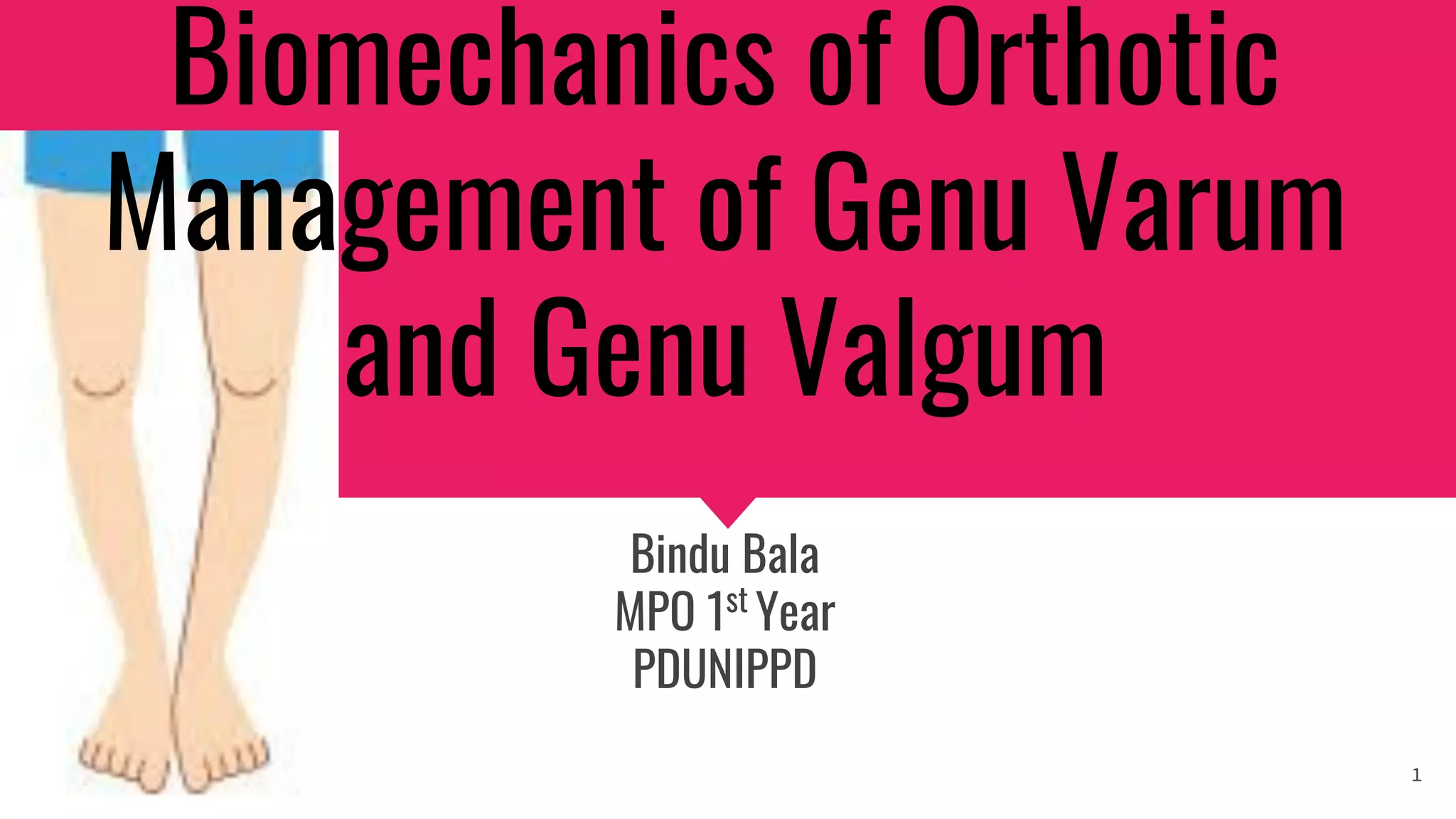
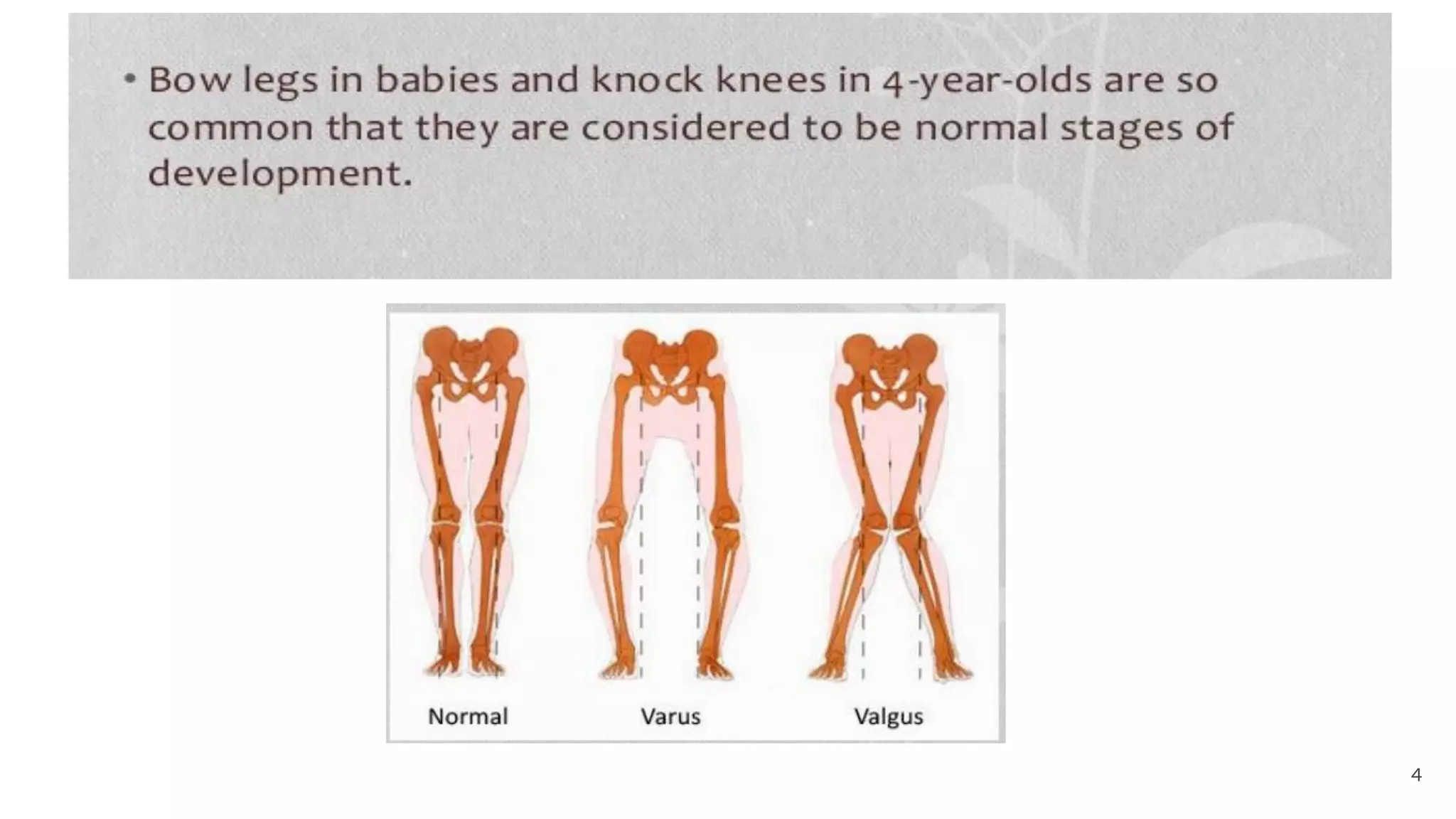
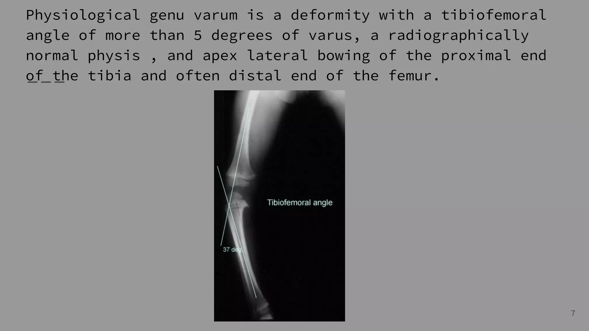
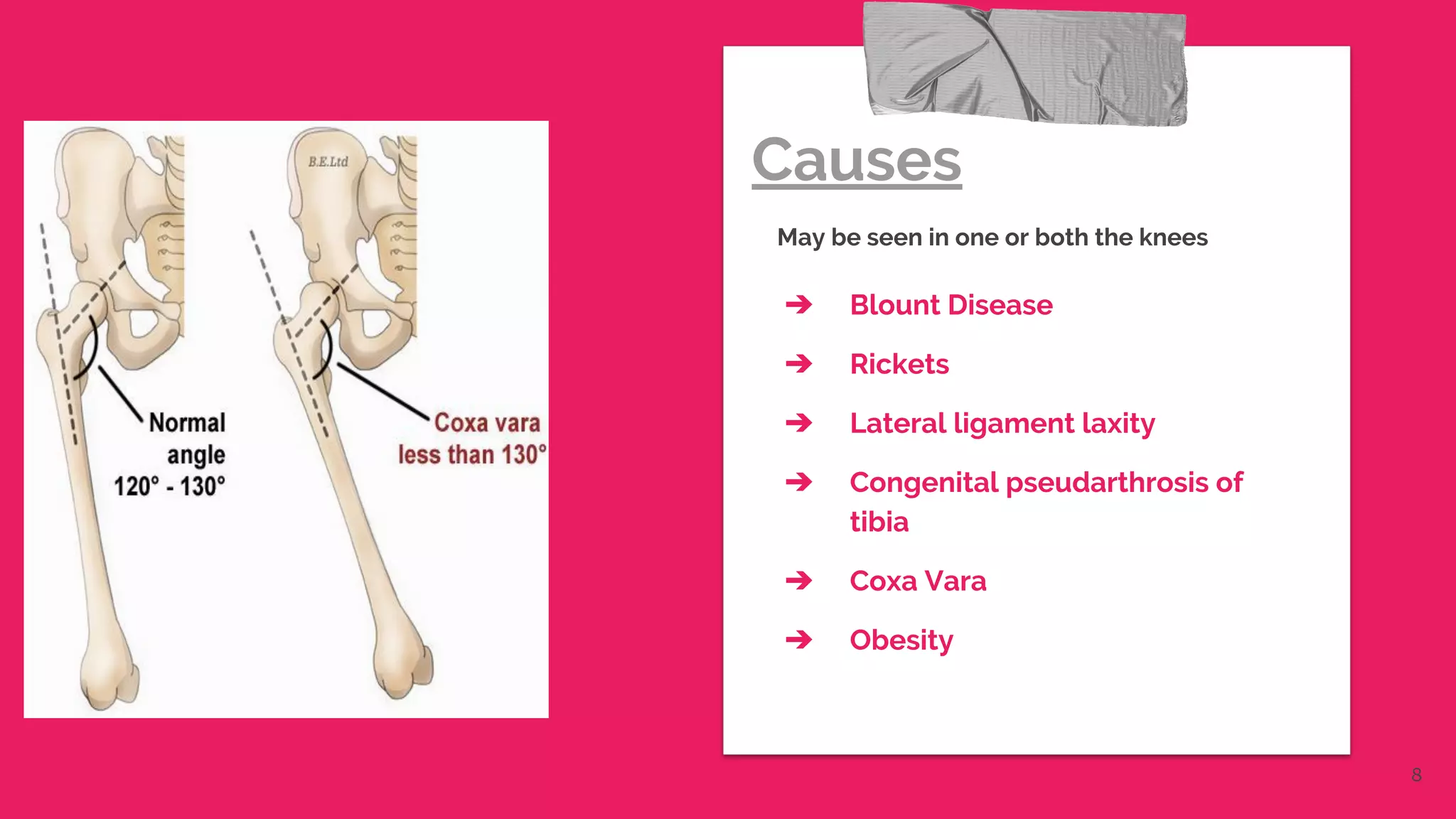
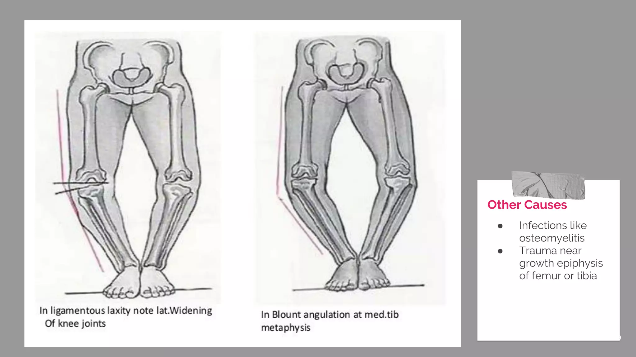
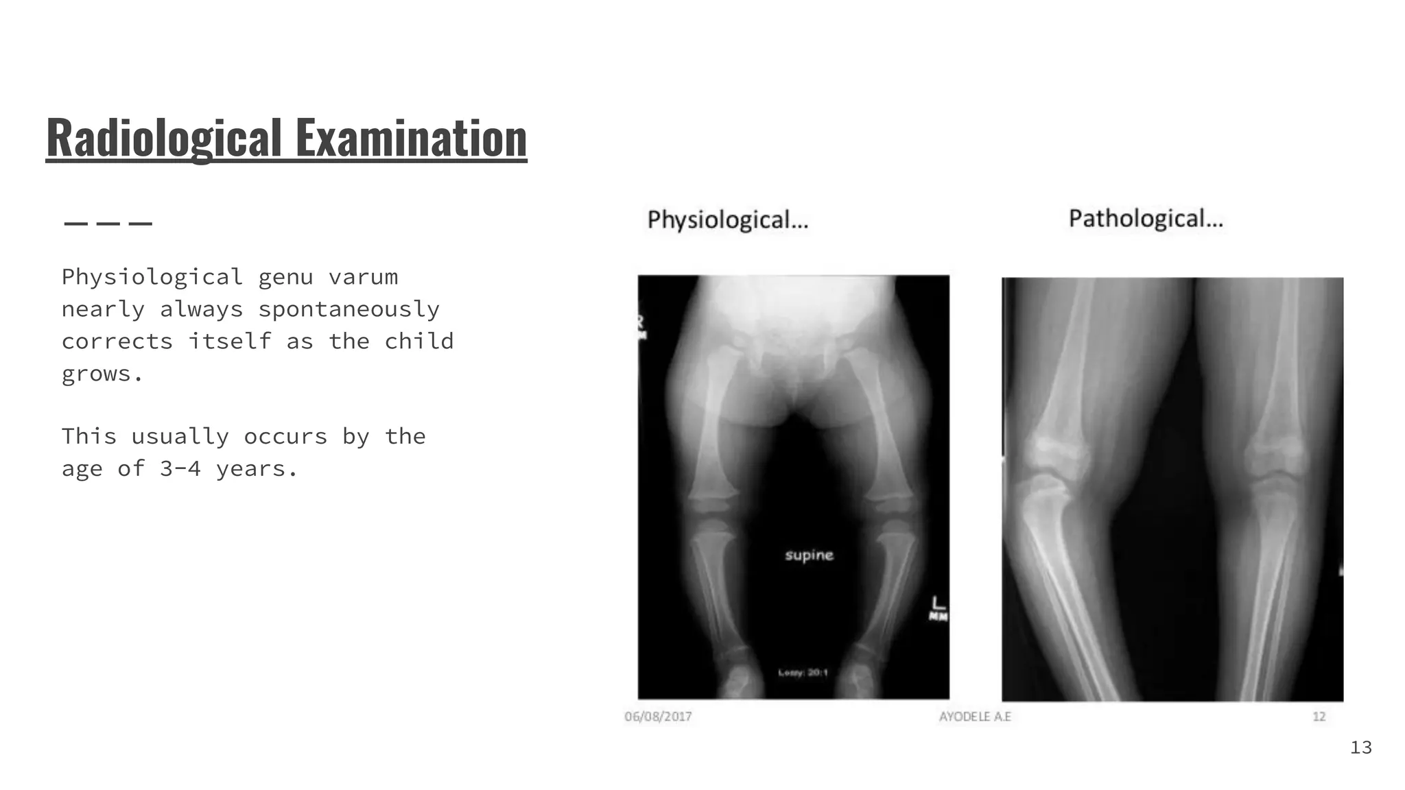
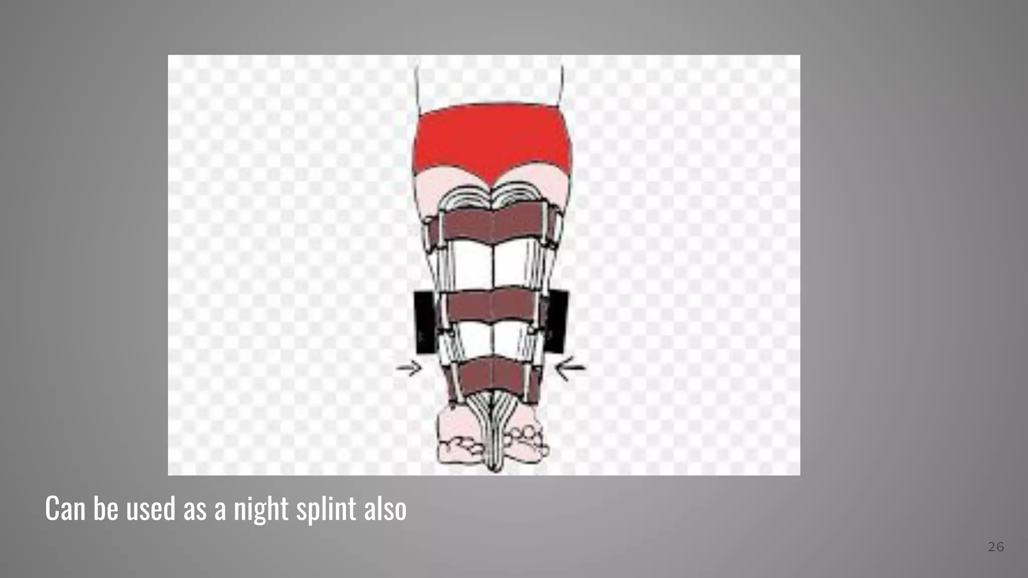
Biomechanics of Orthotic
Management of Genu Varum
and Genu Valgum
Bindu Bala
MPO 1st
Year
PDUNIPPD
1
What is Genu Valgum ?
Genu valgum, commonly called "knock-knee", is a condition
in which the knees angle in and touch each other when the
legs are straightened.
2
What is Genu Varum ?
Also called bow-leggedness, bandiness,
bandy-leg, and tibia vara, is a varus deformity
marked by (outward) bowing at the knee, which
means that the lower leg is angled inward
(medially) in relation to the thigh axis, giving the
limb overall the appearance of an archer's bow.
3
4
How to measure ?
5
The Q-angle is the angle
formed by a line drawn from
the Anterior superior iliac
spine to(ASIS) to the center
of the patella.
A second line is drawn from
the center of the patella to
tibial tubercle.
The angle formed by the two
lines is called Q-Angle.
The normal Q angle in males
is 140
and in females is 170
.
Q-Angle
6
Physiological genu varum is a deformity with a tibiofemoral
angle of more than 5 degrees of varus, a radiographically
normal physis , and apex lateral bowing of the proximal end
of the tibia and often distal end of the femur.
7
Causes
May be seen in one or both the knees
➔ Blount Disease
➔ Rickets
➔ Lateral ligament laxity
➔ Congenital pseudarthrosis of
tibia
➔ Coxa Vara
➔ Obesity
8
Other Causes
● Infections like
osteomyelitis
● Trauma near
growth epiphysis
of femur or tibia
9
In Adults
● Osteoarthritis
● Bone softening
diseases such as
Paget’s Disease
● Malunion of lower
part of femur or
upper part of tibia.
10
Physical Examination
● Bilateral bow leg can be recorded by measuring the
distance between the knees with the child standing and
the heels touching , it should be less than 6 cm.
● McMurray Test-
❏ The patient lies in the supine position with the knee
completely flexed(the heel to the buttock).
❏ The examiner then medially rotates the tibia and extends
the knee. To test the medial meniscus.
❏ Positive test- snap or click is often accompanied by
pain.
11
● Varus Stress Test-
❏ Patient is in supine position with the knee fully
extended.
❏ Stabilise the affected leg in slight external rotation
with one hand on the lateral malleolus.
❏ Place the other hand on the medial aspect of the knee and
apply a laterally directed stress on the medial knee.
❏ Flex their knee to 300
and apply the same pressure on the
lateral side to isolate the lateral collateral ligament.
12
Radiological Examination
Physiological genu varum
nearly always spontaneously
corrects itself as the child
grows.
This usually occurs by the
age of 3-4 years.
13
TREATMENT
14
Different procedures ;two main types
➔ Guided growth
This surgery of the growth plate stops
the growth on the healthy side of the
shinbone which gives the abnormal
side a chance to play catch up,
straightening the leg with the child’s
natural growth.
➔ Tibial Osteotomy
In this procedure, the shinbone is cut
just below the knee and reshaped to
correct the alignment.
After Surgery
A cast may be applied to
protect the bone while it
heals
Surgical
Treatment
15
Mostly used in progressive physiological genu varum but
limited use in pathological genu varum.
Common splints used are- mermaid splint, medial single bar
knee ankle foot orthosis, shoe modification with elevating
inner border of the shoe.
Orthotic Management
16
PREREQUISITES FOR THE APPROPRIATENESS OF AN
ORTHOSIS
● The affected leg should show no contractures at the hip
and the knee-joint. The patient should be able to stretch
the knee-joint passively without problems.
● The iliopsoas and the gluteus maximus should not be too
weak. In any case, the patient must be able to move the
knee joint back and forth actively and easily while in a
standing position.
● The patient, especially children, must be willing to get
used to the orthosis and practice a certain walking
17
Single upright KAFO
● Single upright KAFO with
free knee and ankle joints.
● The upright is on the medial
side of the leg for varum
and lateral side for valgum
18
Story for illustration purposes only
Function
● Used to control genu
Knee movement.
● A ring lock may be used
to control knee flexion
for more smaller,
lightweight patients.
● Ankle varus can be
controlled with the
addition of a T strap.
● Free, limited
motion,dorsiflexion or
plantarflexion may be
incorporated.
19
Force Systems
The first force system to control genu varum
consists of the following
➔ Force One
A laterally directed force on the
medial,proximal thigh band and cuff.
➔ Force Two
A medially directed force on the lateral
aspect of the knee joint(i.e femoral
condyles)
➔ Force Three
A laterally directed force on the medial
side of the calcaneus by the contour of the
shoe 20
Force Systems
The first force system to control genu valgum
consists of the following
➔ Force One
A medially directed force on the lateral
,proximal thigh band and cuff.
➔ Force Two
A laterally directed force on the medial
aspect of the knee joint(i.e femoral
condyles)
➔ Force Three
A medially directed force on the lateral
side of the calcaneus by the contour of the
shoe
21
Due to the knee being involved the ankle is also affected
● In case of valgus the ankle is under a medially directed
force.
● Also in varus the ankle feels a laterally directed force.
22
To correct
this lateral
and medial
wedges are
used inside
the shoes
according to
the deformity.
23
Additional Force
An additional force may
be applied at the ankle
joint with the help of
Lateral T strap or Medial
T strap whichever one is
required.
24
Mermaid Splint
Used to keep both legs
straight.
Two conjoined polypropylene
valves with soft foam
padding.
25
Can be used as a night splint also
26
27

Genu varum orthotic management

  • 1.
    Biomechanics of Orthotic Managementof Genu Varum and Genu Valgum Bindu Bala MPO 1st Year PDUNIPPD 1
  • 2.
    What is GenuValgum ? Genu valgum, commonly called "knock-knee", is a condition in which the knees angle in and touch each other when the legs are straightened. 2
  • 3.
    What is GenuVarum ? Also called bow-leggedness, bandiness, bandy-leg, and tibia vara, is a varus deformity marked by (outward) bowing at the knee, which means that the lower leg is angled inward (medially) in relation to the thigh axis, giving the limb overall the appearance of an archer's bow. 3
  • 4.
  • 5.
  • 6.
    The Q-angle isthe angle formed by a line drawn from the Anterior superior iliac spine to(ASIS) to the center of the patella. A second line is drawn from the center of the patella to tibial tubercle. The angle formed by the two lines is called Q-Angle. The normal Q angle in males is 140 and in females is 170 . Q-Angle 6
  • 7.
    Physiological genu varumis a deformity with a tibiofemoral angle of more than 5 degrees of varus, a radiographically normal physis , and apex lateral bowing of the proximal end of the tibia and often distal end of the femur. 7
  • 8.
    Causes May be seenin one or both the knees ➔ Blount Disease ➔ Rickets ➔ Lateral ligament laxity ➔ Congenital pseudarthrosis of tibia ➔ Coxa Vara ➔ Obesity 8
  • 9.
    Other Causes ● Infectionslike osteomyelitis ● Trauma near growth epiphysis of femur or tibia 9
  • 10.
    In Adults ● Osteoarthritis ●Bone softening diseases such as Paget’s Disease ● Malunion of lower part of femur or upper part of tibia. 10
  • 11.
    Physical Examination ● Bilateralbow leg can be recorded by measuring the distance between the knees with the child standing and the heels touching , it should be less than 6 cm. ● McMurray Test- ❏ The patient lies in the supine position with the knee completely flexed(the heel to the buttock). ❏ The examiner then medially rotates the tibia and extends the knee. To test the medial meniscus. ❏ Positive test- snap or click is often accompanied by pain. 11
  • 12.
    ● Varus StressTest- ❏ Patient is in supine position with the knee fully extended. ❏ Stabilise the affected leg in slight external rotation with one hand on the lateral malleolus. ❏ Place the other hand on the medial aspect of the knee and apply a laterally directed stress on the medial knee. ❏ Flex their knee to 300 and apply the same pressure on the lateral side to isolate the lateral collateral ligament. 12
  • 13.
    Radiological Examination Physiological genuvarum nearly always spontaneously corrects itself as the child grows. This usually occurs by the age of 3-4 years. 13
  • 14.
  • 15.
    Different procedures ;twomain types ➔ Guided growth This surgery of the growth plate stops the growth on the healthy side of the shinbone which gives the abnormal side a chance to play catch up, straightening the leg with the child’s natural growth. ➔ Tibial Osteotomy In this procedure, the shinbone is cut just below the knee and reshaped to correct the alignment. After Surgery A cast may be applied to protect the bone while it heals Surgical Treatment 15
  • 16.
    Mostly used inprogressive physiological genu varum but limited use in pathological genu varum. Common splints used are- mermaid splint, medial single bar knee ankle foot orthosis, shoe modification with elevating inner border of the shoe. Orthotic Management 16
  • 17.
    PREREQUISITES FOR THEAPPROPRIATENESS OF AN ORTHOSIS ● The affected leg should show no contractures at the hip and the knee-joint. The patient should be able to stretch the knee-joint passively without problems. ● The iliopsoas and the gluteus maximus should not be too weak. In any case, the patient must be able to move the knee joint back and forth actively and easily while in a standing position. ● The patient, especially children, must be willing to get used to the orthosis and practice a certain walking 17
  • 18.
    Single upright KAFO ●Single upright KAFO with free knee and ankle joints. ● The upright is on the medial side of the leg for varum and lateral side for valgum 18
  • 19.
    Story for illustrationpurposes only Function ● Used to control genu Knee movement. ● A ring lock may be used to control knee flexion for more smaller, lightweight patients. ● Ankle varus can be controlled with the addition of a T strap. ● Free, limited motion,dorsiflexion or plantarflexion may be incorporated. 19
  • 20.
    Force Systems The firstforce system to control genu varum consists of the following ➔ Force One A laterally directed force on the medial,proximal thigh band and cuff. ➔ Force Two A medially directed force on the lateral aspect of the knee joint(i.e femoral condyles) ➔ Force Three A laterally directed force on the medial side of the calcaneus by the contour of the shoe 20
  • 21.
    Force Systems The firstforce system to control genu valgum consists of the following ➔ Force One A medially directed force on the lateral ,proximal thigh band and cuff. ➔ Force Two A laterally directed force on the medial aspect of the knee joint(i.e femoral condyles) ➔ Force Three A medially directed force on the lateral side of the calcaneus by the contour of the shoe 21
  • 22.
    Due to theknee being involved the ankle is also affected ● In case of valgus the ankle is under a medially directed force. ● Also in varus the ankle feels a laterally directed force. 22
  • 23.
    To correct this lateral andmedial wedges are used inside the shoes according to the deformity. 23
  • 24.
    Additional Force An additionalforce may be applied at the ankle joint with the help of Lateral T strap or Medial T strap whichever one is required. 24
  • 25.
    Mermaid Splint Used tokeep both legs straight. Two conjoined polypropylene valves with soft foam padding. 25
  • 26.
    Can be usedas a night splint also 26
  • 27.