Downloaded 473 times


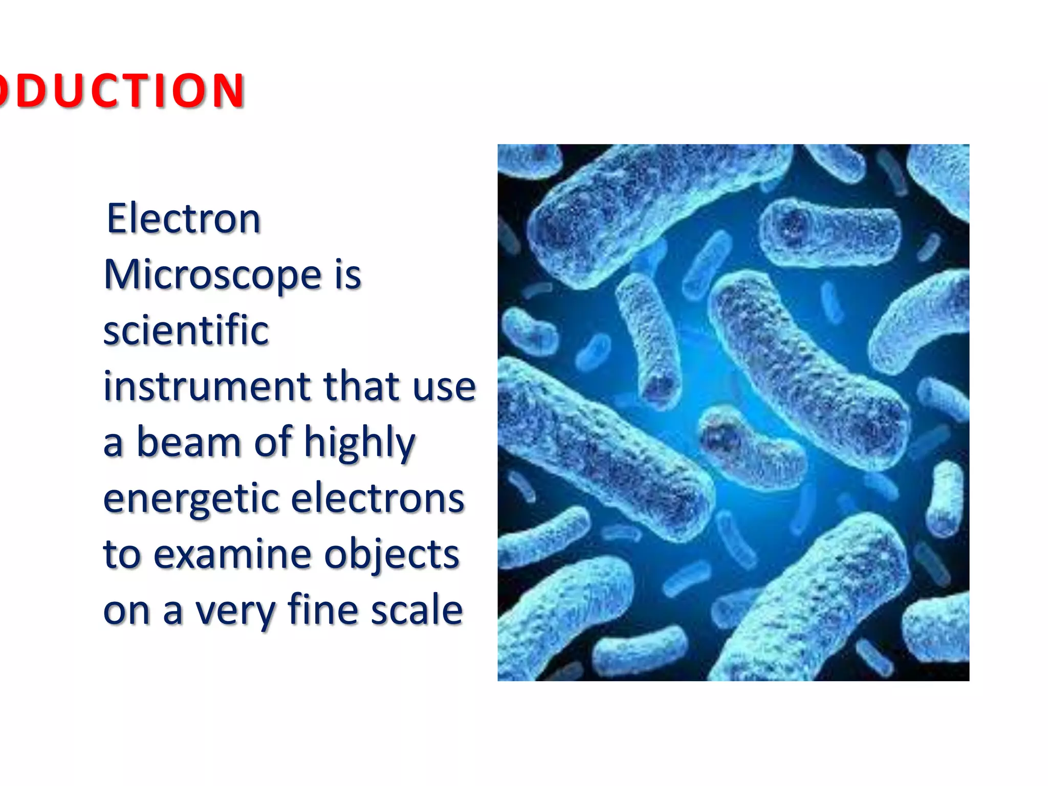


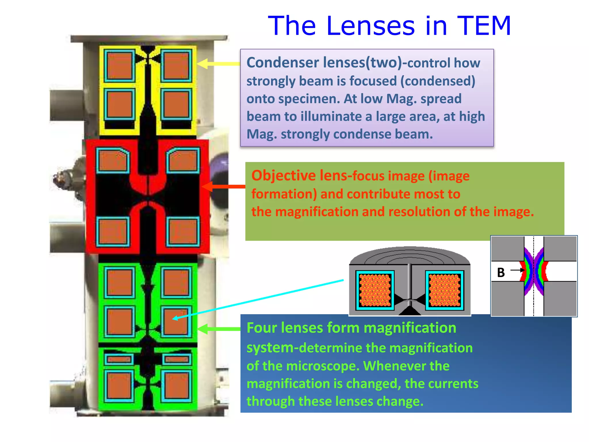
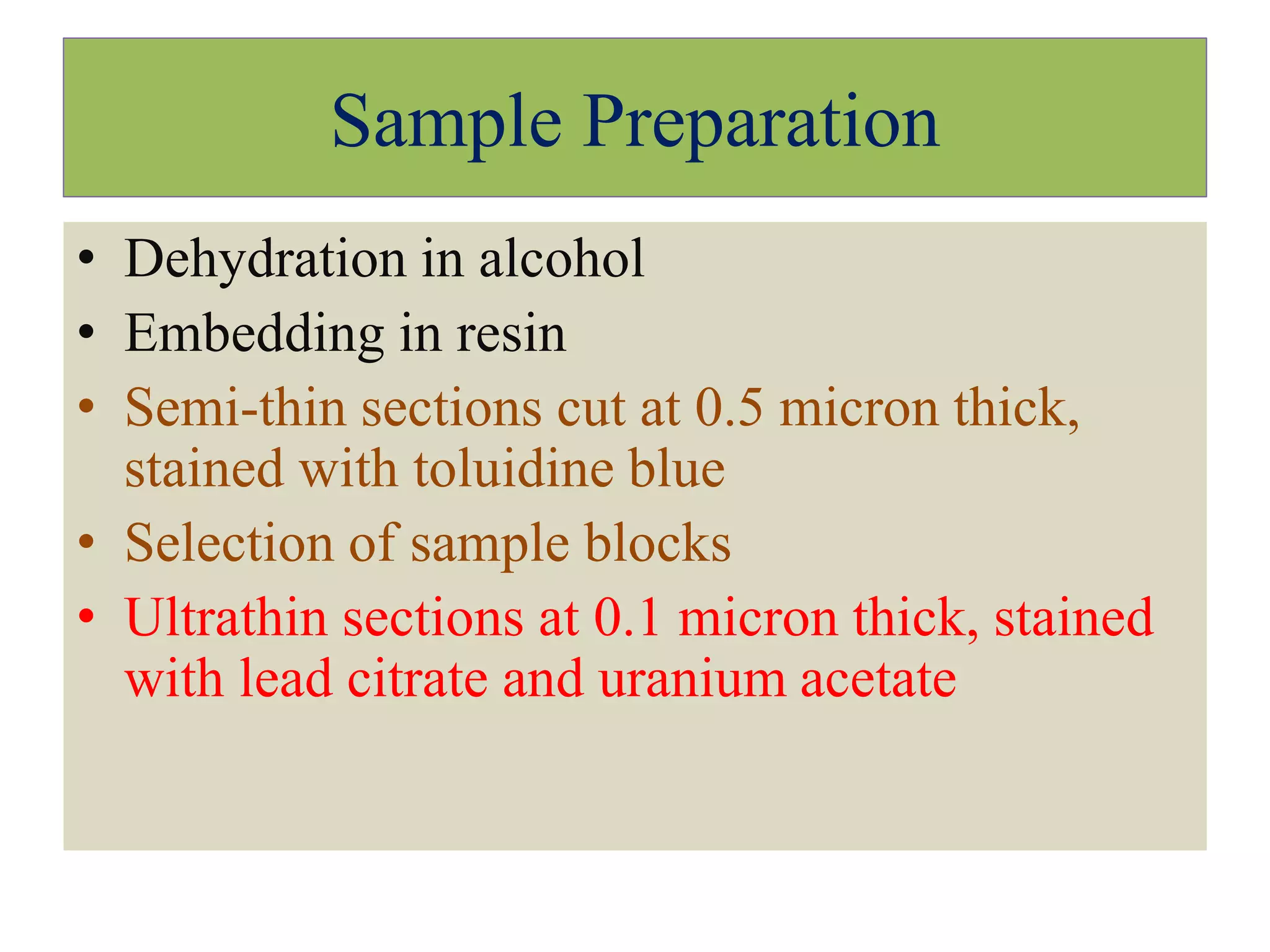

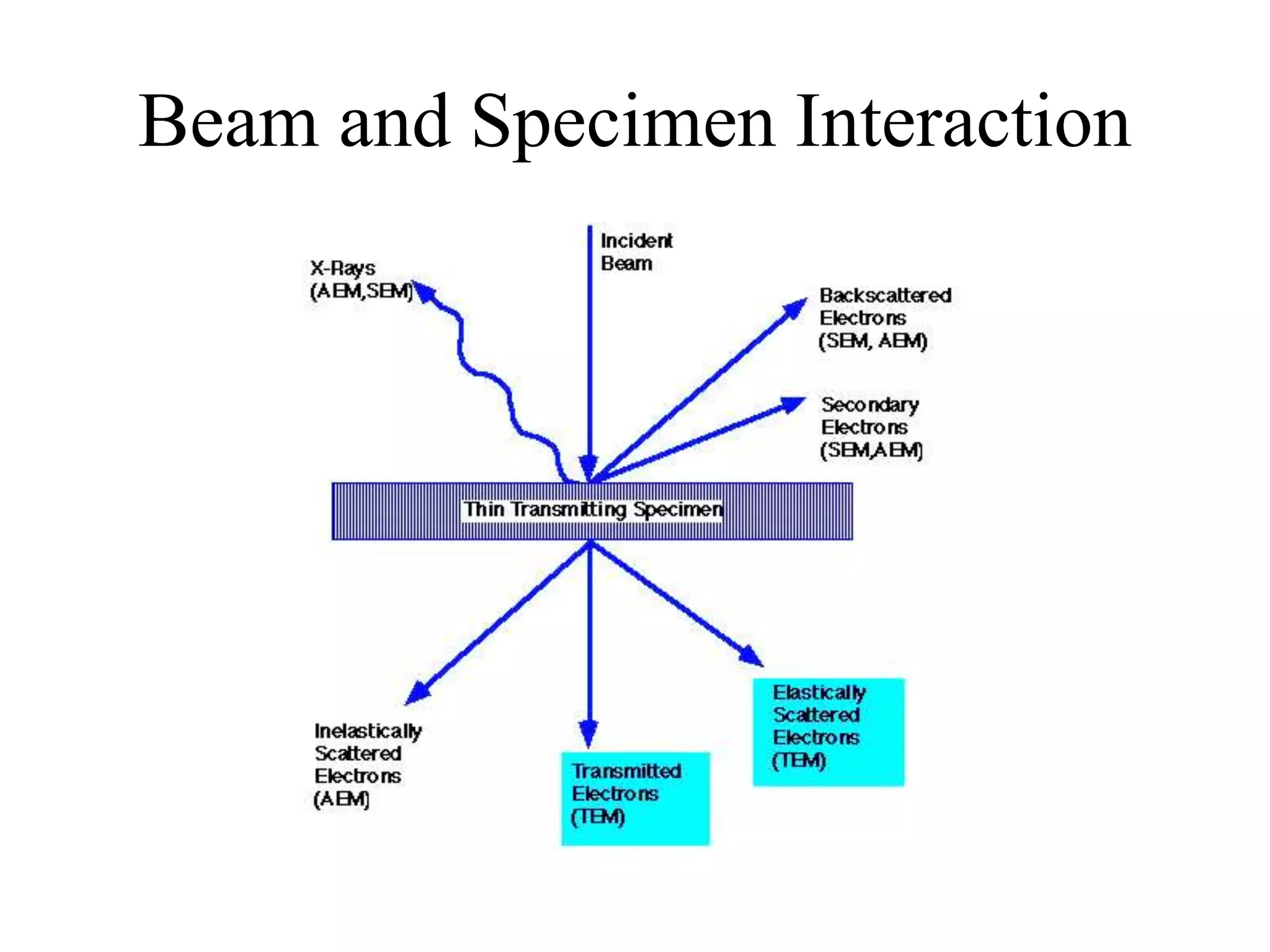

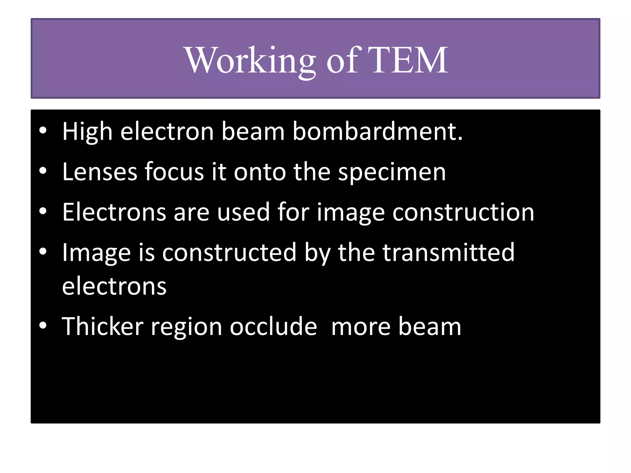


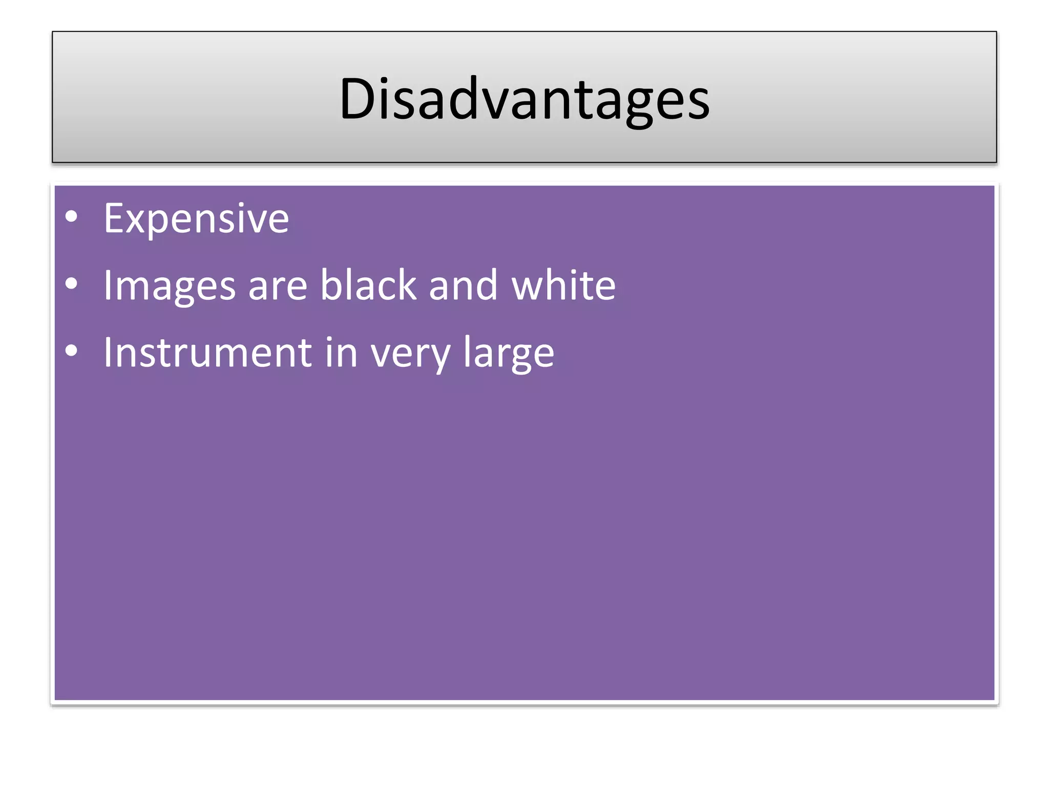


Transmission Electron Microscopy (TEM) is a high-resolution imaging technique that utilizes a beam of energetic electrons to examine ultra-thin specimens. Unlike optical microscopes, TEM requires a vacuum, offers significant magnification up to 10,000x, and provides detailed information on an object's topography, morphology, composition, and crystallographic structure. Despite its advantages, TEM is expensive, operates in challenging conditions, and requires complex sample preparation.


