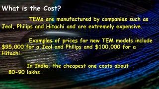
TEM is a type of electron microscope that uses electron beams to produce magnified images of samples. TEMs can magnify up to 1 million times, allowing observation of ultrafine cell structures. Sample preparation is required to make specimens thin enough for electrons to pass through. TEMs are very expensive, ranging from $95,000 to over $100,000, but provide high resolution imaging useful for fields like nanotechnology, biology and materials science.
Transmission electron microscopy (TEM) is a microscopy technique in which a beam of electrons is guided through an ultra thin specimen, interacting with the specimen as it passes through.An image is formed from the fundamental interaction of the electrons transmitted through the specimen; the image is magnified and focused onto an imaging device, such as a fluorescent screen, on a layer of photographic film, or to be observed by a sensor such as a CCD camera.
1) CONTENTS:
Introduction
Construction
Working Principle
The Electron Gun And Condenser System
Image Producing & Recording System
TEM Applications
Advantages
Disadvantages
2) INTRODUCTION:
A Transmission Electron Microscope (TEM) utilizes energetic electron beam to provide morphologic, compositional and crystallographic information on samples.TEM produce High-Resolution, 2D images. The first transmission electron microscope was invented in 1933 by Max Knoll and E. Ruska at the Technical College in Berlin.
3) CONSTRUCTION:
Electron Gun – to produce electrons.
Magnetic condensing lens - to condense the electrons and to adjust the spot size of the electron.The specimen is placed in between the condensing lens and the objective lens.
The magnetic objective lens - to block the high angle diffracted
beam.
Aperture - eliminate the diffracted beam (if any) and in turn
increases the contrast of the image.The magnetic projector lens - to achieve higher magnification.
Fluorescent (Phosphor) screen – To record the image.
4)Working Principle: High voltage electron beam is transmitted through a specimen to form an image. Stream of electrons are produced by the electron gun and is made to fall over the specimen using the magnetic condensing lens.Electrons are made to pass through the specimen and the image is formed on the fluorescent screen.
5) The Electron Gun And Condenser System: The image can be manipulated by adjusting the voltage of the gun to accelerate or decrease the speed of electrons as well as changing the electromagnetic wavelength via the solenoids.
6) Image Producing & Recording System:
Air needs to be pumped out of the vacuum chamber, creating a
space where electrons are able to move.The objective lens is used to produces a image and then further magnified by the projector lens. The lighter areas of the image represent the places where a greater number of electrons were able to pass through the sample and the darker areas reflect the dense areas of the object. Monochromatic image is recorded in fluorescent screen or by capturing the image digitally to display on a computer monitor,basically stored in a TIFF or JPEG format.
7)TEM Applications:
It analyze structure, topographical, morphological, compositional and crystalline information. Can be used in semiconductor analysis and production and the manufacturing of computer and silicon chips. To identify fractures and damages.
8)Advantages:
Powerful magnification . It can produce magnification as high as 1,00,000 times as that of the size of the object.
Images are high-quality and detailed.They are easy to operate with proper training.
9)Disadvantages:
Large and very expensive.
Laborious sample preparation.
TEM require special housing and maintenance.
Samples are limited to those that are electron transparent.
10) Thank You
Beam of electrons is transmitted through an ultra thin specimen,
An image is formed from the interaction of the electrons transmitted through the specimen,
The image is magnified and focused onto an imaging device, such as a fluorescent screen, on a layer of photographic film, or to be detected by a sensor such as a CCD camera
Electron microscope, principle and applicationKAUSHAL SAHU
Introduction
History
Resolution &Magnification of
Electron microscope
Types of electron microscope
1) Transmission electron microscope (TEM)
- Structural parts of TEM
- Principle & Working of TEM
- Sample preparation for TEM
- Advantages & disadvantages of TEM
Scanning electron microscope (SEM)
- Structural parts of SEM
- Principle & Working of SEM
- Sample preparation for SEM
- Advantages & disadvantages of SEM
3) Scanning transmission electron microscope (STEM)
Applications of electron microscope
Conclusion
References
5. Microsocope ELECTRON MICROSCOPE (TEM & SEM ) - BasicsNethravathi Siri
Basics only
Electron beam is the source of illumination.
Image is produced by magnetic field.
Contrasting features between light microscope and electron microscope are
construction, working principle, specimen preparation, cost-expenses and designed
room (vacuum chamber).
Electron Microscopy - Scanning electron microscope, Transmission Electron Mic...Sumer Pankaj
An electron microscope is a microscope that uses a beam of accelerated electrons as a source of illumination. As the wavelength of an electron can be up to 100,000 times shorter than that of visible light photons, electron microscopes have a higher resolving power than light microscopes and can reveal the structure of smaller objects. A transmission electron microscope can achieve better than 50 pm resolution and magnifications of up to about 10,000,000x whereas most light microscopes are limited by diffraction to about 200 nm resolution and useful magnifications below 2000x.
Electron microscopes are used to investigate the ultrastructure of a wide range of biological and inorganic specimens including microorganisms, cells, large molecules, biopsy samples, metals, and crystals. Industrially, electron microscopes are often used for quality control and failure analysis. Modern electron microscopes produce electron micrographs using specialized digital cameras and frame grabbers to capture the image.
Slide 1: Title Slide
Extrachromosomal Inheritance
Slide 2: Introduction to Extrachromosomal Inheritance
Definition: Extrachromosomal inheritance refers to the transmission of genetic material that is not found within the nucleus.
Key Components: Involves genes located in mitochondria, chloroplasts, and plasmids.
Slide 3: Mitochondrial Inheritance
Mitochondria: Organelles responsible for energy production.
Mitochondrial DNA (mtDNA): Circular DNA molecule found in mitochondria.
Inheritance Pattern: Maternally inherited, meaning it is passed from mothers to all their offspring.
Diseases: Examples include Leber’s hereditary optic neuropathy (LHON) and mitochondrial myopathy.
Slide 4: Chloroplast Inheritance
Chloroplasts: Organelles responsible for photosynthesis in plants.
Chloroplast DNA (cpDNA): Circular DNA molecule found in chloroplasts.
Inheritance Pattern: Often maternally inherited in most plants, but can vary in some species.
Examples: Variegation in plants, where leaf color patterns are determined by chloroplast DNA.
Slide 5: Plasmid Inheritance
Plasmids: Small, circular DNA molecules found in bacteria and some eukaryotes.
Features: Can carry antibiotic resistance genes and can be transferred between cells through processes like conjugation.
Significance: Important in biotechnology for gene cloning and genetic engineering.
Slide 6: Mechanisms of Extrachromosomal Inheritance
Non-Mendelian Patterns: Do not follow Mendel’s laws of inheritance.
Cytoplasmic Segregation: During cell division, organelles like mitochondria and chloroplasts are randomly distributed to daughter cells.
Heteroplasmy: Presence of more than one type of organellar genome within a cell, leading to variation in expression.
Slide 7: Examples of Extrachromosomal Inheritance
Four O’clock Plant (Mirabilis jalapa): Shows variegated leaves due to different cpDNA in leaf cells.
Petite Mutants in Yeast: Result from mutations in mitochondrial DNA affecting respiration.
Slide 8: Importance of Extrachromosomal Inheritance
Evolution: Provides insight into the evolution of eukaryotic cells.
Medicine: Understanding mitochondrial inheritance helps in diagnosing and treating mitochondrial diseases.
Agriculture: Chloroplast inheritance can be used in plant breeding and genetic modification.
Slide 9: Recent Research and Advances
Gene Editing: Techniques like CRISPR-Cas9 are being used to edit mitochondrial and chloroplast DNA.
Therapies: Development of mitochondrial replacement therapy (MRT) for preventing mitochondrial diseases.
Slide 10: Conclusion
Summary: Extrachromosomal inheritance involves the transmission of genetic material outside the nucleus and plays a crucial role in genetics, medicine, and biotechnology.
Future Directions: Continued research and technological advancements hold promise for new treatments and applications.
Slide 11: Questions and Discussion
Invite Audience: Open the floor for any questions or further discussion on the topic.
This pdf is about the Schizophrenia.
For more details visit on YouTube; @SELF-EXPLANATORY;
https://www.youtube.com/channel/UCAiarMZDNhe1A3Rnpr_WkzA/videos
Thanks...!
Seminar of U.V. Spectroscopy by SAMIR PANDASAMIR PANDA
Spectroscopy is a branch of science dealing the study of interaction of electromagnetic radiation with matter.
Ultraviolet-visible spectroscopy refers to absorption spectroscopy or reflect spectroscopy in the UV-VIS spectral region.
Ultraviolet-visible spectroscopy is an analytical method that can measure the amount of light received by the analyte.
THE IMPORTANCE OF MARTIAN ATMOSPHERE SAMPLE RETURN.Sérgio Sacani
The return of a sample of near-surface atmosphere from Mars would facilitate answers to several first-order science questions surrounding the formation and evolution of the planet. One of the important aspects of terrestrial planet formation in general is the role that primary atmospheres played in influencing the chemistry and structure of the planets and their antecedents. Studies of the martian atmosphere can be used to investigate the role of a primary atmosphere in its history. Atmosphere samples would also inform our understanding of the near-surface chemistry of the planet, and ultimately the prospects for life. High-precision isotopic analyses of constituent gases are needed to address these questions, requiring that the analyses are made on returned samples rather than in situ.
Richard's aventures in two entangled wonderlandsRichard Gill
Since the loophole-free Bell experiments of 2020 and the Nobel prizes in physics of 2022, critics of Bell's work have retreated to the fortress of super-determinism. Now, super-determinism is a derogatory word - it just means "determinism". Palmer, Hance and Hossenfelder argue that quantum mechanics and determinism are not incompatible, using a sophisticated mathematical construction based on a subtle thinning of allowed states and measurements in quantum mechanics, such that what is left appears to make Bell's argument fail, without altering the empirical predictions of quantum mechanics. I think however that it is a smoke screen, and the slogan "lost in math" comes to my mind. I will discuss some other recent disproofs of Bell's theorem using the language of causality based on causal graphs. Causal thinking is also central to law and justice. I will mention surprising connections to my work on serial killer nurse cases, in particular the Dutch case of Lucia de Berk and the current UK case of Lucy Letby.
Multi-source connectivity as the driver of solar wind variability in the heli...Sérgio Sacani
The ambient solar wind that flls the heliosphere originates from multiple
sources in the solar corona and is highly structured. It is often described
as high-speed, relatively homogeneous, plasma streams from coronal
holes and slow-speed, highly variable, streams whose source regions are
under debate. A key goal of ESA/NASA’s Solar Orbiter mission is to identify
solar wind sources and understand what drives the complexity seen in the
heliosphere. By combining magnetic feld modelling and spectroscopic
techniques with high-resolution observations and measurements, we show
that the solar wind variability detected in situ by Solar Orbiter in March
2022 is driven by spatio-temporal changes in the magnetic connectivity to
multiple sources in the solar atmosphere. The magnetic feld footpoints
connected to the spacecraft moved from the boundaries of a coronal hole
to one active region (12961) and then across to another region (12957). This
is refected in the in situ measurements, which show the transition from fast
to highly Alfvénic then to slow solar wind that is disrupted by the arrival of
a coronal mass ejection. Our results describe solar wind variability at 0.5 au
but are applicable to near-Earth observatories.
Observation of Io’s Resurfacing via Plume Deposition Using Ground-based Adapt...Sérgio Sacani
Since volcanic activity was first discovered on Io from Voyager images in 1979, changes
on Io’s surface have been monitored from both spacecraft and ground-based telescopes.
Here, we present the highest spatial resolution images of Io ever obtained from a groundbased telescope. These images, acquired by the SHARK-VIS instrument on the Large
Binocular Telescope, show evidence of a major resurfacing event on Io’s trailing hemisphere. When compared to the most recent spacecraft images, the SHARK-VIS images
show that a plume deposit from a powerful eruption at Pillan Patera has covered part
of the long-lived Pele plume deposit. Although this type of resurfacing event may be common on Io, few have been detected due to the rarity of spacecraft visits and the previously low spatial resolution available from Earth-based telescopes. The SHARK-VIS instrument ushers in a new era of high resolution imaging of Io’s surface using adaptive
optics at visible wavelengths.
Earliest Galaxies in the JADES Origins Field: Luminosity Function and Cosmic ...Sérgio Sacani
We characterize the earliest galaxy population in the JADES Origins Field (JOF), the deepest
imaging field observed with JWST. We make use of the ancillary Hubble optical images (5 filters
spanning 0.4−0.9µm) and novel JWST images with 14 filters spanning 0.8−5µm, including 7 mediumband filters, and reaching total exposure times of up to 46 hours per filter. We combine all our data
at > 2.3µm to construct an ultradeep image, reaching as deep as ≈ 31.4 AB mag in the stack and
30.3-31.0 AB mag (5σ, r = 0.1” circular aperture) in individual filters. We measure photometric
redshifts and use robust selection criteria to identify a sample of eight galaxy candidates at redshifts
z = 11.5 − 15. These objects show compact half-light radii of R1/2 ∼ 50 − 200pc, stellar masses of
M⋆ ∼ 107−108M⊙, and star-formation rates of SFR ∼ 0.1−1 M⊙ yr−1
. Our search finds no candidates
at 15 < z < 20, placing upper limits at these redshifts. We develop a forward modeling approach to
infer the properties of the evolving luminosity function without binning in redshift or luminosity that
marginalizes over the photometric redshift uncertainty of our candidate galaxies and incorporates the
impact of non-detections. We find a z = 12 luminosity function in good agreement with prior results,
and that the luminosity function normalization and UV luminosity density decline by a factor of ∼ 2.5
from z = 12 to z = 14. We discuss the possible implications of our results in the context of theoretical
models for evolution of the dark matter halo mass function.
3. INTRODUCTION
ELECTRON MICROSCOPE - System of
electromagnetic coils where electron beams are used as a
source of illumination.
Electron microscope = Magnification is
high.
Magnification of 2000 times than that
of light microscope.
4. TYPES OF ELECTRON MICROSCOPE
1. Transmission Electron Microscope (TEM)
2. Scanning Electron Microscope (SEM)
5. TRANSMISSION ELECTRON MICROSCOPE
TEM is a special type of microscope that uses
electron for magnification.
Electrons - smaller wavelength.
Achieve extreme magnification.
Average TEM - Magnification of 1,000,000x
6. DEFINITION :
Electron Microscope in which electron beam is
passed through the specimen to produce its image.
First TEM was designed by Max knoll and Ernest
ruska in 1931.
TEM is a powerful tool for Material science.
Reveals the finest details of internal structures.
7. PRINCIPLE
Basic principle of electron microscope is
similar to that of the ordinary compound microscope.
Electron Beam - Light Beam
Electromagnetic coils - Optical lenses.
When light voltage current is passed through
a filament of cathode ray tube, electron beams are
produced from the filament.
8. If some voltage of current is applied to
electromagnetic coils kept around the path of electron
beam, the direction of electron beam can be changed
suitably to focus on the specimen.
When an electron beam is passed through a
specimen stained with metallic gold or osmium, the
specimen absorbs some rays and reflects some rays to pass
through it.
9.
10. Interaction between electrons and specimen in
the beam produces the image of the specimen.
Image of the specimen can be collected by an
objective lens (an electromagnetic coil)
Magnified by an Amplifier (another coil)
Due to electron distribution, image cannot be
seen with naked eyes.
Image - Recorded on a Screen or Camera.
11. INSTRUMENTATION
TEM consists of electron gun, condenser lens,
objective lens, amplifier lens, projector lens & fluorescent
screen.
ELECTRON GUN - Source of electron beam
CONDENSER LENS - Located below the electron gun
Collect and concentrate the
electron into a strong beam
before focusing onto specimen.
12. SPECIMEN STAGE - Below the condenser lens
OBJECTIVE LENS - Another electromagnetic coil,
placed below the specimen stage.
It collects the images of the
specimen and focuses it towards
the amplifier lens.
AMPLIFIER LENS - Magnifies image produced by the
objective lens to several 1000
Times.
PROJECTOR LENS - Collects the magnified image &
focus it onto fluorescent screen.
13.
14. SAMPLE PREPARATION
The biological samples have to be loaded
with heavy atoms like gold or osmonium.
SAMPLE PREPARATION AND EXAMINATION :
1. Wet specimen is dehydrated using ethanol or acetone.
2. Fixation is done by Osmonium tetroxide,
glutaraldehyde, potassium permanganate,formalin etc.
15. 3. The fixed specimen is embedded in hard embedding
medium like aradilite and cut into thin section of 50-
100nm thickness using ultramicrotome.
4. Thin sections subjected to metallic staining and placed
on the specimen stage between the condenser lens coil
and objective coil.
16. IMAGING
A Transmission Electron Microscope produces a
high-resolution, black and white image from the
interaction that takes place between prepared samples
and energetic electrons in the vacuum chamber.
Air needs to be pumped out of the vacuum
chamber, creating a space where electrons are able to
move.
The electrons then pass through multiple
electromagnetic lenses.
17. These solenoids are tubes with coil wrapped around
them.
The beam passes through the solenoids, down the
column, makes contact with the screen where the
electrons are converted to light and form an image.
The image can be manipulated by adjusting the
voltage of the gun to accelerate or decrease the speed of
electrons as well as changing the electromagnetic
wavelength via the solenoids.
The coils focus images onto a screen or photographic
plate.
18. During transmission, the speed of electrons directly
correlates to electron wavelength; the faster electrons
move, the shorter wavelength and the greater the quality
and detail of the image.
The lighter areas of the image represent the places
where a greater number of electrons were able to pass
through the sample and the darker areas reflect the dense
areas of the object.
These differences provide information on the
structure, texture, shape and size of the sample.
19. SAMPLE PROPERTIES :
Samples need to have certain properties.
They need to be sliced thin enough for electrons to
pass through, a property known as electron transparency.
Samples need to be able to withstand the vacuum
chamber and often require special preparation before
viewing.
Types of preparation include dehydration, sputter
coating of non-conductive materials, cryofixation,
sectioning and staining.
20. APPLICATIONS
● Ideal tool for study of ultra structure of cells.
● Identification of plant and animal viruses based on
their structural features.
● Employed in the localization of nucleic acid, enzymes
and proteins in cell and cell organelles.
● Used in cancer research for the cytological
observation of cancer cells.
21. ● Used in various fields such as nanotechnology, life
sciences, medical, biological, material research,
forensic analysis, metallurgy as well as in industries
and education.
● Used in the production and manufacturing of
computer and silicon chips.
● Provide topographical, morphological, compositional
and crystalline informations.
22. Advantages
A Transmission Electron Microscope is an
impressive instrument with a number of advantages such
as:
● TEMs offer the most powerful magnification,
potentially over one million times or more.
● TEMs have a wide-range of applications and can be
utilized in a variety of different scientific, educational
and industrial fields
23. ● TEMs provide information on element and
compound structure
● Images are high-quality and detailed
● TEMs are able to yield information of surface
features, shape, size and structure
● They are easy to operate with proper training
24. Disadvantages
Some limitations of electron microscopes include:
● TEMs are large and very expensive
● Laborious sample preparation
● Operation and analysis requires special training
● Samples are limited to those that are electron
transparent, able to tolerate the vacuum chamber and
small enough to fit in the chamber.
● TEMs require special housing and maintenance
● Images are black and white
25. What is the Cost?
TEMs are manufactured by companies such as
Jeol, Philips and Hitachi and are extremely expensive.
Examples of prices for new TEM models include
$95,000 for a Jeol and Philips and $100,000 for a
Hitachi.
In India, the cheapest one costs about
80-90 lakhs.
26. SUMMARY
TEM is useful for small, nanoscale analytes.
TEM can create 3D images of samples.
TEM can be modified for different atoms and
molecules.
TEM is not cheap. It is Good!