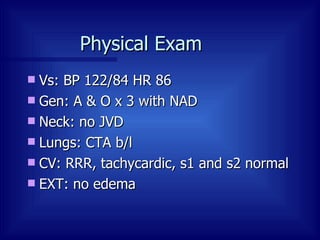
Case Management Zahir Rahman  4.12.04
H & P 56 y.o with h/o of HTN and WPW Presented to the ED with c/o of palpitations for 1 hour with mild chest discomfort.  Denied dizziness or SOB In ED, pt found to be tachycardic with HR 190 and bp 92/50 Pt had a h/o of multiple admissions (5-6 episodes in the last 6 years) for tachycardia always treated with IV medications, never had EPS, never been cardioverted and had always refused ablation
H & P PMHx HTN WPW DM ? Hole in the heart SHx 1ppd x 40 y Occ Etoh  No drugs Medications Inderal LA 160 qd Quinidine 325 po qd Amaryl Labs: H/H: 17.9/51.4 Bun/Cr: 10/1.4
Physical Exam  Vs: BP 122/84 HR 86 Gen: A & O x 3 with NAD Neck: no JVD Lungs: CTA b/l CV: RRR, tachycardic, s1 and s2 normal EXT: no edema
EKG
H & P  Patient was given adenosine without success or change in the rhythm Patients bp dropped to 76/40 with adenosine and responded to IVF increasing bp 100/60.  After discussion with EP, patient was electively cardioverted with 50 joule biphasic synchronized converting to NSR  BP increased to 122/84 post cardioversion
EKG with defibrillation
EKG post defib
Echo
Echo NL LV function NL RV function, RV not dilated Moderate eccentric MR Septal leaflet displacement TV (35 mm) Displacement index (17 mm/m 2 ) VSD (? Flow) Trace TR Mild LV diastolic dysfunction ?ASD
W sign: direct atrial to ventricular conduction seen with accessory bypass tracts
Atrial PES generating orthodromic tachycardia  Refractory accessory bypass tract and conduction through AV node to generate tachycardia
Ebstein’s Anomaly of the Tricuspid Valve
Ebstein’s Anomaly First described by Wilhelm Ebstein in 1866 19 y.o. boy with pulmonary TB c/o SOB and palpitations since childhood At autopsy Tricuspid valve was markedly abnormal with valvular tissue attached partially to the tricuspid annulus and adherent to the right ventricular myocardium The Foramen Ovale was patent and his right heart chambers were dilated  In 1927, Alfred Arnstein suggested the name Ebstein's anomaly for these defects  In 1937, Yates and Shapiro described the first case of the anomaly with associated radiographic and electrocardiographic data.  Graham TP et al. Roberts Congenital Heart Disease in adults.  Complex cyanotic heart disease in adults: 395-99.
Ebstein’s Anomaly The prevalence of Ebstein’s is estimated to be 5.2/100,000 live births Accounts for < 1 % of Congenital heart disease Case control analyses suggest genetic, reproductive and environmental risk factors: FHx White Race Previous miscarriages Maternal exposure to BZD and Lithium Ferencz et al.  Ebsteins malformation of the tricuspid valve: genetic and environmental factors.  The Baltimore-Washington Infant Study Group.  Teratology 1994;50:137-47.
Ebstein’s Anomaly Varying degrees of apical displacement or dysplasia of the septal and posterior leaflet of T.V. Atrialization of the inflow tract of the right ventricle (loss of coarse trabeculae) A variably small functional (muscular) right ventricle Varying degrees of malformation and displacement of the anterior leaflet Usually elongated and sail-like  1.  Freedom RM et al.  Topol Textbook of CV Medicine.  Congenital Heart Disease: Chapter 30. 2. Friedman WF et al.  Braunwald Heart Disease.  Congenital Heart Diesease: Chapter 43.
Ebstein’s anomaly and distal linear attachments A:  Moderate displacement of septal and posterior leaflets ( closed arrow ) with mobile anterior leaflet ( open arrow ).  B: Marked displacement of septal and posterior leaflets with mobile anterior leaflet  C:  Nonmobile anterior leaflet with chordal tethering ( arrows ) to ventricle  D:  Severely stenotic or imperforate Ebstein’s anomaly ( arrows ).  Freedom RM et al.  Topol Textbook of CV Medicine.  Congenital Heart Disease: Chapter 30.
Carpentier’s Classification Type A – RV volume is adequate Type B – Large atrialized RV segment; mobile anterior leaflet Type C – Restricted motion of anterior leaflet; may cause infindubular obstruction Type D – Near complete atrialization of RV
Ebstein’s Anomaly : Clinical Presentation  The degree of impairment of RV function is primarily dependant on: 1 Extent of atrialized RV inflow Magnitude of T.R. With mild T.V. displacement, patients have survived to 7 th  and 8 th  decade of life. 2 Fetuses with florid T.R. develop severe hydrops with intrauterine death. 2 Friedman WF et al.  Braunwald Heart Disease.  Congenital Heart Diesease: Chapter 43. 2.  Freedom RM et al.  Topol Textbook of CV Medicine.  Congenital Heart Disease: Chapter 30.
Ebstein’s: Associated Anomalies Interatrial communication is found in over 50% of Ebstein’s patients PFO or Secundum ASD Pulmonic Stenosis or atresia is the most common important associated defect Neonatal elevated PVR increases TR, increasing R to L shunt, worsening cyanosis ASD primum and VSD alone or in combination RVOTO Tetralogy of Fallot Ebsteins is commonly observed associated with congenitally corrected TGA (or L-type) Freedom RM et al.  Topol Textbook of CV Medicine.  Congenital Heart Disease: Chapter 30.
Ebsteins: Clinical Presentation and outcome Celermajer et al.  reviewed 220 cases of Ebsteins anomaly from fetal to adult life between 1958 and 1991 with 1 – 34 years of follow up. Study was conducted to investigate the presentation and outcome of patients with Ebstein’s anomaly. Celermajer et al. Ebstein’s anomaly: presentation and outcome from fetus to adult.  JACC 1994;23:170-6.
Ebstein’s: MC presentation in each age group Fetuses  86% - abnormal routine prenatal scan Neonates (0-1 mos) 74% with cyanosis Infants (2mos – 2yr) 43% with heart failure 35% with cyanosis Children (3-10 y) 63% with incidental murmur Adolescent (11-18 y) 40% with arrythmia 33% with incidental murmur Adult (> 18 y) 43% with arrythmia 26% - HF with DOE Celermajer et al. Ebstein’s anomaly: presentation and outcome from fetus to adult.  JACC 1994;23:170-6.
Ebstein’s  Celermajer et al. Ebstein’s anomaly: presentation and outcome from fetus to adult.  JACC 1994;23:170-6 .
Ebstein’s: outcome  Early presentation usually associated with pulmonary stenosis or atresia Actuarial survival for a all liveborn patients 67% at 1 y and 61% at 10 y 58 total deaths 25 HF, 19 periop, 8 sudden Predictors of death: Echocardiographic grade of severity at presentation Fetal presentation RVOTO Morbidity was mainly related to arrhythmias and late hemodynamic deterioration Celermajer et al. Ebstein’s anomaly: presentation and outcome from fetus to adult.  JACC 1994;23:170-6.
Ebsteins: Physical exam  S1 – abnormal with loud second component Secondary to abrupt cessation of motion of the large sail-like anterior tricuspid leaflet (sail-sound) Split S2 – due to RBBB or single S2 if decreased PBF S3 and S4 to produce quadruple and quintuple rhythms Cyanosis +/-  Central if R to L shunt Peripheral if low output failure Peripheral pulses normal or slightly diminished with low output heart failure TR - HSM which increase with inspiration Even with severe TR – JV pulsations are rarely abnormal probably because of the damping effect of a large right atrium  Friedman WF et al.  Braunwald Heart Disease.  Congenital Heart Diesease: Chapter 43.
Ebstein’s: CXR “ Balloon shaped” enlarged heart with a narrow vascular pedicle Right atrium is usually quite prominent virtually filling the right chest Pulmonary vascularity correlates with degree of atrial shunting: in cyanotic patients pulmonary flow is diminished and lung fields are oligemic  Cardiomegaly, highly variable is the rule Friedman WF et al.  Braunwald Heart Disease.  Congenital Heart Diesease: Chapter 43.
Ebstein’s: EKG Tall, broad “Himalayan” P waves 1 Prolonged PR interval in pts w/o WPW 1 Commonly a distinctive RBBB (50-75%) 1 Splintered v4R, v1 with rsR’ or rsR’S’ with usually low voltage R’ SVT: Afib, Aflutter, PAT, Junctional Tachycardia in 25-50% pts 1 WPW with short PR in 25% pts 2 Low voltage is typical 3 Alternatively, ECG is normal 3 1. Graham TP et al. Roberts Congenital Heart Disease in adults.  Complex cyanotic heart disease in adults: 395-99. 2. Bialostozky et al.  Ebstein’s malformation of tricuspid valve.  Am. J. Cardiology. 1972:29;826. 3. Friedman WF et al.  Braunwald Heart Disease.  Congenital Heart Diesease: Chapter 43.
Ebstein’s : WPW Right sided accessory pathways Failure of the AV annulus to fuse with the hypoplastic central cardiac skeleton allowing the fetal AV accessory pathways to presist. Usually a band like area of accessory tracts which is difficult to ablate in adults Orthodromic re-entry tachycardia is the most common mechanism ( AV node anterograde conduction and accessory pathway retrograde conduction) Graham TP et al. Roberts Congenital Heart Disease in adults.  Complex cyanotic heart disease in adults: 395-99.
Ebsteins: Catheterization Quantify R to L shunting Estimate degree of T.R.  Estimate size of atrialized and distal RV cavities. Measure C.O. Identify other associated anomalies Rarely, needed unless assessing coronary anatomy Simultaneous intracardiac electrogram and pressure recording obtained from the atrialized portion of the RV: An atrial pressure waveform An ventricular electrogram   Graham TP et al. Roberts Congenital Heart Disease in adults.  Complex cyanotic heart disease in adults: 395-99.
Ebsteins - echocardiography Gold standard for diagnosis of Ebsteins anomaly 4 chamber views shows downward displacement of the septal leaflet and motion or tethering of leaflets Subcostal view defines the dysplasia of the leaflets of the valve, RAE Degree of TR or TS can also be determined by color flow doppler Friedman WF et al.  Braunwald Heart Disease.  Congenital Heart Diesease: Chapter 43.
Ebsteins: Echo Shina et al found that a displacement index of > 8 mm/m2 was a sensitive predictor for ebsteins anomaly . Shina et al. 2D echocardiographic spectrum of Ebsteins anomaly: Detailed anatomic assessment.  JACC 1984: 3.
Ebstein’s: Echocardiographic Severity index Celermajer et al. developed a new echocardiographic grade (1 to 4) in order of increasing severity Cardiac death by grade in infants: I: 0 of 4 II: 1 of 10 (10%) III: 4 of 9 (44%) IV: 5 of 5 (100%) Celermajer et al.  Outcome in neonates with Ebstein’s anomaly.  JACC 1992; 19:1047-8.
Recommendations for Diagnostic Evaluation of Ebstein’s Anomaly in the Adolescent or Young Adult CLASS I ECG Chest X-ray Echo - Doppler Study Pulse Oximetry at rest and/ or during exercise CLASS IIa EPS if documented or suspected atrial arrythmia Above studies at initial evaluation and every 1 – 3 years, depending upon severity BONOW ET AL., ACC/AHA TASK FORCE REPORT JACC Vol. 32, No. 5, November 1998:1486-1588 ACC/AHA Guidelines for the Management of Patients With Valvular Heart Disease
Ebstein’s: Medical Care All patients need lifetime bacterial endocarditis. 1 In Ductal-dependent Ebsteins (due to a functional pulmonary stenosis) E-type prostaglandin dilates the arterial duct improving oxygenation and cardiac output for infants who are dangerously hypoxemic. 1 For SVTs and accessory pathways, RFA should be performed prior to TV surgery to avoid the need for intraoperative AV disconnection. 2 In the pediatric radiofrequency ablation registry’s experience: Acute success rate has been reported between 27-90% with recurrence rates as high as 32%. 3 Freedom RM et al.  Topol Textbook of CV Medicine.  Congenital Heart Disease: Chapter 30. Graham TP et al. Roberts Congenital Heart Disease in adults.  Complex cyanotic heart disease in adults: 395-99 Reich et al. The Pediatric Radiofrequency Ablation Registry’s experience with Ebstein’s anomaly.  J Cardiovasc Electrophysiol 1998;9:1370-7.
Recommendations for Surgery in the Adolescent or Young Adult with Ebstein’s Anomaly with Severe Tricuspid Regurgitation Class I CHF Deteriorating exercise capacity – NYHA Class III or IV Progressive cyanosis with arterial sat < 80% at rest or with exercise Progressive cardiac enlargement with a cardiothoracic ratio > 60% Class IIa Systemic emboli despite adequate anticoagulation NYHA class II with valve reparable Atrial Fibrillation Deteriorating exercise tolerance (NYHA II) Asymptomatic patients with increasing heart size Class IIb Asymptomatic patients with stable heart size BONOW ET AL., ACC/AHA TASK FORCE REPORT JACC Vol. 32, No. 5, November 1998:1486-1588 ACC/AHA Guidelines for the Management of Patients With Valvular Heart Disease
Ebstein’s: Surgery  In the most severe form (RV hypoplastic and pulmonic atresia) Close the tricuspid valve  Creating a tricuspid atresia physiology (Starnes procedure) Create a systemic artery to pulmonary artery shunt  Cardiothoracic Surgery. www.ctsnet.org
Ebstein’s: Surgery At 6 mos of age: Glenn procdure is done At 2-4 years a fontan completion is usually performed Cardiothoracic Surgery. www.ctsnet.org
Ebstein’s: Surgery Goal of surgery is to increase pulmonary blood flow, minimize TR, eliminate interatrial shunting and improve right ventricular function For moderate to severe forms:  Atrialized ventricle plication Tricuspid valve reconstruction Either repair Bioprosthesis Mechanical prosthesis DeVega tricuspid annuloplasty Renfu et al. Experience in corrective surgery for Ebstein’s anomaly in 139 patients.  J Heart Valve Dis 2001;10:396-8.
Ebstein’s: Outcomes Mortality in neonates (n=46) has improved from 81% (1954-1985) to 47% (1986-1996).1 Perioperative mortality (n=139) has improved from 8.6% (1980-1990) to 3.3% (1990-2000).2 In unoperated adult patients (n=72), mean age (23.9 +/- 10.4 y) followed for 1.6 to 22 years, 42% mortality.3 MC cause of death was heart failure Actuarial survival (n=220) 67% at 1 y and 61% at 10 y.4 Yetman AT, Freedom et al. Outcome of cyanotic neonates with Ebstein’s Anomaly.  Am J Cardio 1998:81:749-54. Renfu et al. Experience in corrective surgery for Ebstein’s anomaly in 139 patients.  J Heart Valve Dis 2001;10:396-8. Attie et al. The adult patient with Ebstein anomaly. Outcome of 72 unoperated patients.  Medicine 2000;79:27-36. 4. Celermajer et al. Ebstein’s anomaly: presentation and outcome from fetus to adult.  JACC 1994;23:170-6.
Ebstein’s Anomaly: Conclusions Displacement index of > 8 mm/m2 is a sensitive predictor of Ebstein’s RV impairment dependant on:  Extent of atrialized inflow Magnitude of TR Infants MC present with cyanosis Adults MC present with arrhthmia Mortality in unoperated adults is high, most commonly from heart failure Predictors of Death  Male sex CT ratio > 0.65 NYHA III or IV Breathlessness

Ebsteins Anomaly

  • 1.
    Case Management ZahirRahman 4.12.04
  • 2.
    H & P56 y.o with h/o of HTN and WPW Presented to the ED with c/o of palpitations for 1 hour with mild chest discomfort. Denied dizziness or SOB In ED, pt found to be tachycardic with HR 190 and bp 92/50 Pt had a h/o of multiple admissions (5-6 episodes in the last 6 years) for tachycardia always treated with IV medications, never had EPS, never been cardioverted and had always refused ablation
  • 3.
    H & PPMHx HTN WPW DM ? Hole in the heart SHx 1ppd x 40 y Occ Etoh No drugs Medications Inderal LA 160 qd Quinidine 325 po qd Amaryl Labs: H/H: 17.9/51.4 Bun/Cr: 10/1.4
  • 4.
    Physical Exam Vs: BP 122/84 HR 86 Gen: A & O x 3 with NAD Neck: no JVD Lungs: CTA b/l CV: RRR, tachycardic, s1 and s2 normal EXT: no edema
  • 5.
  • 6.
    H & P Patient was given adenosine without success or change in the rhythm Patients bp dropped to 76/40 with adenosine and responded to IVF increasing bp 100/60. After discussion with EP, patient was electively cardioverted with 50 joule biphasic synchronized converting to NSR BP increased to 122/84 post cardioversion
  • 7.
  • 8.
  • 9.
  • 10.
    Echo NL LVfunction NL RV function, RV not dilated Moderate eccentric MR Septal leaflet displacement TV (35 mm) Displacement index (17 mm/m 2 ) VSD (? Flow) Trace TR Mild LV diastolic dysfunction ?ASD
  • 11.
    W sign: directatrial to ventricular conduction seen with accessory bypass tracts
  • 12.
    Atrial PES generatingorthodromic tachycardia Refractory accessory bypass tract and conduction through AV node to generate tachycardia
  • 13.
    Ebstein’s Anomaly ofthe Tricuspid Valve
  • 14.
    Ebstein’s Anomaly Firstdescribed by Wilhelm Ebstein in 1866 19 y.o. boy with pulmonary TB c/o SOB and palpitations since childhood At autopsy Tricuspid valve was markedly abnormal with valvular tissue attached partially to the tricuspid annulus and adherent to the right ventricular myocardium The Foramen Ovale was patent and his right heart chambers were dilated In 1927, Alfred Arnstein suggested the name Ebstein's anomaly for these defects In 1937, Yates and Shapiro described the first case of the anomaly with associated radiographic and electrocardiographic data. Graham TP et al. Roberts Congenital Heart Disease in adults. Complex cyanotic heart disease in adults: 395-99.
  • 15.
    Ebstein’s Anomaly Theprevalence of Ebstein’s is estimated to be 5.2/100,000 live births Accounts for < 1 % of Congenital heart disease Case control analyses suggest genetic, reproductive and environmental risk factors: FHx White Race Previous miscarriages Maternal exposure to BZD and Lithium Ferencz et al. Ebsteins malformation of the tricuspid valve: genetic and environmental factors. The Baltimore-Washington Infant Study Group. Teratology 1994;50:137-47.
  • 16.
    Ebstein’s Anomaly Varyingdegrees of apical displacement or dysplasia of the septal and posterior leaflet of T.V. Atrialization of the inflow tract of the right ventricle (loss of coarse trabeculae) A variably small functional (muscular) right ventricle Varying degrees of malformation and displacement of the anterior leaflet Usually elongated and sail-like 1. Freedom RM et al. Topol Textbook of CV Medicine. Congenital Heart Disease: Chapter 30. 2. Friedman WF et al. Braunwald Heart Disease. Congenital Heart Diesease: Chapter 43.
  • 17.
    Ebstein’s anomaly anddistal linear attachments A: Moderate displacement of septal and posterior leaflets ( closed arrow ) with mobile anterior leaflet ( open arrow ). B: Marked displacement of septal and posterior leaflets with mobile anterior leaflet C: Nonmobile anterior leaflet with chordal tethering ( arrows ) to ventricle D: Severely stenotic or imperforate Ebstein’s anomaly ( arrows ). Freedom RM et al. Topol Textbook of CV Medicine. Congenital Heart Disease: Chapter 30.
  • 18.
    Carpentier’s Classification TypeA – RV volume is adequate Type B – Large atrialized RV segment; mobile anterior leaflet Type C – Restricted motion of anterior leaflet; may cause infindubular obstruction Type D – Near complete atrialization of RV
  • 19.
    Ebstein’s Anomaly :Clinical Presentation The degree of impairment of RV function is primarily dependant on: 1 Extent of atrialized RV inflow Magnitude of T.R. With mild T.V. displacement, patients have survived to 7 th and 8 th decade of life. 2 Fetuses with florid T.R. develop severe hydrops with intrauterine death. 2 Friedman WF et al. Braunwald Heart Disease. Congenital Heart Diesease: Chapter 43. 2. Freedom RM et al. Topol Textbook of CV Medicine. Congenital Heart Disease: Chapter 30.
  • 20.
    Ebstein’s: Associated AnomaliesInteratrial communication is found in over 50% of Ebstein’s patients PFO or Secundum ASD Pulmonic Stenosis or atresia is the most common important associated defect Neonatal elevated PVR increases TR, increasing R to L shunt, worsening cyanosis ASD primum and VSD alone or in combination RVOTO Tetralogy of Fallot Ebsteins is commonly observed associated with congenitally corrected TGA (or L-type) Freedom RM et al. Topol Textbook of CV Medicine. Congenital Heart Disease: Chapter 30.
  • 21.
    Ebsteins: Clinical Presentationand outcome Celermajer et al. reviewed 220 cases of Ebsteins anomaly from fetal to adult life between 1958 and 1991 with 1 – 34 years of follow up. Study was conducted to investigate the presentation and outcome of patients with Ebstein’s anomaly. Celermajer et al. Ebstein’s anomaly: presentation and outcome from fetus to adult. JACC 1994;23:170-6.
  • 22.
    Ebstein’s: MC presentationin each age group Fetuses 86% - abnormal routine prenatal scan Neonates (0-1 mos) 74% with cyanosis Infants (2mos – 2yr) 43% with heart failure 35% with cyanosis Children (3-10 y) 63% with incidental murmur Adolescent (11-18 y) 40% with arrythmia 33% with incidental murmur Adult (> 18 y) 43% with arrythmia 26% - HF with DOE Celermajer et al. Ebstein’s anomaly: presentation and outcome from fetus to adult. JACC 1994;23:170-6.
  • 23.
    Ebstein’s Celermajeret al. Ebstein’s anomaly: presentation and outcome from fetus to adult. JACC 1994;23:170-6 .
  • 24.
    Ebstein’s: outcome Early presentation usually associated with pulmonary stenosis or atresia Actuarial survival for a all liveborn patients 67% at 1 y and 61% at 10 y 58 total deaths 25 HF, 19 periop, 8 sudden Predictors of death: Echocardiographic grade of severity at presentation Fetal presentation RVOTO Morbidity was mainly related to arrhythmias and late hemodynamic deterioration Celermajer et al. Ebstein’s anomaly: presentation and outcome from fetus to adult. JACC 1994;23:170-6.
  • 25.
    Ebsteins: Physical exam S1 – abnormal with loud second component Secondary to abrupt cessation of motion of the large sail-like anterior tricuspid leaflet (sail-sound) Split S2 – due to RBBB or single S2 if decreased PBF S3 and S4 to produce quadruple and quintuple rhythms Cyanosis +/- Central if R to L shunt Peripheral if low output failure Peripheral pulses normal or slightly diminished with low output heart failure TR - HSM which increase with inspiration Even with severe TR – JV pulsations are rarely abnormal probably because of the damping effect of a large right atrium Friedman WF et al. Braunwald Heart Disease. Congenital Heart Diesease: Chapter 43.
  • 26.
    Ebstein’s: CXR “Balloon shaped” enlarged heart with a narrow vascular pedicle Right atrium is usually quite prominent virtually filling the right chest Pulmonary vascularity correlates with degree of atrial shunting: in cyanotic patients pulmonary flow is diminished and lung fields are oligemic Cardiomegaly, highly variable is the rule Friedman WF et al. Braunwald Heart Disease. Congenital Heart Diesease: Chapter 43.
  • 27.
    Ebstein’s: EKG Tall,broad “Himalayan” P waves 1 Prolonged PR interval in pts w/o WPW 1 Commonly a distinctive RBBB (50-75%) 1 Splintered v4R, v1 with rsR’ or rsR’S’ with usually low voltage R’ SVT: Afib, Aflutter, PAT, Junctional Tachycardia in 25-50% pts 1 WPW with short PR in 25% pts 2 Low voltage is typical 3 Alternatively, ECG is normal 3 1. Graham TP et al. Roberts Congenital Heart Disease in adults. Complex cyanotic heart disease in adults: 395-99. 2. Bialostozky et al. Ebstein’s malformation of tricuspid valve. Am. J. Cardiology. 1972:29;826. 3. Friedman WF et al. Braunwald Heart Disease. Congenital Heart Diesease: Chapter 43.
  • 28.
    Ebstein’s : WPWRight sided accessory pathways Failure of the AV annulus to fuse with the hypoplastic central cardiac skeleton allowing the fetal AV accessory pathways to presist. Usually a band like area of accessory tracts which is difficult to ablate in adults Orthodromic re-entry tachycardia is the most common mechanism ( AV node anterograde conduction and accessory pathway retrograde conduction) Graham TP et al. Roberts Congenital Heart Disease in adults. Complex cyanotic heart disease in adults: 395-99.
  • 29.
    Ebsteins: Catheterization QuantifyR to L shunting Estimate degree of T.R. Estimate size of atrialized and distal RV cavities. Measure C.O. Identify other associated anomalies Rarely, needed unless assessing coronary anatomy Simultaneous intracardiac electrogram and pressure recording obtained from the atrialized portion of the RV: An atrial pressure waveform An ventricular electrogram Graham TP et al. Roberts Congenital Heart Disease in adults. Complex cyanotic heart disease in adults: 395-99.
  • 30.
    Ebsteins - echocardiographyGold standard for diagnosis of Ebsteins anomaly 4 chamber views shows downward displacement of the septal leaflet and motion or tethering of leaflets Subcostal view defines the dysplasia of the leaflets of the valve, RAE Degree of TR or TS can also be determined by color flow doppler Friedman WF et al. Braunwald Heart Disease. Congenital Heart Diesease: Chapter 43.
  • 31.
    Ebsteins: Echo Shinaet al found that a displacement index of > 8 mm/m2 was a sensitive predictor for ebsteins anomaly . Shina et al. 2D echocardiographic spectrum of Ebsteins anomaly: Detailed anatomic assessment. JACC 1984: 3.
  • 32.
    Ebstein’s: Echocardiographic Severityindex Celermajer et al. developed a new echocardiographic grade (1 to 4) in order of increasing severity Cardiac death by grade in infants: I: 0 of 4 II: 1 of 10 (10%) III: 4 of 9 (44%) IV: 5 of 5 (100%) Celermajer et al. Outcome in neonates with Ebstein’s anomaly. JACC 1992; 19:1047-8.
  • 33.
    Recommendations for DiagnosticEvaluation of Ebstein’s Anomaly in the Adolescent or Young Adult CLASS I ECG Chest X-ray Echo - Doppler Study Pulse Oximetry at rest and/ or during exercise CLASS IIa EPS if documented or suspected atrial arrythmia Above studies at initial evaluation and every 1 – 3 years, depending upon severity BONOW ET AL., ACC/AHA TASK FORCE REPORT JACC Vol. 32, No. 5, November 1998:1486-1588 ACC/AHA Guidelines for the Management of Patients With Valvular Heart Disease
  • 34.
    Ebstein’s: Medical CareAll patients need lifetime bacterial endocarditis. 1 In Ductal-dependent Ebsteins (due to a functional pulmonary stenosis) E-type prostaglandin dilates the arterial duct improving oxygenation and cardiac output for infants who are dangerously hypoxemic. 1 For SVTs and accessory pathways, RFA should be performed prior to TV surgery to avoid the need for intraoperative AV disconnection. 2 In the pediatric radiofrequency ablation registry’s experience: Acute success rate has been reported between 27-90% with recurrence rates as high as 32%. 3 Freedom RM et al. Topol Textbook of CV Medicine. Congenital Heart Disease: Chapter 30. Graham TP et al. Roberts Congenital Heart Disease in adults. Complex cyanotic heart disease in adults: 395-99 Reich et al. The Pediatric Radiofrequency Ablation Registry’s experience with Ebstein’s anomaly. J Cardiovasc Electrophysiol 1998;9:1370-7.
  • 35.
    Recommendations for Surgeryin the Adolescent or Young Adult with Ebstein’s Anomaly with Severe Tricuspid Regurgitation Class I CHF Deteriorating exercise capacity – NYHA Class III or IV Progressive cyanosis with arterial sat < 80% at rest or with exercise Progressive cardiac enlargement with a cardiothoracic ratio > 60% Class IIa Systemic emboli despite adequate anticoagulation NYHA class II with valve reparable Atrial Fibrillation Deteriorating exercise tolerance (NYHA II) Asymptomatic patients with increasing heart size Class IIb Asymptomatic patients with stable heart size BONOW ET AL., ACC/AHA TASK FORCE REPORT JACC Vol. 32, No. 5, November 1998:1486-1588 ACC/AHA Guidelines for the Management of Patients With Valvular Heart Disease
  • 36.
    Ebstein’s: Surgery In the most severe form (RV hypoplastic and pulmonic atresia) Close the tricuspid valve Creating a tricuspid atresia physiology (Starnes procedure) Create a systemic artery to pulmonary artery shunt Cardiothoracic Surgery. www.ctsnet.org
  • 37.
    Ebstein’s: Surgery At6 mos of age: Glenn procdure is done At 2-4 years a fontan completion is usually performed Cardiothoracic Surgery. www.ctsnet.org
  • 38.
    Ebstein’s: Surgery Goalof surgery is to increase pulmonary blood flow, minimize TR, eliminate interatrial shunting and improve right ventricular function For moderate to severe forms: Atrialized ventricle plication Tricuspid valve reconstruction Either repair Bioprosthesis Mechanical prosthesis DeVega tricuspid annuloplasty Renfu et al. Experience in corrective surgery for Ebstein’s anomaly in 139 patients. J Heart Valve Dis 2001;10:396-8.
  • 39.
    Ebstein’s: Outcomes Mortalityin neonates (n=46) has improved from 81% (1954-1985) to 47% (1986-1996).1 Perioperative mortality (n=139) has improved from 8.6% (1980-1990) to 3.3% (1990-2000).2 In unoperated adult patients (n=72), mean age (23.9 +/- 10.4 y) followed for 1.6 to 22 years, 42% mortality.3 MC cause of death was heart failure Actuarial survival (n=220) 67% at 1 y and 61% at 10 y.4 Yetman AT, Freedom et al. Outcome of cyanotic neonates with Ebstein’s Anomaly. Am J Cardio 1998:81:749-54. Renfu et al. Experience in corrective surgery for Ebstein’s anomaly in 139 patients. J Heart Valve Dis 2001;10:396-8. Attie et al. The adult patient with Ebstein anomaly. Outcome of 72 unoperated patients. Medicine 2000;79:27-36. 4. Celermajer et al. Ebstein’s anomaly: presentation and outcome from fetus to adult. JACC 1994;23:170-6.
  • 40.
    Ebstein’s Anomaly: ConclusionsDisplacement index of > 8 mm/m2 is a sensitive predictor of Ebstein’s RV impairment dependant on: Extent of atrialized inflow Magnitude of TR Infants MC present with cyanosis Adults MC present with arrhthmia Mortality in unoperated adults is high, most commonly from heart failure Predictors of Death Male sex CT ratio > 0.65 NYHA III or IV Breathlessness