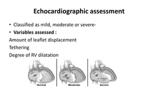
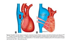
This document provides an overview of Ebstein's anomaly, a congenital heart defect where the tricuspid valve is displaced downward into the right ventricle. It discusses the history, anatomy, pathophysiology, clinical presentation, diagnosis using echocardiography, associated abnormalities, and surgical management. Surgical techniques include biventricular repair to reconstruct the tricuspid valve, single ventricle palliation with right ventricle exclusion, and cardiac transplantation for the most severe cases with significant biventricular dysfunction.
2. INTRODUCTION
⢠a congenital malformation, in which there is downward (apical)
displacement of insertion of septal and posterior leaflets
⢠âEbstein anomaly,â also referred to as âEbstein malformation,â
was clinically recognized in 1950
3. HISTORY
Wilhelm Ebstein
⢠The anomaly was first described in 1866 in an autopsy of a 19-
year-old labourer who had cyanosis and dyspnea since early
childhood
⢠first reported case in a live patient in 1949
4. PREVALANCE
⢠1 per 20,000 live births
⢠0.7 percent of all congenital heart defects
⢠Equal male : female incidence
⢠Represents 40% of congenital malformations of TV
5. ANATOMY- NORMAL TV
⢠Anterior leaflet â semicircular âlargest
⢠Posterior leaflet- scalloped
⢠Septal leaflet- attached to ventricular septum ,but some part
attached to posterior wall
⢠Septal leaflet normally exhibits slight apical displacement at
base compared to MV -15mm in children ,20mm in adults
6.
7. EMBRYOLOGY
⢠Leaflets and tensile apparatus formed by a process of delamination
of the inner layers of the inlet zone of the ventricles
⢠Delamination begins at the tips of the leaflets and toward the AV
junction
⢠In EA - failure of this delamination occurs ď Mechanism unknown
8. Septum formation of the Atrioventricular Canal
ď§ At the end of the 4th week, two mesenchymal cushions, the atrioventricular endocardial cushions,
appear at the superior and inferior borders of the atrioventricular canal, two additional lateral cushions appear
at the left and right borders.
ď§ At the end of the 5th week there is complete fusion of the superior and inferior cushions with complete
division of the canal into left and right orifices.
9. Atrioventricular Valves
ď§ After the endocardial cushions fuse, each AV orifice is surrounded by local proliferations of mesenchymal tissue.
ď§ Bloodstream hollows and thins out tissue on the ventricular surface of these proliferations, forming valves that
remain attached to the ventricular wall by muscular cords.
ď§ Finally, muscular tissue in the cords degenerates and is replaced by dense connective tissue.
13. PATHOLOGY
1. Displacement of the septal and posterior leaflets towards apex of
the right ventricle (RV)
2. Anterior leaflet is usually attached at the annular level and is large
and sail-like with multiple attachments to ventricular wall
:fenestrated
3. âatrialized RVâ - thin and dysplastic
4. âfunctional RVâ - smaller, lacks inlet portion and has a small
trabecular portion
5. Infundibular portion of RV is sometimes obstructed by redundant
anterior leaflet or its chordal attachments
14. Valve Leaflets and Valve Orifice
⢠Septal and posterior leaflets -adherent ,redundant and
dysplastic Nodular appearance of the free edge
⢠Anterior leaflet - large and âsaillikeâ â fenestrated- additional
tricuspid regurgitation
⢠Rarely, concomitant tricuspid stenosis or even atresia
⢠Redundancy of tricuspid valve tissue may encroach RV outflow
tract and cause sub pulmonary stenosis
15. Right atrium (RA) â
dilated and hypertrophied - depends on
⢠degree of displacement of TV leaflets
⢠resultant tricuspid regurgitation
Right Ventricle -
⢠In mild and moderate cases- normal with mild RV hypertrophy
⢠severe cases-near-normal thickness to very thin and dysplastic
16. Max point of displacement
⢠Commissure between posterior and septal leaflets
⢠Normal displacement - < 8mm/m2
Gross anatomy depicting
â˘Dilatation of the atrialized portion of the RV
Electrical potentials of ventricle, but pressure waveform that
of atrium.
â˘Displaced posterior leaflet
â˘Septum pushed towards LV and compressing it
17. Conduction System
⢠AV node is in triangle of Koch , is compressed and closer than usual to coronary sinus ostium
⢠Downward displacement of septal tricuspid leaflet is accompanied by discontinuity between
the central fibrous body and the septal atrioventricular ring âcreate substrate for WPW
⢠Accessory pathway ,usually seen around the tricuspid annulus :wolff Parkinson white
syndrome -5-25%
25. Pathophysiology
moderate to severe forms-
with each atrial contractionď blood
is propelled into the atrialized RV
With ventricular contraction that
follows- blood is forced back into the
right atrium
more pronounced in children with
significant TR
With the next atrial contraction, this
blood is forced back into the
atrialized RV
main determinants - degree of displacement of TV and
degree of TR
mild Ebstein â
tricuspid valve function is
close to normal
26. âping-pong effectâ
causes right atrial dilatation and increases right atrial pressure
right to left shunt across ASD/ PFO
arterial desaturation and pulmonary oligemia.
27. ⢠Newborn babies with severe Ebstein anomaly - cyanotic secondary to Rď L
shunt across atrial septum
⢠This is further accentuated by the usual high pulmonary vascular resistance
that exists at this age
⢠Significant TR causes severe RA enlargement in utero as well as after birth
inadequate RV myocardium Lead to inadequate forward flow via the
pulmonary valve
high PVR :inability of the pulmonary valve leaflets to open
âfunctionalâ pulmonary atresia
28. PVR DECREASE
⢠pulmonary valve may open
⢠R ď L shunting at atrial septal level decreaseď cyanosis
decreases
⢠establishment of forward flow via the pulmonary valve
⢠Cyanosis may return in later childhood or adolescence when
tricuspid valve function deteriorates (causing regurgitation)
This clinical course is described as âtransientâ or âintermittentâ
cyanosis
29. SYMPTOMS
⢠Around 35% patients present in infancy and 65% present in
older age groups (adolescence and adulthood)
⢠Of those patients presenting in infancy, majority present with
heart failure
30. Fetal life
⢠Incidental diagnosis on routine antenatal echo
⢠Hydrops fetalis is an ominous sign
⢠Cardiomegaly and tachyarrhythmias â also detectable
Neonatal life
⢠Severe cyanosis or heart failure
⢠Cyanosis improves with decreasing pulmonary vascular resistance
31. Adult life
⢠Fatigue
⢠Exertional dyspnea
⢠Cyanosis
⢠Worsening TR
⢠Right heart failure
⢠Left heart failure
⢠Palpitations/ arrhythmias
⢠Paradoxical embolism
⢠Incidental murmur
32. Physical examination
⢠Cyanosis and clubbing
⢠Usually left parasternal prominence and occasionally right
parasternal prominence Absence of left parasternal heave
importantly rules out the diagnosis
⢠Arterial pulses Normal to diminished in volume
33. Heart sounds
â S1 Widely split with tricuspid component loud( the SAIL SOUND)
Mitral component soft â (long PR interval)
â S2 Usually is normal
â In presence of RBBB â may produce a wide variable split
â S3 and S4 Usually heard (triple or quadruple rhythm)
â Origin of S3 is debated â probably due to the fluttering of the tricuspid
leaflet during diastole
⢠TR murmur âearly systolic decrescendo murmur(low pressure TR)
34. CHEST XRAY
⢠Severe cardiomegaly (box like contour)
⢠right atrial silhouette almost always enlargement
⢠Lung fields are either normal or oligemic
⢠Infundibulum either straightens left cardiac border or forms a
conspicuous convex shoulder
⢠Cardiomegaly in milder cases is commensurate with the
severity of tricuspid regurgitation.
35.
36.
37. ECHOCARDIOGRAPHY
⢠âDisplacement Indexâ is measured in systole or diastole from the
insertion point of the anterior mitral leaflet to the hinge point of
the tricuspid septal leaflet
⢠displacement index >8 mm/m2 -diagnosis of Ebstein anomaly
⢠determination of forward flow through RVOT and across the
pulmonary valve
⢠difficult in the neonatal setting with elevated pulmonary vascular
resistance
⢠pulmonary regurgitation -differentiate anatomic atresia from the
more common âfunctionalâ atresia of the pulmonary valve
44. MORTALITY
⢠NEONATAL DEATH- severe hypoxemia , congestive heart failure or both
⢠Younger patients- congestive heart failure
⢠Older patients(20%) âwith hemodynamically mild lesions âfrom atrial or
ventricular arrhythmias
⢠Brain abscess or paradoxical embolism and infective endocarditis-rare
⢠Mortality in neonatal period is 20%-40% and less than 50% survive to 5 yrs
⢠Mortality at all ages is 12.5%
48. NEONATAL EBSTEIN
⢠Poor prognosis
⢠Reported survival only 68%
Indication for surgery
⢠Heart failure
⢠Profound cyanosis
49. NEWBORN
⢠Asymptomatic cyanotic - no active treatment
⢠cyanosis severe PG E1 infusion (0.05â0.1 mcg/kg/min) until PVR
drops.(temporarily keeping the PDA open )
⢠inhaled nitric oxide to reduce PVR
⢠surgical systemic-pulmonary shunt to maintain adequate pulmonary blood
flow
⢠Furosemide and Digoxin- heart failure
⢠Supraventricular tachycardia or atrial flutter -anti-arrhythmic medications
⢠Restrictive PFO/ASD may require balloon atrial septostomy
50.
51.
52.
53. SURGICAL TECHNIQUES
⢠A. Biventricular repair (Knott approach)
⢠B. Single ventricle pathway with right ventricular exclusion
(starnes approach)
⢠C. Cardiac transplant
54. Biventricular repair (Knott approach)
⢠TV is repaired and the atrial septum is partially closed
⢠Repair typically a mono cusp type based on a satisfactory
anterior leaflet
⢠Right atrial reduction With vent repair
⢠Survival to hospital discharge was 74%
55.
56. Single ventricle pathway with right ventricular
exclusion (starnes approach)
Performed in anatomic RVOT obstruction
Objectives : exclude right ventricle and establish
a reliable source of pulmonary blood flow
⢠closing the tricuspid annular orifice using a
pericardial patch, removing the entire
septum primum and placing either a central
aorto-pulmonary shunt or Blalock-Taussig
shunt from innominate artery to right or left
pulmonary artery
⢠These neonates will subsequently need
bidirectional Glenn procedure and eventually
Fontan operation
57.
58. Cardiac transplantation
⢠With the improved results of the biventricular and single
ventricular approaches ,transplantation rarely is performed in
the current era
⢠Cardiac transplantation remains an option in the most severe
forms of ebsteins anomaly particularly when there is significant
left ventricular dysfunction
60. Indication for surgery
⢠Decreased exercise tolerance
⢠Cyanosis (80% or less; Hemoglobin 16 gm% or more)
⢠Paradoxical embolization
⢠Progressive right ventricular dilatation (Cardiothoracic ratio >60%)
⢠Prior to significant right ventricular dysfunction
⢠Onset or progression of atrial arrhythmias
⢠Prior to left ventricular dysfunction , NYHA class III or IV or
significant symptoms
61. PRINCIPLES OF SURGERY FOR Ebsteins anomaly
a. Closure of any intra cardiac communications
b. TV repair or replacement
c. Ablation of arrhythmias
d. Selective plication of the atrialized RV from apex to base
e. Reduction right atrioplasty
f. Repair of associated defects(eg. VSD closure)
62. Repair of tricuspid valve
The goals of operation is:
⢠To obtain a competent TV
⢠Preserve right ventricular contractility
⢠Decrease the risk of late rhythm disturbances
63. Relative contraindication to the reconstruction
technique
(1) patient age >60 years
(2) moderate pulmonary hypertension
(3) severe LV dysfunction (EF <30%)
(4) absent septal leaflet
(5) heavy muscularization of the anterior leaflet
(6) severe tricuspid valve annular dilation with massive RV
enlargement and systolic dysfunction (usually in older adults)
64. DANIELSONS REPAIR
⢠Creation of a monocusp valve using the anterior leaflet
This consisted of â
⢠Plication of the free wall of the atrialized RV
⢠Posterior tricuspid annuloplasty
⢠Right reduction atrioplasty
65.
66. Carpentier Repair
⢠anterior leaflet and the adjacent part of the inferior leaflet
were detached followed by longitudinal plication of the
atrialized RV and RA
⢠The anterior and inferior leaflets were repositioned to cover
the orifice area at the normal level
⢠Remodeling and reinforcement of the tricuspid annulus was
performed with a Artificial ring
67. DA SILVA APPROACH
⢠Reconstruct Tricuspid valve and RV
⢠Leaflet to leaflet coaptation forming longitudinal cone shaped
TV
68.
69. BDCPA
Two mechanism-
⢠Reduces venous return to the enlarged , dysfunctional RV by
approx. one third
⢠Provides sufficient preload to the LV to sutain adequate
systemic perfusion when right sided output is low
Preferred in the following situation
⢠LVEDP is < 12 mmHg
⢠Transpulmonary gradient < 10mm Hg
70. Indications for the BDCPA include
1. Severe RV enlargement or /and dysfunction
2. Squashed LV (D shaped LV)
3. Moderate degree of TV stenosis )
4. RA:LA pressure ratio > 1.5 indicates poor RV function
5. Preoperatve cyanosis at rest or with exercise
disadvantages:
⢠(1) pulsatility of the head and neck veins, (2) facial swelling, (3) potential
development of venoâ veno collaterals and/or pulmonary arteriovenous
fistulae, and (4) limitation of access to the heart from the internal jugular
vein
71. Arrhythmia management
⢠Atrial fibrillation and flutter âMC
⢠Most surgeons used successfully the right sided cut and sew
lesions of cox âmaze III procedure in ebsteins anomaly
⢠Radiofrequency ablation âprocedure time for maze procedure
is shortened significantly
⢠A biatrial maze procedure , performed particularly when there
is chronic atrial fibrillation , left atrial dilation or
concomiatriant mitral regurgitation
72. Cardiac Transplantation
⢠rarely required for Ebstein anomaly
⢠transplantation - presence of severe biventricular dysfunction
with an LV EF <25%
⢠Other indications: severe LV dilation and dysfunction
particularly when associated with mitral regurgitation
Scores of 0 to 10 are possible. There were no survivors when the score was greater than or equal to 5. Conversely, when the scores were less than or equal to 3, survival was 91 percent.
Thus, tricuspid valve repair techniques continue to evolve, and there is no universally
correct technique because of the anatomic variation observed in patients with Ebstein anomaly.
Concept of the cone reconstruction in Ebstein malformation. A: Adherent segments of tricuspid
valve tissue being separated from the anatomic annulus and the underlying right ventricular myocardium. B:
Sheet of tricuspid of tissue after it has been released. This tissue is used to create a cone, often attaching the
anterior leaflet to the remnants of the septal leaflet (see suture line, C). Once the cone is created, the base is
attached to the atrioventricular junction, restoring the hinge points to a nondisplaced position (C). When dilated,
thin, or significantly dyskinetic, the atrialized right ventricle can be reduced in size by either elliptical resection or
plication (C). The annuloplasty reduces the size of the intraventricular junction to what is appropriate to the size
of the reconstructed cone