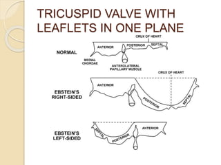
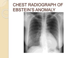
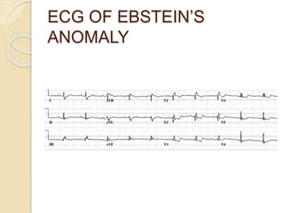
Wilhelm Ebstein first described Ebstein's anomaly in 1866. It is a rare congenital heart defect where the tricuspid valve is displaced downward into the right ventricle. Echocardiography is the primary diagnostic tool and shows apical displacement of the tricuspid valve leaflets. Treatment depends on severity but may include surgical repair of the tricuspid valve or exclusion of the dysfunctional right ventricle. Prognosis varies from critical in severe neonatal cases to relatively mild in some adults.
preop TEE assessment of atrial septal defect is very important for making decision for device closure, properly assessed adequate rims of ASD will reduce risk of device embolization to almost nil.
Ebstein’s anomaly is a rare congenital heart disorder occurring in ≈1 per 200 000 live births and accounting for <1% of all cases of congenital heart disease. This anomaly was described by Wilhelm Ebstein in 1866 in a report titled, “Concerning a very rare case of insufficiency of the tricuspid valve caused by a congenital malformation.” The patient was a 19-year-old cyanotic man with dyspnea, palpitations, jugular venous distension, and cardiomegaly. At autopsy, Ebstein described an enlarged and fenestrated anterior leaflet of the tricuspid valve. The posterior and septal leaflets were hypoplastic, thickened, and adherent to the right ventricle. There was also a thinned and dilated atrialized portion of the right ventricle, an enlarged right atrium, and a patent foramen ovale. By 1950, only 3 cases of this anomaly had been published.
A lecture on the echocardiographic evaluation of hypertrophic cardiomyopathy. Starts with an overview of the topic then a systematic approach to diagnosis and then a differential diagnosis followed by take-home messages and conclusion.
TGA is a complex congenital heart disease.Understanding the anatomy,physiology,surgery and anaesthetic management is very important for patient's better outcome.This ppt explains all these points in detail.
TAPVC defines the anomaly in which the pulmonary veins have no connection with the left atrium. Rather, the pulmonary veins connect directly to one of the systemic veins (TAPVC) or drain in to right atrium.
A PFO or ASD is present essentially in those who survive after birth
When pulmonary veins drain anomalously into the right atrium either because of complete absence of the interatrial septum or malattachment of the septum primum , then it is known as total anomalous pulmonary venous drainage.
When some or all of the pulmonary veins drain anomalously in to RA or its tributaries without being abnormally connected, the terms partially anomalous pulmonary venous drainage (PAPVD) or totally anomalous pulmonary venous drainage (TAPVD) with normal pulmonary venous connections are used.
Generally occurs secondary to pulmonary atresia with intact IVS .
Pathophysiology- it develops because of a reduction in the blood flow secondary to inflow impedence from tricuspid atresia or outflow impedence from pulmonary arterial atresia .
Typical findings- a small , hypertrophic RV and a small or absent pulmonary artery
Tetralogy of Fallot
Tetralogy of Fallot with Pulmonary
Stenosis
TETRALOGY OF FALLOT WITH CONGENITAL PULMONARY ATRESIA
Tetralogy of Fallot with Absent Pulmonary Valve
TEST BANK for Operations Management, 14th Edition by William J. Stevenson, Ve...kevinkariuki227
TEST BANK for Operations Management, 14th Edition by William J. Stevenson, Verified Chapters 1 - 19, Complete Newest Version.pdf
Knee anatomy and clinical tests 2024.pdfvimalpl1234
This includes all relevant anatomy and clinical tests compiled from standard textbooks, Campbell,netter etc..It is comprehensive and best suited for orthopaedicians and orthopaedic residents.
NVBDCP.pptx Nation vector borne disease control programSapna Thakur
NVBDCP was launched in 2003-2004 . Vector-Borne Disease: Disease that results from an infection transmitted to humans and other animals by blood-feeding arthropods, such as mosquitoes, ticks, and fleas. Examples of vector-borne diseases include Dengue fever, West Nile Virus, Lyme disease, and malaria.
- Video recording of this lecture in English language: https://youtu.be/lK81BzxMqdo
- Video recording of this lecture in Arabic language: https://youtu.be/Ve4P0COk9OI
- Link to download the book free: https://nephrotube.blogspot.com/p/nephrotube-nephrology-books.html
- Link to NephroTube website: www.NephroTube.com
- Link to NephroTube social media accounts: https://nephrotube.blogspot.com/p/join-nephrotube-on-social-media.html
Tom Selleck Health: A Comprehensive Look at the Iconic Actor’s Wellness Journeygreendigital
Tom Selleck, an enduring figure in Hollywood. has captivated audiences for decades with his rugged charm, iconic moustache. and memorable roles in television and film. From his breakout role as Thomas Magnum in Magnum P.I. to his current portrayal of Frank Reagan in Blue Bloods. Selleck's career has spanned over 50 years. But beyond his professional achievements. fans have often been curious about Tom Selleck Health. especially as he has aged in the public eye.
Follow us on: Pinterest
Introduction
Many have been interested in Tom Selleck health. not only because of his enduring presence on screen but also because of the challenges. and lifestyle choices he has faced and made over the years. This article delves into the various aspects of Tom Selleck health. exploring his fitness regimen, diet, mental health. and the challenges he has encountered as he ages. We'll look at how he maintains his well-being. the health issues he has faced, and his approach to ageing .
Early Life and Career
Childhood and Athletic Beginnings
Tom Selleck was born on January 29, 1945, in Detroit, Michigan, and grew up in Sherman Oaks, California. From an early age, he was involved in sports, particularly basketball. which played a significant role in his physical development. His athletic pursuits continued into college. where he attended the University of Southern California (USC) on a basketball scholarship. This early involvement in sports laid a strong foundation for his physical health and disciplined lifestyle.
Transition to Acting
Selleck's transition from an athlete to an actor came with its physical demands. His first significant role in "Magnum P.I." required him to perform various stunts and maintain a fit appearance. This role, which he played from 1980 to 1988. necessitated a rigorous fitness routine to meet the show's demands. setting the stage for his long-term commitment to health and wellness.
Fitness Regimen
Workout Routine
Tom Selleck health and fitness regimen has evolved. adapting to his changing roles and age. During his "Magnum, P.I." days. Selleck's workouts were intense and focused on building and maintaining muscle mass. His routine included weightlifting, cardiovascular exercises. and specific training for the stunts he performed on the show.
Selleck adjusted his fitness routine as he aged to suit his body's needs. Today, his workouts focus on maintaining flexibility, strength, and cardiovascular health. He incorporates low-impact exercises such as swimming, walking, and light weightlifting. This balanced approach helps him stay fit without putting undue strain on his joints and muscles.
Importance of Flexibility and Mobility
In recent years, Selleck has emphasized the importance of flexibility and mobility in his fitness regimen. Understanding the natural decline in muscle mass and joint flexibility with age. he includes stretching and yoga in his routine. These practices help prevent injuries, improve posture, and maintain mobilit
ARTIFICIAL INTELLIGENCE IN HEALTHCARE.pdfAnujkumaranit
Artificial intelligence (AI) refers to the simulation of human intelligence processes by machines, especially computer systems. It encompasses tasks such as learning, reasoning, problem-solving, perception, and language understanding. AI technologies are revolutionizing various fields, from healthcare to finance, by enabling machines to perform tasks that typically require human intelligence.
Pulmonary Thromboembolism - etilogy, types, medical- Surgical and nursing man...VarunMahajani
Disruption of blood supply to lung alveoli due to blockage of one or more pulmonary blood vessels is called as Pulmonary thromboembolism. In this presentation we will discuss its causes, types and its management in depth.
These lecture slides, by Dr Sidra Arshad, offer a quick overview of physiological basis of a normal electrocardiogram.
Learning objectives:
1. Define an electrocardiogram (ECG) and electrocardiography
2. Describe how dipoles generated by the heart produce the waveforms of the ECG
3. Describe the components of a normal electrocardiogram of a typical bipolar leads (limb II)
4. Differentiate between intervals and segments
5. Enlist some common indications for obtaining an ECG
Study Resources:
1. Chapter 11, Guyton and Hall Textbook of Medical Physiology, 14th edition
2. Chapter 9, Human Physiology - From Cells to Systems, Lauralee Sherwood, 9th edition
3. Chapter 29, Ganong’s Review of Medical Physiology, 26th edition
4. Electrocardiogram, StatPearls - https://www.ncbi.nlm.nih.gov/books/NBK549803/
5. ECG in Medical Practice by ABM Abdullah, 4th edition
6. ECG Basics, http://www.nataliescasebook.com/tag/e-c-g-basics
The prostate is an exocrine gland of the male mammalian reproductive system
It is a walnut-sized gland that forms part of the male reproductive system and is located in front of the rectum and just below the urinary bladder
Function is to store and secrete a clear, slightly alkaline fluid that constitutes 10-30% of the volume of the seminal fluid that along with the spermatozoa, constitutes semen
A healthy human prostate measures (4cm-vertical, by 3cm-horizontal, 2cm ant-post ).
It surrounds the urethra just below the urinary bladder. It has anterior, median, posterior and two lateral lobes
It’s work is regulated by androgens which are responsible for male sex characteristics
Generalised disease of the prostate due to hormonal derangement which leads to non malignant enlargement of the gland (increase in the number of epithelial cells and stromal tissue)to cause compression of the urethra leading to symptoms (LUTS
New Drug Discovery and Development .....NEHA GUPTA
The "New Drug Discovery and Development" process involves the identification, design, testing, and manufacturing of novel pharmaceutical compounds with the aim of introducing new and improved treatments for various medical conditions. This comprehensive endeavor encompasses various stages, including target identification, preclinical studies, clinical trials, regulatory approval, and post-market surveillance. It involves multidisciplinary collaboration among scientists, researchers, clinicians, regulatory experts, and pharmaceutical companies to bring innovative therapies to market and address unmet medical needs.
2. History
Wilhelm Ebstein – German
physician(1866) presented account
titled “On a Very Rare Case of
Insufficiency of the Tricuspid Valve
Caused by a Severe Congenital
Malformation of the Same”.
By 1950 only 3 case reports had been
published.
4. Prevalence
1 in 200,000 live births. 1% of all
cases of CHD.
40% of congenital malformations of
tricuspid valve.
5. Pathologic
Anatomy
Normally 3 leaflets : anterior,
inferior(posterior) and septal.
Anterior : semicircular or quadrangular
and
Posterior: scalloped
Septal : attaches ventricular septum and
posterior wall of right ventricle. Apical
displacement of basal attachment
compared to mitral valve : 15mm in
children, and 20mm in adults.Normally <
8mm/m2.
6. Contd…
Leaflets develop equally from
endocardial cushion and myocardium.
Leaflets and tensile apparatus develop
from delamination of the inner layer of
inlet zone of ventricles.
Ebstein ‘s anomaly : DELAMINATION
FAILURE.
Mechanism unknown probably TV
leaflets displacement.
7. Ebstein’s
characteristics
Adherence of septal and inferior
leaflets to myocardium.
Apical displacement of functional
annulus(septal>inferior>anterior).
Dilatation of “atrialized” RV
(ARV).Devoid of muscular tissue.
Redundancy,fenestrations, and
tethering of the anterior leaflet.
Dilation of the right AV junction.
9. Tricuspid Valve
TV morphology in ebstein’s is variable.
Tricuspid orifice is typically
incompetent,occasionally stenotic and
rarely imperforate.
Anterior Leaflet: In ebstein’s redundant
and may contain several fenestrations
and chordae tendineae are short and
malformed.Anterior leaflet mobile
tissue may get displaced into RVOT.
10. Inferior and septal leaflet development
is variable.
Apical displacement is maximal at
inferior and septal leaflets
commisure.TV annulus is dilated but
not displaced.
RCA demarcates true annulus and is
vulnerable to kinking during RV
plication,annuloplasty or TV
replacement.
THE RANGE OF VARIABILITY IS
13. Right Ventricle
RV in ebstein’s divided into 2 regions:
1. Inlet portion : involved in
malformation
2. Trabecular and outlet portions: not
involved
ARV dilated > half RV volume instead
of usual < one- third.
>2/3 of ebstein’s heart have RV
dilatation involving both portions
leading in some cases to D shaped or
crescent shaped LV.
14. Classification
s
Difficult as no two hearts are alike.
Echocardiographic appearance: mild,
moderate and severe according to :
Extent of apical displacement, degree of
TR, degree of RV dilatation and
dysfunction.
Four types based on anatomic findings
during surgery.
15. 1988 Carpentier et al. 4 types:
Type A : true RV volume adequate
Type B: large ARV but mobile anterior
leaflet
Type C: anterior leaflet movement
restricted causing RVOT obstruction.
Type D: complete atrialization
16. Celermajer et al.ECHO
GRADING
For neonates with ebstein’s anomaly the
Great Ormond Street Echocardiography
(GOSE)score
Ratio of combined area of the RA and
ARV is compared to the functional RV
and left heart.
o Grade 1 : ratio < 0.5
o Grade 2 : ratio of 0.5 to 0.99
o Grade 3 : ratio of 1.0 to 1.49
o Grade 4 : ratio ≥ 1.5
17. Genetic
Factors
Most cases are sporadic hence males
and females are equally affected;
familial ebstein’s anomaly is rare.
Cardiac transcription factors: NKX2.5
mutation, 10p13-p14 deletion and
1p34.3-p36.11 deletion.
MYH 7 mutation in subset with LV
noncompaction and this warrants
genetic testing and family evaluation.
18. Environmental and reproductive factors
More common in twins
Family history of congenital heart disease
Maternal exposure to lithium( 28 fold
increased risk) and benzodiazepines.
19. Associated Cardiac Defects
Interatrial septal defects: patent PFO
or ASD mostly secundum type present
in 80% to 94%
VSD with or without pulmonary
atresia.
RVOT obstruction: pulmonary valve
stenosis or pulmonary atresia, branch
pulmonary artery stenosis and rarely
displaced TV.
Patent ductus arteriosus
Coarctation of aorta
20. Accessory pathways:15% to 20%
patients.Majority around orifice of
malformed TV.
Left side heart lesions: 39% patients
o Diastolic and systolic dysfunction
o Mitral valve prolapse
o Subaortic stenosis
o Bicuspid or atretic aortic valve
o Muscle bands of LV
o Myocardial changes resembling LV
noncompaction
21. Congenitally corrected transposition of
the great arteries(cc-TGA):
o 15% to 50% of cc-TGA have
ebstenoid displacement of
morphologic TV but lack ARV and
dilated morphologic RV unlike right
sided ebstein anomaly.
22. Pathophysiology
Functional impairement of RV and TR
retard forward flow.
ARV acts as a passive reservoir during
atrial systole and ballons out decreasing
volume of ejected blood.
RA dilates increasing size of interatrial
communication.TR increases with
annular dilatation.
Symptomatic neonates have massive
cardiomegaly with lung hypoplasia.
23. Contd…
Decreased forward flow leads to
physiologic pulmonary atresia and
child is duct dependent.
LV output is compromised in sick
neonates with severe cyanosis and
acidosis.
Mild cyanosis may not be noted till
adult life .
24. Clinical Presentation
Varies widely
Symptoms are related to anatomic
severity
Most common presentation varies with
age at presentation
o Fetuses- abnormal routine prenatal
scan(86%). Ebstein’s anomaly is
common lesion referred for fetal
echocardiography because severe forms
lead to cardiomegaly,hydrops and
tachyarrhythmias.
25. Contd..
o Neonates- cyanosis(74%)
o Infants-heart failure(43%)
o Children- incidental murmur(63%)
o Adolescents and adults-
arrhythmia(42%),decrease exercise
tolerance , fatigue or right sided heart
failure
26. Symptoms
o Cyanosis and Heart failure
From TR
TRANSIENT NEONATAL CYANOSIS:
appears soon after birth due to high
pulmonary vascular resistance and
improves as pulmonary vascular
resistance decreases.
May appear first during pregnancy
27. o Exertional Dyspnea,
Fatigue,Cyanosis(50% cases), AND
Palpitations
May occur at a later age
May recur and may be insidious in
onset
o Palpitations
Due to atrial tachyarrhythmias
Present in 20%-30% cases
Some may be due to WPW
28. Paradoxical Embolization:leading to
brain abscesses and sudden death
Chest pain –
enigma.Retrosternal,sharp,shooting
suggesting serous origin (fibrinous
pericardium over ARV.
29. Physical Examination
o Vary with severity of pathology and
magnitude of right to left shunting.
Precordial asymmetry usually left
parasternal prominence occasionally
right anterior chest wall because of
enlarged RA
Murmur and click
Cyanosis: digital clubbing will depend
on degree of cyanosis
30. JVP: prominent “a” wave but large V
wave is rare despite severe TR
because large RA engulfs increased
volume ,thin walled toneless ARV and
as TR is low pressure and hypokinetic.
Hepatomegaly:passive hepatic
congestion and is pulsatile .
Palpable prominent diffuse apical
impulse
Systolic thrill at the left lower sternal
border
31. First heart sound: widely split due to
RBBB,hypokinetic RV,large size and
increased excursion of anterior leaflet
requiring longer time to reach fully
tensed closed position.Single S1 (T1)
due to prolonged PR and preexcited
RV.Loud T1 (sail sound) sign of anterior
leaflet mobility.
Second heart sound: widely split due to
RBBB. Single S2 (A2) due to low
pressure in pulmonary trunk.Paradoxical
split due to preexcited RV
32. Prominent S3 and/or a loud S4 give the
impression of multiple heart sounds
(triple or quadruple gallop)
Systolic murmur: from TR
,decresendo,grade 2/6 to 3/6 ,prominent
in leftward location towards apex,
intensity increases with inspiration . Mid-
diastolic murmur due to high diastolic
flow volume.Soft or absent in adults
because of low velocity to- and –fro flow
and rapid equalization of pressure
across TV
33. Diagnostic
Evaluation
Chest Radiography: normal to globe
shaped with narrow waist(pulmonary
trunk is not border forming and
ascending aorta is small or absent.
CT ratio > 0.65 indicates poor prognosis.
Infundibulum straightens the left cardiac
border or forms a convex border
Lungs fields may be normal or
decreased due to hypoplasia
MOST CONSISTENT FEATURE IS
ENLARGED RA SILHOUETTE
35. Electrocardiograph
y
Ebstein’s anomaly may be diagnosed
using ECG
Rarely normal even with a mild
anomaly.
Rarely and oddly ECG may be normal
even when the anomaly is severe
36. Major findings are:
Intraatrial conduction disturbance: right
atrial P wave(Himalayan)and prolonged
PR
Atrioventricular nodal conduction:
prolonged PR
Infranodal conduction
Intra-His or infra-His conduction
abnormality
RBBB
Bizarre second QRS attached to
preceding normal QRS
38. WPW preexcitation
Supraventricular tachycardia
Atrial fibrillation or flutter
Arrhythmogenic ARV
Deep Q waves in leads V1-V4 and
inferior leads
Complete heart block is rare but first
degree AV block occurs in 42%
patients.
39. Echocardiograph
y
2D echocardiography is the diagnostic
test of choice
3D echocardiography provides
additional details of TV anatomy and
size and function of different cardiac
chambers.
Apical displacement of septal leaflet
by at least 8mm/m2 body surface area
is the diagnostic feature
41. Presence of at least three accessory
attachments of leaflets to the
ventricular wall confirms tethering.
Marked enlargement of RA and ARV is
present when combined area of RA
and ARV is larger than combined area
of functional RV ,LA and LV in apical 4
chamber view in end diastole.
42. Helps assess site and degree of TR
and feasibility of its repair.
Ebstein’s anomaly can be diagnosed
in utero as early as 18th week of
pregnancy.
Patency of RVOT and GOSE score
can predict outcome in neonatal
ebstein’s anomaly
43. Cardiac Catheterization
Rarely required apart from
preoperative coronary angiography in
older patients
Required in patients with LV
dysfunction and suspected elevated
pulmonary artery pressure and
elevated LVEDP. Important if
bidirectional cavopulmonary
anastomosis is being considered.
44. Magnetic Resonance Imaging
Quantitative measurement of RA and
ventricular size and function in
presence of distorted RV anatomy.
Axial imaging provides new
expressions of disease severity such
as atrialized RV volume.
3D images provide greater dilineation
of disease severity
45. MANAGEMEN
T
Neonatal Ebstein
Poor prognosis: 2o% to 40% do not
survive 1 month and less than 50%
survive to 5 years.
Surgery required in presence of heart
failure or cyanosis. Options include:
(a)biventricular repair(Knott-Craig
approach), (b) single ventricle pathway
with right ventricular exclusion ( Starnes’
approach), and , rarely, (c) cardiac
transplantation
46. Biventricular repair( Knott-Craig
approach):TV repaired and atrial septum
partially closed along with routine right
atrial reduction.High early mortality
(25%) intermediate outcome is
promising.
Right ventricular exclusion
Starnes approach: useful with RVOT
obstruction (a) fenestrated patch closure
of the TV orifice, (b) enlarging the
interatrial communication, (c) right atrial
reduction, (d) placing systemic to
47. Modified Starnes Repair( Total
Ventricular Exclusion): RV free wall is
resected and closed primarily.
Cardiac Transplantation:rarely
performed in current era.Option in
patients with significant LV
dysfunction.
48. Management in Children and
Adults
Medical
Bacterial endocarditis prophylaxis
Those not candidates for surgery
managed with ACEI’s and heart failure
regimes.
Arrhythmias managed medically with
operative or catheter based
interventions
49. Catheter Ablation: success rate is
lower.Atrial fibrillation and flutter are
treated surgically at the time of operation.
Surgery
Indications:symptoms ,cyanosis and
paradoxical embolization, decreased
exercise performance, progressive
increase in cardiothoracic ratio,
progressive right ventricular dilatation and
dysfunction, onset and progression of
arrhythmias,NYHA class III or IV
symptoms and echocardiographic
probability of successful valve repair.
50. Principles of surgery
Closure of intracardiac
communications
TV repair or replacement
Ablation of arrhythmias
Plication of ARV
Reduction right atrioplasty
Repair of associated defects
51. TV Repair
Mayo Clinic Experience
o Danielson Repair
o Modified Danielson repair
BRAZIL Experience(da Silva
Approach):cone repair;most anatomic
repair.
FRENCH EXPERIENCE (BROUSSAIS
Approach)
Ventriculization procedure
52. Tricuspid Valve
Replacement
If TV repair not feasible
Porcine bioprosthetic valve is good
alternative over mechanical valve due
to good durability and lack of need for
anticoagulation.
54. o One and Half Ventricle repair
BDCPA: reduces venous return to
dysfunctional enlarged RV and
provides preload to LV.
Indications for BDCPA:
Severe RV enlargement
D shaped LV
Moderate TS(mean gradient
>6mmHg)
RA/LA pressure ratio > 1.5
Preoperative cyanosis at rest or with
exercise
55. o Disadvantages of BDCPA:
Pulsations of head and neck veins
Facial swelling
Development of pulmonary AV fistulae
Compromised access to the heart
using internal jugular vein approach
56. o Plication of ARV
ADVANTAGES: (a) decreases size of
nonfunctioning RV, (b) minimizes
pancake effect of RV on LV.
Potential risk of RCA injury
o Right reduction atrioplasty: performed
routinely. Avoid suture in crista
terminalis.
57. Surgical Treatment of
Arrhythmias
In atrial fibrillation right sided cut and
sew lesions of Cox-maze III procedure
Bilateral maze done in chronic AF, LA
dilation and MR.
In atrial flutter right atrial isthmus
lesion is added.
AVNRT unsuccessful ablation in
preoperative EP laboratory treated by
perinodal cryoablation
58. Heart Transplantation
o In presence of severe biventricular
failure ( LV Ejection Fraction < 25%),
severe LV dilatation and severe MR
with LV dysfunction.