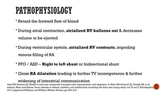
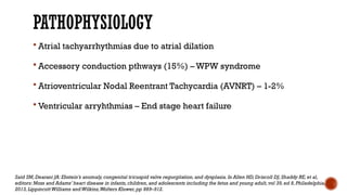
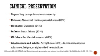
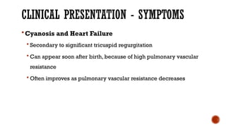
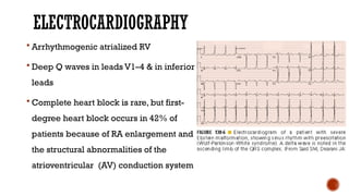
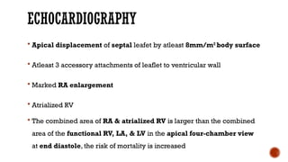
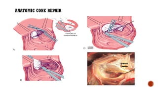
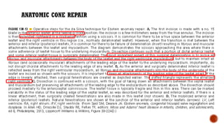
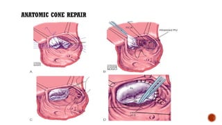
EBSTEIN’S ANOMAY
DR. MD WAHAJ ALI
HISTORY
 1866 – Dr.Wilhelm Ebstein (German
Physician) described cardiac findings
of 19 year old patient (Joseph
Prescher) who had died of cyanotic
heart disease
EbsteinW: Ueber einen sehr seltenen Fall von Insuffi cienz derValvula tricuspidalis, bedingt durch eine angeborene hochgradige
Missbildung derselben.Arch Anat Physiol 238–254,1866.
HISTORY
 1950 – Helen Taussig - First clinical syndrome analysis
 1950’s – BT shunt for neonatal Ebstein ( functional tricuspid or
pulmonary atresia)
 1958 – Tricuspid valve reconstruction – Hunter & Lillehei – Attempt to
create competent valve by repositioning of displaced leaflet &
excluding atrialised chamber ( 2 patients – both didn’t survive)
HISTORY
 Barnard (1963); Lillehei (1967) – Tricuspid valve replacement
 Danielson (1972); Carpenter (1988) – TV repair based on use of
anterior leaflet
 Starnes (1991) – Single ventricle palliation of neonatal Ebstein
 Knott-Craig (1994) – Biventricular repair of neonatal Ebstein anomaly
PATHOLOGICAL ANATOMY
1. Failure of delamination of the septal, inferior, & anterior leaflets of
the TV with subsequent adherence of the leaflets to the underlying
myocardium
2. Apical displacement of the functional TV annulus
(Septal > Inferior > Anterior)
3. Dilation of the atrialised portion of the right ventricle (RV)
4. Anterior leaflet fenestrations, redundancy or tethering
5. Dilation of the true anatomic TV annulus
LamersWH,Viragh S,Wessels A,et al: Formation of the tricuspid valve in the human heart.Circulation 91:111–121,1995.
DOWNWARD DISPLACEMENT
TRICUSPID VALVE
 Highly variable morphology
 Almost always incompetent
 Anterior leaflet may be large & fenestrated
 Chordae tendinea may be short & poorly formed
 Inferior & Septal leaflet may be redundant or absent due to failure of
delamination
 Downward displacement of septal & posterior leaflet in relation to
AML > 0.8mm/m2
RIGHT VENTRICLE
 Divided into 2 regions
 Atrialised RV (aRV) – Functionally integrated with RA
 Thin walled & devoid of muscle tissue
 May be disproportionately dilated
 Funtional RV
 Apical & infundibular component may be thinner
 RV dilation (>2/3)
 Septum is deviated leftwards
 Circular RV & D-shaped LV
ASSOCIATED CARDIAC DEFECTS
 Atrial septal defects - PFO or OS ASD (80-94%)
 VSD +/- Pulmonary atresia
 RVOT obstruction – Structural (PS, PA, branch PA stenosis)
 PDA
 CoA
 Accessory conduction pathways – WPW (15-20%)
ASSOCIATED CARDIAC DEFECTS
 Left sided lesions (Uncommon)
 Mitral valve prolapse
 Accessory mitral valve tissue
 Subaortic stenosis
 Bicuspid or atretic AoV
 Muscle bundles in LV cavity
 Congenitally corrected TGA (cc-TGA)
 Lack of atrialization of RV free wall
CLASSOFICATION
 CARPENTIER CLASSIFICATION (1988)
 Type A – Volume of true RV is adequate
 Type B – Large atrialized RV, anterior leaflet
moves freely
 Type C – Severe restriction of anterior
leaflet movement causing significant RVOTO
 Type D – Near complete atrialization
Carpentier A,Chauvaud S,Mace L,et al:A new reconstructive operation for Ebstein’s anomaly of the tricuspid valve.J Thorac
Cardiovasc Surg 96:92–101,1988.
CLASSIFICATION
GENETIC FACTORS
 Most cases are sporadic
 Cardiac transcription factor NKX2.5 mutations
 10p13-p14 deletion
 1p34.3-p36.11 deletion
 Mutation in gene MYH7
Benson DW,Silberbach GM,Kavanaugh-McHugh A,et al: Mutations in the cardiac transcription factor NKX2.5 affect diverse
cardiac developmental pathways.J Clin Invest 104:1567–1573,1999.
Yatsenko SA,Yatsenko AN,Szigeti K,et al: Interstitial deletion of 10p and atrial septal defect in DiGeorge 2 syndrome.Clin Genet 66:128–136,2004.
Postma AV,van Engelen K,van de Meerakker J,et al: Mutations in the sarcomere gene MYH7 in Ebstein anomaly.Circ Cardiovasc Genet 4:43–50,
2011.
PATHOPHYSIOLOGY
 Retard the forward flow of blood
 During atrial contraction, atrialized RV balloons out & decreases
volume to be ejected
 During ventricular systole, atrialized RV contracts, impeding
venous filling of RA
 PFO / ASD – Right to left shunt or bidirectional shunt
 Gross RA dilatation leading to further TV incompetence & further
widening of interatrial communication
Said SM,Dearani JA: Ebstein’s anomaly,congenital tricuspid valve regurgitation,and dysplasia.In Allen HD, Driscoll DJ,Shaddy RE,et al,
editors: Moss and Adams’ heart disease in infants,children,and adolescents including the fetus and young adult,vol 39,ed 8,Philadelphia,
2013,LippincottWilliams andWilkins,Wolters Kluwer,pp 889–912.
PATHOPHYSIOLOGY
 Atrial tachyarrhythmias due to atrial dilation
 Accessory conduction pthways (15%) – WPW syndrome
 Atrioventricular Nodal Reentrant Tachycardia (AVNRT) – 1-2%
 Ventricular arryhthmias – End stage heart failure
Said SM,Dearani JA: Ebstein’s anomaly,congenital tricuspid valve regurgitation,and dysplasia.In Allen HD, Driscoll DJ,Shaddy RE,et al,
editors: Moss and Adams’ heart disease in infants,children,and adolescents including the fetus and young adult,vol 39,ed 8,Philadelphia,
2013,LippincottWilliams andWilkins,Wolters Kluwer,pp 889–912.
CLINICAL PRESENTATION
 Depending on age & anatomic severity
 Fetuses: Abnormal routine prenatal scan (86%)
 Neonates: Cyanosis (74%)
 Infants: heart failure (43%)
 Children: Incidental murmur (63%)
 Adolescents and adults: Arrhythmia (42%), decreased exercise
tolerance, fatigue, or right-sided heart failure
Celermajer DS,Bull C,Till JA,et al: Ebstein’s anomaly: presentation and outcome from fetus to adult.J Am Coll Cardiol 23:170–176, 1994.
CLINICAL PRESENTATION - SYMPTOMS
Cyanosis and Heart Failure
 Secondary to significant tricuspid regurgitation
 Can appear soon after birth, because of high pulmonary vascular
resistance
 Often improves as pulmonary vascular resistance decreases
CLINICAL PRESENTATION - SYMPTOMS
Exertional Dyspnea, Fatigue, Cyanosis, & Palpitations
 Can occur at a later age
 Can recur and can be insidious in onset
CLINICAL PRESENTATION - SYMPTOMS
Palpitations
 Secondary to atrial tachyarrhythmia (atrial flutter and fibrillation
most common)
 May be present in 20% - 30% of cases
 Some of these arrhythmias are due to WPW syndrome
CLINICAL PRESENTATION - SYMPTOMS
Paradoxic Embolization
 In the presence of an interatrial communication, patients with
Ebstein malformation are at risk for paradoxic embolization, brain
abscesses, and sudden death
PHYSICAL EXAMINATION
 Murmur & thrill
 Cyanosis
 Prominent “a” waves on JVP
 Hepatomegaly
 Palpable, prominent diffuse apical impulse
 Split heart sound
DIAGNOSIS – CHEST RADIOGRAPH
 Markedly enlarged cardiac
silhouette
 CTR > 0.65 (Poor prognosis)
 RA enlargement
ELECTROCARDIOGRAPHY
 PR interval prolongation & tall P waves
 RBBB
 WPW preexcitation
 Supraventricular tachycardia
 Atrial flutter or fibrillation
ELECTROCARDIOGRAPHY
 Arrhythmogenic atrialized RV
 Deep Q waves in leads V1–4 & in inferior
leads
 Complete heart block is rare, but first-
degree heart block occurs in 42% of
patients because of RA enlargement and
the structural abnormalities of the
atrioventricular (AV) conduction system
ECHOCARDIOGRAPHY
 Diagnostic test of choice
 Site & degree of TR & feasibility of repair
 Diagnose in utero – 18 weeks
 Can predict outcomes in neonates - RVOTO
ECHOCARDIOGRAPHY
 Apical displacement of septal leafet by atleast 8mm/m2
body surface
 Atleast 3 accessory attachments of leaflet to ventricular wall
 Marked RA enlargement
 Atrialized RV
 The combined area of RA & atrialized RV is larger than the combined
area of the functional RV, LA, & LV in the apical four-chamber view
at end diastole, the risk of mortality is increased
ECHOCARDIOGRAPHY
CELERMAJER INDEX SCORE
CARDIAC CATHETERIZATION
 Rarely necessary
 Coronary angiography
 PA pressures - Usually normal
 RVEDP – Elevated
 RA pressure – Normal
 LV dysfunction – Suspicion of elevated LA pressure or elevated LVEDP
CARDIAC MRI
 Quantitative measurement of RA & RV size & systolic function
 Axial imaging provides most reliable volume of atrialized RV
 Detailed visualization of pathological anatomy (3D images)
 Technique for precise volumetric analysis of ventricular function and
intracardiac blood flow, without any geometric assumptions
 CMR delayed contrast enhancement image is a potential tool to
recognize areas of right ventricle dysplasia
CARDIAC MRI
The following acquisitions were performed parallel to the long axis of
the heart (horizontal and vertical long-axis planes) & perpendicular to it
(short-axis planes)
1. Balanced SSFP ( b-SSFP= steady-state free precession) sequences
using retrospective ECG gating & parallel imaging technique with an
acceleration factor of two and reconstruction algorithm GRAPPA
(generalized auto calibrating partially parallel acquisition)
2. Dark-blood techniques, being the single-shot inversion-recovery fast
SE sequence with breath holding the most commonly used
3. Phase contrast velocity map of the aorta and pulmonary arteries
4. At last delayed contrast enhanced images performed ten minutes
after injection of gadolinium-DTPA using a segmented inversion-
recovery spoiled GRE sequence
CARDIAC MRI
 Findings
 Morphology
 Tricuspid valve
 Degree of apical displacement of postero-septal leaflet - the grade of leaflet tethering
to the ventricular wall can be calculated according to their extension (modified
Becker´s dysplasia classification):
 Grade I - Up to 25% of the distance from the atrioventricular junction to the apex,
 Grade II - From 25-50%
 Grade III - More than 50% of the distance
MODIFIED BECKER’S DYSPLASTIC CLASSIFICATION
NATURAL HISTORY
 Survival was related to severity & presence of RV or LV dysfunction
 The estimated cumulative overall survival rates were 89%, 76%, 53%, & 41% at 1, 10,
15, and 20 years of follow-up, respectively
NATURAL HISTORY
 Predictors of cardiac-related death
1. Cardiothoracic ratio of 0.65 or greater
2. Increasing severity of TV displacement on echocardiography
3. NYHA class III or IV
4. Cyanosis
5. Severe TR
6. Younger age at diagnosis
MANAGEMENT
1. Neonates & Infants
2. Children & Adults
NEONATES & INFANTS
 Indications for Operation
1. Congestive heart failure
2. Profound cyanosis
 Three pathways
1. Biventricular repair (Knott – Craig approach)
2. Single ventricular repair (RV exclusion technique / Starnes
approach)
3. Cardiac Transplantation
BIVENTRICULAR REPAIR (KNOTT - CRAIG APPROACH)
 TV is repaired – Monocusp type of valve repair based on satisfactory
antrior leaflet
 ASD is partially closed – Right to left shunting in early postoperative
period
 RA reduction
 Delayed sternal closure
 Inhaled Nitric oxide
 Prophylactic Peritoneal dialysis
BIVENTRICULAR REPAIR (KNOTT - CRAIG APPROACH)
RV EXCLUSION TECHNIQUE / STARNES APPROACH)
 Fenestrated patch closure of TV orifice (4-5mm)
 Enlarging the ASD
 Systemic to pulmonary artery shunt
 Particularly appealing in
 Anatomic pulmonary atresia
 RVOT obstruction
 Preparing for Fontan procedure
RV EXCLUSION TECHNIQUE / STARNES APPROACH)
MODIFIED STARNES APPROACH (TOTAL VENTRICULAR EXCLUSION)
 Total RV exclusion - Sano
 Free wall of RV is resected & closed primarily or with PTFE patch
 Acts like a large RV plication & RV volume reduction procedure
 May improve LV filling
MODIFIED STARNES APPROACH (TOTAL VENTRICULAR EXCLUSION)
HEART TRANSPLANTATION
 Most severe cases
 Rarely necessarily
 Limitations
 Scarcity of donor organs
 Long term immunosuppression
CHILDREN & ADULTS
 Observation alone
1. Asymptomatic
2. No right to left shunt
3. Mild cardiomegaly
4. Normal exercise tolerance
5. NYHA – I / II – Managed medically – Diuretics & Antiarrhythmics
CHILDREN & ADULTS
 Indications for Operation
1. Symptomatic – Fatigue, Cyanosis, Paradoxical embolism
2. Objevtive evidence
 Decreasing exercise performance (Exercise testing)
 Progressive increase in heart size (CXR)
 Progressive RV dilation or reduction in RVSP (ECHO)
 Atrial or Ventricular arrhythmias
3. Between 2 and 5 years of age
CHILDREN & ADULTS
 Operative Managment
 Closure of ASD
 Correction of associated anomalies
 Any indicated antiarrhythmia procedures
 Internal plication of atrialized RV
 Repair of TV
 Right reduction atrioplasty
CHILDREN & ADULTS
 Anatomic Cone Repair
 Concept of monocusp repair
 Depends on adequate anterior leaflet
 Danielson monocusp repair (Mayo clinic) – Sebening stich (Anterior
papillary muscle to ventricular septum)
 French experience (Carpentier) – Mobilization (surgical
delamination) of anterior leaflet & annular reattachment
 Brazilian experience (da Silva) – Surgical delamination of all leaflets
ANATOMIC CONE REPAIR
ADJUNCTS TO CONE REPAIR
1. Leaflet augmentation
Cor Matrix membrane or
autologus pericardium to
increase leaflet height
ADJUNCTS TO CONE REPAIR
2. Cone augmentation
 Small triangular patch
 To avoid tricuspid stenosis
ADJUNCTS TO CONE REPAIR
3. Leaflet plication
 To increase leaflet height
4. Surgical fenestration
 To create autologus
neochordae when linear
attachment is present
ADJUNCTS TO CONE REPAIR
5. Sebening stich
 Approximation of mobilized “base
intact” anterior papillary muscle to
ventricular septum
6. Modified Sebening stich
 Approximation of mobilized “base
intact” RV free wall papillary muscle to
corresponding head of papillary
muscle arising from ventricular septum
ADJUNCTS TO CONE REPAIR
7. Artificial Gore-Tex chordae
RELATIVE CONTRAINDICATIONS TO CONE REPAIR
 Age > 60 years
 Moderate pulmonary hypertension
 Significant LV dysfunction (EF < 30%)
 Absent septal leaflet
 Poor delamination or poor quality of anterior leaflet (< 50%)
 Severe muscularization of anterior leaflet
 Severe RV enlargement
 Severe dilation of right AV junction
TRICUSPID VALVE REPLACEMENT
 Cannot undergo valve repair
 Inability to obtain a satisfactory cone repair
 Muscularization of anterior leaflet
 Absent septal leaflet
 Age > 60 years
 Massive RV or annular dilation
TRICUSPID VALVE REPLACEMENT
 Bioprosthetic (Porcine) valve replacement
 Good durability in tricuspid positon
 Lack the need of Warfarin anticoagulation (Require in first 3-6 months)
 Mechaical valves
 Higher frequency of valve malfunction & thrombotic complications when
RV function is poor
TRICUSPID VALVE REPLACEMENT
THE 1.5 VENTRICLE - REPAIR
 Bidirectional Cavopulmonary shunts
 When RV is severely dilated poorly functioning
 Mild – moderate TS (mean grad > 6mmHg) with LV dysfunction
 PA & LA pressures must be low
 LVEDP < 12mmHg
 Transpulmonary grad < 10mmHg
 PA mean < 18mmHg
 LVEF 35-40%
THE 1.5 VENTRICLE - REPAIR
 Disadvantages
 Pulsations of head & neck vessels
 Facial swelling
 Development of arteriovenous fistulae in pulmonary vasculature
 Compromises access to pacemaker lead placement
HEART TRANSPLANTATION
 Severe biventricular dysfunction (LVEF < 25%)
 Significant LV dilation & dysfunction
 Severe nonstructural mitral regurgitation
POSTOPERATIVE CARE
 Discontinuation of CPB
 Epinephrine & Milrinone
 Higher heart rates (100-120bpm) are preffered
 Temporary atrial pacing if needed
 Low dose Vasopressin
 Cautious volume administration
 RA pressures < 10-12 mmHg
 Nitric oxide
POSTOPERATIVE CARE
 At Hospital Discharge
 Beta blockers / ACE inhibitors
 Sildenafil for 6-8 weeks
 Amiodarone for 2-3 months
Presentation on Ebstein's Anomaly A concise view.pptx

Presentation on Ebstein's Anomaly A concise view.pptx

  • 1.
  • 2.
    HISTORY  1866 –Dr.Wilhelm Ebstein (German Physician) described cardiac findings of 19 year old patient (Joseph Prescher) who had died of cyanotic heart disease EbsteinW: Ueber einen sehr seltenen Fall von Insuffi cienz derValvula tricuspidalis, bedingt durch eine angeborene hochgradige Missbildung derselben.Arch Anat Physiol 238–254,1866.
  • 3.
    HISTORY  1950 –Helen Taussig - First clinical syndrome analysis  1950’s – BT shunt for neonatal Ebstein ( functional tricuspid or pulmonary atresia)  1958 – Tricuspid valve reconstruction – Hunter & Lillehei – Attempt to create competent valve by repositioning of displaced leaflet & excluding atrialised chamber ( 2 patients – both didn’t survive)
  • 4.
    HISTORY  Barnard (1963);Lillehei (1967) – Tricuspid valve replacement  Danielson (1972); Carpenter (1988) – TV repair based on use of anterior leaflet  Starnes (1991) – Single ventricle palliation of neonatal Ebstein  Knott-Craig (1994) – Biventricular repair of neonatal Ebstein anomaly
  • 5.
    PATHOLOGICAL ANATOMY 1. Failureof delamination of the septal, inferior, & anterior leaflets of the TV with subsequent adherence of the leaflets to the underlying myocardium 2. Apical displacement of the functional TV annulus (Septal > Inferior > Anterior) 3. Dilation of the atrialised portion of the right ventricle (RV) 4. Anterior leaflet fenestrations, redundancy or tethering 5. Dilation of the true anatomic TV annulus LamersWH,Viragh S,Wessels A,et al: Formation of the tricuspid valve in the human heart.Circulation 91:111–121,1995.
  • 8.
  • 9.
    TRICUSPID VALVE  Highlyvariable morphology  Almost always incompetent  Anterior leaflet may be large & fenestrated  Chordae tendinea may be short & poorly formed  Inferior & Septal leaflet may be redundant or absent due to failure of delamination  Downward displacement of septal & posterior leaflet in relation to AML > 0.8mm/m2
  • 10.
    RIGHT VENTRICLE  Dividedinto 2 regions  Atrialised RV (aRV) – Functionally integrated with RA  Thin walled & devoid of muscle tissue  May be disproportionately dilated  Funtional RV  Apical & infundibular component may be thinner  RV dilation (>2/3)  Septum is deviated leftwards  Circular RV & D-shaped LV
  • 12.
    ASSOCIATED CARDIAC DEFECTS Atrial septal defects - PFO or OS ASD (80-94%)  VSD +/- Pulmonary atresia  RVOT obstruction – Structural (PS, PA, branch PA stenosis)  PDA  CoA  Accessory conduction pathways – WPW (15-20%)
  • 13.
    ASSOCIATED CARDIAC DEFECTS Left sided lesions (Uncommon)  Mitral valve prolapse  Accessory mitral valve tissue  Subaortic stenosis  Bicuspid or atretic AoV  Muscle bundles in LV cavity  Congenitally corrected TGA (cc-TGA)  Lack of atrialization of RV free wall
  • 14.
    CLASSOFICATION  CARPENTIER CLASSIFICATION(1988)  Type A – Volume of true RV is adequate  Type B – Large atrialized RV, anterior leaflet moves freely  Type C – Severe restriction of anterior leaflet movement causing significant RVOTO  Type D – Near complete atrialization Carpentier A,Chauvaud S,Mace L,et al:A new reconstructive operation for Ebstein’s anomaly of the tricuspid valve.J Thorac Cardiovasc Surg 96:92–101,1988.
  • 15.
  • 16.
    GENETIC FACTORS  Mostcases are sporadic  Cardiac transcription factor NKX2.5 mutations  10p13-p14 deletion  1p34.3-p36.11 deletion  Mutation in gene MYH7 Benson DW,Silberbach GM,Kavanaugh-McHugh A,et al: Mutations in the cardiac transcription factor NKX2.5 affect diverse cardiac developmental pathways.J Clin Invest 104:1567–1573,1999. Yatsenko SA,Yatsenko AN,Szigeti K,et al: Interstitial deletion of 10p and atrial septal defect in DiGeorge 2 syndrome.Clin Genet 66:128–136,2004. Postma AV,van Engelen K,van de Meerakker J,et al: Mutations in the sarcomere gene MYH7 in Ebstein anomaly.Circ Cardiovasc Genet 4:43–50, 2011.
  • 17.
    PATHOPHYSIOLOGY  Retard theforward flow of blood  During atrial contraction, atrialized RV balloons out & decreases volume to be ejected  During ventricular systole, atrialized RV contracts, impeding venous filling of RA  PFO / ASD – Right to left shunt or bidirectional shunt  Gross RA dilatation leading to further TV incompetence & further widening of interatrial communication Said SM,Dearani JA: Ebstein’s anomaly,congenital tricuspid valve regurgitation,and dysplasia.In Allen HD, Driscoll DJ,Shaddy RE,et al, editors: Moss and Adams’ heart disease in infants,children,and adolescents including the fetus and young adult,vol 39,ed 8,Philadelphia, 2013,LippincottWilliams andWilkins,Wolters Kluwer,pp 889–912.
  • 18.
    PATHOPHYSIOLOGY  Atrial tachyarrhythmiasdue to atrial dilation  Accessory conduction pthways (15%) – WPW syndrome  Atrioventricular Nodal Reentrant Tachycardia (AVNRT) – 1-2%  Ventricular arryhthmias – End stage heart failure Said SM,Dearani JA: Ebstein’s anomaly,congenital tricuspid valve regurgitation,and dysplasia.In Allen HD, Driscoll DJ,Shaddy RE,et al, editors: Moss and Adams’ heart disease in infants,children,and adolescents including the fetus and young adult,vol 39,ed 8,Philadelphia, 2013,LippincottWilliams andWilkins,Wolters Kluwer,pp 889–912.
  • 19.
    CLINICAL PRESENTATION  Dependingon age & anatomic severity  Fetuses: Abnormal routine prenatal scan (86%)  Neonates: Cyanosis (74%)  Infants: heart failure (43%)  Children: Incidental murmur (63%)  Adolescents and adults: Arrhythmia (42%), decreased exercise tolerance, fatigue, or right-sided heart failure Celermajer DS,Bull C,Till JA,et al: Ebstein’s anomaly: presentation and outcome from fetus to adult.J Am Coll Cardiol 23:170–176, 1994.
  • 20.
    CLINICAL PRESENTATION -SYMPTOMS Cyanosis and Heart Failure  Secondary to significant tricuspid regurgitation  Can appear soon after birth, because of high pulmonary vascular resistance  Often improves as pulmonary vascular resistance decreases
  • 21.
    CLINICAL PRESENTATION -SYMPTOMS Exertional Dyspnea, Fatigue, Cyanosis, & Palpitations  Can occur at a later age  Can recur and can be insidious in onset
  • 22.
    CLINICAL PRESENTATION -SYMPTOMS Palpitations  Secondary to atrial tachyarrhythmia (atrial flutter and fibrillation most common)  May be present in 20% - 30% of cases  Some of these arrhythmias are due to WPW syndrome
  • 23.
    CLINICAL PRESENTATION -SYMPTOMS Paradoxic Embolization  In the presence of an interatrial communication, patients with Ebstein malformation are at risk for paradoxic embolization, brain abscesses, and sudden death
  • 24.
    PHYSICAL EXAMINATION  Murmur& thrill  Cyanosis  Prominent “a” waves on JVP  Hepatomegaly  Palpable, prominent diffuse apical impulse  Split heart sound
  • 25.
    DIAGNOSIS – CHESTRADIOGRAPH  Markedly enlarged cardiac silhouette  CTR > 0.65 (Poor prognosis)  RA enlargement
  • 26.
    ELECTROCARDIOGRAPHY  PR intervalprolongation & tall P waves  RBBB  WPW preexcitation  Supraventricular tachycardia  Atrial flutter or fibrillation
  • 27.
    ELECTROCARDIOGRAPHY  Arrhythmogenic atrializedRV  Deep Q waves in leads V1–4 & in inferior leads  Complete heart block is rare, but first- degree heart block occurs in 42% of patients because of RA enlargement and the structural abnormalities of the atrioventricular (AV) conduction system
  • 28.
    ECHOCARDIOGRAPHY  Diagnostic testof choice  Site & degree of TR & feasibility of repair  Diagnose in utero – 18 weeks  Can predict outcomes in neonates - RVOTO
  • 29.
    ECHOCARDIOGRAPHY  Apical displacementof septal leafet by atleast 8mm/m2 body surface  Atleast 3 accessory attachments of leaflet to ventricular wall  Marked RA enlargement  Atrialized RV  The combined area of RA & atrialized RV is larger than the combined area of the functional RV, LA, & LV in the apical four-chamber view at end diastole, the risk of mortality is increased
  • 30.
  • 31.
  • 32.
    CARDIAC CATHETERIZATION  Rarelynecessary  Coronary angiography  PA pressures - Usually normal  RVEDP – Elevated  RA pressure – Normal  LV dysfunction – Suspicion of elevated LA pressure or elevated LVEDP
  • 33.
    CARDIAC MRI  Quantitativemeasurement of RA & RV size & systolic function  Axial imaging provides most reliable volume of atrialized RV  Detailed visualization of pathological anatomy (3D images)  Technique for precise volumetric analysis of ventricular function and intracardiac blood flow, without any geometric assumptions  CMR delayed contrast enhancement image is a potential tool to recognize areas of right ventricle dysplasia
  • 36.
    CARDIAC MRI The followingacquisitions were performed parallel to the long axis of the heart (horizontal and vertical long-axis planes) & perpendicular to it (short-axis planes) 1. Balanced SSFP ( b-SSFP= steady-state free precession) sequences using retrospective ECG gating & parallel imaging technique with an acceleration factor of two and reconstruction algorithm GRAPPA (generalized auto calibrating partially parallel acquisition) 2. Dark-blood techniques, being the single-shot inversion-recovery fast SE sequence with breath holding the most commonly used 3. Phase contrast velocity map of the aorta and pulmonary arteries 4. At last delayed contrast enhanced images performed ten minutes after injection of gadolinium-DTPA using a segmented inversion- recovery spoiled GRE sequence
  • 37.
    CARDIAC MRI  Findings Morphology  Tricuspid valve  Degree of apical displacement of postero-septal leaflet - the grade of leaflet tethering to the ventricular wall can be calculated according to their extension (modified Becker´s dysplasia classification):  Grade I - Up to 25% of the distance from the atrioventricular junction to the apex,  Grade II - From 25-50%  Grade III - More than 50% of the distance
  • 38.
  • 39.
    NATURAL HISTORY  Survivalwas related to severity & presence of RV or LV dysfunction  The estimated cumulative overall survival rates were 89%, 76%, 53%, & 41% at 1, 10, 15, and 20 years of follow-up, respectively
  • 40.
    NATURAL HISTORY  Predictorsof cardiac-related death 1. Cardiothoracic ratio of 0.65 or greater 2. Increasing severity of TV displacement on echocardiography 3. NYHA class III or IV 4. Cyanosis 5. Severe TR 6. Younger age at diagnosis
  • 41.
    MANAGEMENT 1. Neonates &Infants 2. Children & Adults
  • 42.
    NEONATES & INFANTS Indications for Operation 1. Congestive heart failure 2. Profound cyanosis  Three pathways 1. Biventricular repair (Knott – Craig approach) 2. Single ventricular repair (RV exclusion technique / Starnes approach) 3. Cardiac Transplantation
  • 43.
    BIVENTRICULAR REPAIR (KNOTT- CRAIG APPROACH)  TV is repaired – Monocusp type of valve repair based on satisfactory antrior leaflet  ASD is partially closed – Right to left shunting in early postoperative period  RA reduction  Delayed sternal closure  Inhaled Nitric oxide  Prophylactic Peritoneal dialysis
  • 44.
    BIVENTRICULAR REPAIR (KNOTT- CRAIG APPROACH)
  • 45.
    RV EXCLUSION TECHNIQUE/ STARNES APPROACH)  Fenestrated patch closure of TV orifice (4-5mm)  Enlarging the ASD  Systemic to pulmonary artery shunt  Particularly appealing in  Anatomic pulmonary atresia  RVOT obstruction  Preparing for Fontan procedure
  • 46.
    RV EXCLUSION TECHNIQUE/ STARNES APPROACH)
  • 47.
    MODIFIED STARNES APPROACH(TOTAL VENTRICULAR EXCLUSION)  Total RV exclusion - Sano  Free wall of RV is resected & closed primarily or with PTFE patch  Acts like a large RV plication & RV volume reduction procedure  May improve LV filling
  • 48.
    MODIFIED STARNES APPROACH(TOTAL VENTRICULAR EXCLUSION)
  • 49.
    HEART TRANSPLANTATION  Mostsevere cases  Rarely necessarily  Limitations  Scarcity of donor organs  Long term immunosuppression
  • 50.
    CHILDREN & ADULTS Observation alone 1. Asymptomatic 2. No right to left shunt 3. Mild cardiomegaly 4. Normal exercise tolerance 5. NYHA – I / II – Managed medically – Diuretics & Antiarrhythmics
  • 51.
    CHILDREN & ADULTS Indications for Operation 1. Symptomatic – Fatigue, Cyanosis, Paradoxical embolism 2. Objevtive evidence  Decreasing exercise performance (Exercise testing)  Progressive increase in heart size (CXR)  Progressive RV dilation or reduction in RVSP (ECHO)  Atrial or Ventricular arrhythmias 3. Between 2 and 5 years of age
  • 52.
    CHILDREN & ADULTS Operative Managment  Closure of ASD  Correction of associated anomalies  Any indicated antiarrhythmia procedures  Internal plication of atrialized RV  Repair of TV  Right reduction atrioplasty
  • 53.
    CHILDREN & ADULTS Anatomic Cone Repair  Concept of monocusp repair  Depends on adequate anterior leaflet  Danielson monocusp repair (Mayo clinic) – Sebening stich (Anterior papillary muscle to ventricular septum)  French experience (Carpentier) – Mobilization (surgical delamination) of anterior leaflet & annular reattachment  Brazilian experience (da Silva) – Surgical delamination of all leaflets
  • 54.
  • 55.
  • 56.
  • 57.
  • 58.
  • 59.
  • 60.
  • 61.
    ADJUNCTS TO CONEREPAIR 1. Leaflet augmentation Cor Matrix membrane or autologus pericardium to increase leaflet height
  • 62.
    ADJUNCTS TO CONEREPAIR 2. Cone augmentation  Small triangular patch  To avoid tricuspid stenosis
  • 63.
    ADJUNCTS TO CONEREPAIR 3. Leaflet plication  To increase leaflet height 4. Surgical fenestration  To create autologus neochordae when linear attachment is present
  • 64.
    ADJUNCTS TO CONEREPAIR 5. Sebening stich  Approximation of mobilized “base intact” anterior papillary muscle to ventricular septum 6. Modified Sebening stich  Approximation of mobilized “base intact” RV free wall papillary muscle to corresponding head of papillary muscle arising from ventricular septum
  • 65.
    ADJUNCTS TO CONEREPAIR 7. Artificial Gore-Tex chordae
  • 66.
    RELATIVE CONTRAINDICATIONS TOCONE REPAIR  Age > 60 years  Moderate pulmonary hypertension  Significant LV dysfunction (EF < 30%)  Absent septal leaflet  Poor delamination or poor quality of anterior leaflet (< 50%)  Severe muscularization of anterior leaflet  Severe RV enlargement  Severe dilation of right AV junction
  • 67.
    TRICUSPID VALVE REPLACEMENT Cannot undergo valve repair  Inability to obtain a satisfactory cone repair  Muscularization of anterior leaflet  Absent septal leaflet  Age > 60 years  Massive RV or annular dilation
  • 68.
    TRICUSPID VALVE REPLACEMENT Bioprosthetic (Porcine) valve replacement  Good durability in tricuspid positon  Lack the need of Warfarin anticoagulation (Require in first 3-6 months)  Mechaical valves  Higher frequency of valve malfunction & thrombotic complications when RV function is poor
  • 69.
  • 70.
    THE 1.5 VENTRICLE- REPAIR  Bidirectional Cavopulmonary shunts  When RV is severely dilated poorly functioning  Mild – moderate TS (mean grad > 6mmHg) with LV dysfunction  PA & LA pressures must be low  LVEDP < 12mmHg  Transpulmonary grad < 10mmHg  PA mean < 18mmHg  LVEF 35-40%
  • 71.
    THE 1.5 VENTRICLE- REPAIR  Disadvantages  Pulsations of head & neck vessels  Facial swelling  Development of arteriovenous fistulae in pulmonary vasculature  Compromises access to pacemaker lead placement
  • 72.
    HEART TRANSPLANTATION  Severebiventricular dysfunction (LVEF < 25%)  Significant LV dilation & dysfunction  Severe nonstructural mitral regurgitation
  • 73.
    POSTOPERATIVE CARE  Discontinuationof CPB  Epinephrine & Milrinone  Higher heart rates (100-120bpm) are preffered  Temporary atrial pacing if needed  Low dose Vasopressin  Cautious volume administration  RA pressures < 10-12 mmHg  Nitric oxide
  • 74.
    POSTOPERATIVE CARE  AtHospital Discharge  Beta blockers / ACE inhibitors  Sildenafil for 6-8 weeks  Amiodarone for 2-3 months

Editor's Notes

  • #43 74% survival to hospital dismissal All patients were in NYHA -I