Downloaded 42 times
![322
Infection imaging has long been an important area for
nuclear scintigraphy. Gallium-67 citrate was the first
clinical infection-seeking radiopharmaceutical and is
still used today, although in a more limited role. Radiola-
beled leukocytes (white blood cells [WBCs]) are usually
the preferred radiopharmaceutical. Fluorine-18 fluorode-
oxyglucose (FDG) is increasingly used (Box 14-1). New
radiopharmaceuticals with various mechanisms of uptake
are under active investigation.
PATHOPHYSIOLOGY OF INFLAMMATION
AND INFECTION
Inflammation is a tissue response to injury that attracts cells
of the immune system, specialized serum proteins, and
chemical mediators to the site of damage. Infection implies
the presence of microorganisms. Although infection is usu-
ally associated with inflammation, the reverse is not always
true. The inflammatory reaction is triggered by the prod-
ucts of tissue injury. which may result from trauma, foreign
particles, ischemia, and neoplasm. Infection without inflam-
mation occurs in severely immunosuppressed patients.
The inflammatory response is associated with increased
blood flow, increased vascular permeability, and emigra-
tion of leukocytes out of blood vessels into the tissues
(chemotaxis). Plasma carries proteins, antibodies, and
chemical mediators that modulate the inflammatory
response to the site of infection (Fig. 14-1).
RADIOPHARMACEUTICALS
Gallium-67 Citrate
The radiopharmaceutical Ga-67 citrate was originally
developed as a bone-seeking radiopharmaceutical, used
initially as a tumor-imaging agent and subsequently for its
infection-seeking properties.
Gallium is a group III element in the Periodic Table of the
Elements (Fig. 1-1) with atomic structure and biological
behavior similar to those of iron. The radionuclide Ga-67 is
produced by cyclotron. It decays by electron capture, emits
a spectrum of gamma rays (93, 185, 288, 394 keV), and has a
physical half-life of 78 hours (Table 14-1). With each decay,
it emits four photopeaks ranging from 93 to 394 keV, all
with low abundance (% likelihood of emission with each
decay) (Table 14-1). The lower-energy photons cause
considerable scatter; the higher-energy photons are difficult
to collimate and not efficiently detected by the thin crystal
(three-eighth inch) of present-day gamma cameras.
Mechanism of Uptake, Pharmacokinetics,
and Distribution
After intravenous injection, Ga-67 citrate circulates in
plasma bound to transferrin. The complex is transported
to the inflammatory site by locally increased blood flow
and vascular permeability (Table 14-2). Its ferric ion–like
properties allow it to bind to lactoferrin released from
dying leukocytes (higher binding affinity than to transfer-
rin) and bacterial siderophores.
Ga-67 clears slowly from the blood pool. By 48 hours after
injection, 10% is still bound to plasma proteins and total
body clearance is slow (biological half-life of 25 days). Bind-
ing at the site of infection occurs by 12 to 24 hours. Excretion
is primarily via the kidneys the first 24 hours. Subsequently,
the colon becomes the major route of excretion.
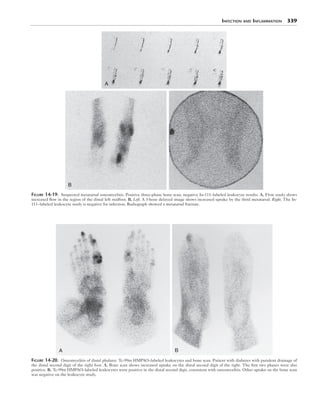
Ga-67 has widespread organ uptake and soft tissue dis-
tribution (Table 14-3). Greatest uptake is seen in the liver,
followed by bone and bone marrow and spleen (Fig. 14-2).
Renal clearance is seen during the initial 24 hours. By 48
hours, the kidneys are only faintly seen.
Sites of variable uptake include the nasopharynx, lacri-
mal and salivary glands, and breast (Fig. 14-2); all elabo-
rate lactoferrin. Inflammatory or stimulatory processes
increase lactoferrin production and uptake, thus increased
uptake occurs in the salivary glands in Sjögren’s syndrome,
in lacrimal glands in sarcoidosis, and breast during lacta-
tion. Breast uptake varies according to the menstrual cycle
phase and is seen prominently postpartum. Thymic
uptake is normal in children.
Postoperative sites may have increased Ga-67 uptake for
2 to 3 weeks. Uptake occurs at healing fractures (Fig. 14-3)
and in sterile abscesses associated with frequent intramus-
cular injections (e.g., insulin or iron-depot injections). Sali-
vary gland uptake is increased after local external beam
irradiation or chemotherapy. Normal distribution is altered
by whole-body irradiation, multiple blood transfusions
(excess ferric ions), or recent gadolinium MRI (Fig. 14-4).
Methodology
An imaging protocol for Ga-67 is described in Box 14-2.
Bowel preparation with laxatives and enemas to facilitate
more rapid clearance is no longer recommended because it
is not usually effective and may cause mucosal irritation
and inflammation, producing increased Ga-67 uptake.
Chapter 14
Infection and Inflammation
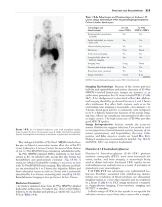
Box 14-1. Radiopharmaceuticals for Infection
Imaging Approved for Clinical Use
Ga-67 citrate
In-111 oxine–labeled leukocytes
Tc-99m HMPAO–labeled leukocytes
F-18 FDG
Tc-99m fanolesomab (NeutroSpec) (FDA approved,
then withdrawn from U.S. market)
Tc-99m sulesomab (LeuTech) (approved and used in
Europe)
FDA, U.S. Food and Drug Administration; FDG, fluorodeoxyglucose; HMPAO,
hexamethylpropyleneamine oxime.](https://image.slidesharecdn.com/14infectioninflammation-141117030350-conversion-gate01/85/14-infection-inflammation-1-320.jpg)


![326 Nuclear Medicine: The Requisites
function. The radiolabel usually remains stable in vivo for
more than 24 hours.
Details of the radiolabeling process are detailed
(Box 14-3). The patient’s blood is drawn, it is allowed to
settle, and then the majority of the erythrocytes removed.
In-111 oxine labels granulocytes, lymphocytes, mono-
cytes, platelets, and erythrocytes.
Radiolabeling cannot be performed in plasma. It must
be separated and retained for later resuspension with the
WBCs before reinfusion. The leukocyte pellet is sus-
pended in saline and incubated with In-111 oxine. The
lipid solubility of the In-111 oxine complex allows it to dif-
fuse through cell membranes. Intracellularly, the complex
dissociates, oxine diffuses back out of the cell, and In-111
binds to nuclear and cytoplasmic proteins.
Pure granulocyte preparations have been radiolabeled
and used clinically. However, they require elaborate den-
sity gradient separation techniques and have not shown a
clinical advantage over mixed leukocytes and thus are not
generally used.
Standard quality control measures, such as testing for ste-
rility and pyrogenicity, cannot be performed because of the
need for prompt reinfusion after labeling to ensure cell via-
bility. However, the final radiopharmaceutical preparation
is routinely examined for abnormal morphology, clumping,
excessive red cell contamination, and percent labeling effi-
ciency, which typically ranges from 75% to 90% (Box 14-2).
When labeling is less than 50%, the cells should not be rein-
fused. The final preparation contains radiolabeled granulo-
cytes, lymphocytes, and monocytes, but there will also be
10% to 20% platelets and erythrocytes.
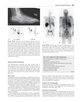
After infusion of the radiolabeled leukocytes, no signifi-
cant elution of the In-111 from the leukocytes occurs. The
effective half-life of clearance from the blood circulation is
7.5 hours. Initial distribution after reinfusion is to the blood
pool, lungs, liver, and spleen. Early lung uptake is the
result of cellular activation from in vitro cell manipulation.
By 4 hours after reinjection, lung and blood pool activity
decrease, although not always completely (Fig. 14-5).
By 24 hours, blood-pool activity is normally no longer
present. Persistent blood pool at 24 hours suggests a high
percentage of labeled erythrocytes or platelets. The highest
uptake is in the spleen, followed by liver and then bone
marrow (Fig. 14-5). Neither genitourinary, hepatobiliary, nor
intestinal clearance is normally seen (Table 14-3).
The ultimate test of viability of leukocytes is in vivo
function manifested by a normal distribution within the
body and the ability to detect infection. If the infused leu-
kocytes become nonviable, increased liver and lung uptake
may be seen on scintigraphy. With excessive erythrocyte
and platelet labeling, blood pool is prominent.
Radiation Dosimetry
The adult spleen receives 15 to 20 rads/500 μCi (15-20
cGy/18.5 MBq); however, in small children the spleen may
Box 14-3. Radiolabeling Autologous Leukocytes
with Indium-111 Oxine
PREPARATION
Patient’s peripheral leukocyte count should be
greater than 5000 cells/mm3.
PROCEDURE
1. Collect autologous blood
Draw 30 to 50 mL into an ACD anticoagulated
syringe using a 19-gauge needle.
2. Isolate leukocytes:
Separate red blood cells (RBCs) by gravity
sedimentation and 6% hetastarch, a settling
agent.
Centrifuge the leukocyte-rich plasma at 300 to
350 g for 5 minutes to remove platelets and
proteins.
A white blood cell (WBC) button forms at the
bottom of the tube.
Draw off and save the leukocyte-poor plasma
(LRP) for later washing and resuspension.
3. Label leukocytes
Suspend WBCs (LRP) in saline (includes
neutrophils, lymphocytes, monocytes, some
RBCs).
Incubate with In-111 oxine for 30 minutes at
room temperature and gently agitate.
Remove unbound In-111 by centrifugation. Save
wash for calculation of labeling efficiency.
4. Prepare injectate
Resuspend 500 μmCi In-111–labeled leuko-
cytes in saved plasma (LPP).
Inject via peripheral vein within 2 to 4 hours.
5. Perform quality control
Microscopic examination of cells.
Calculate labeling efficiency:
Assay the cells and wash in dose calibrator.
Labeling efficiency = C/([C+ W] × 100%)
where C is activity associated with the cells
and W is activity associated with the wash.
Table 14-4 Radiation Dosimetry for Gallium-67
Citrate, Indium-111 Oxine, and Technetium-99m
HMPAO Leukocytes
Organ
Ga-67 citrate
rads/5 mCi
(cGy/185
MBq)
In-111 oxine
WBCs rads/500
μCi (cGy/18.5
MBq)
Tc-99m
HMPAO WBCs
rads/10 mCi
(cGy/370 MBq)
Bladder wall 2.8
Large
intestine
3.7 3.6
Liver 2.2 2.66 1.5
Bone marrow 3.5 1.99 1.6
Spleen 1.8 20.00 2.2
Ovaries 1.5 0.20 0.3
Testes 1.0 0.014 1.9
Total body
Effective
dose
2.2
1.9
0.37
0.7
0.3
0.4
HMPAO, Hexamethylpropyleneamine oxime; WBCs, white blood cells.
Target organ dose is in bold.](https://image.slidesharecdn.com/14infectioninflammation-141117030350-conversion-gate01/85/14-infection-inflammation-5-320.jpg)


![344 Nuclear Medicine: The Requisites
If In-111–labeled leukocyte scintigraphy is used, images
should be acquired at 4 hours rather than 24 hours because
of shedding of the inflamed leukocytes into the bowel
lumen from the inflammatory sites and subsequent peri-
stalsis, which can result in incorrect assignment of disease
to sites distal to the true lesion. Tc-99m HMPAO imaging
should be performed by 2 hours.
Renal Disease
Ga-67 has been used to diagnose diffuse interstitial nephri-
tis and localized renal infection. Delayed 48-hour imaging
is required because of early urinary tract clearance. Renal
parenchymal infection, (e.g., pyelonephritis, diffuse inter-
stitial nephritis, lobar nephronia [focal interstitial nephri-
tis], and perirenal infections) can be diagnosed. Generally,
Ga-67 has been superseded by radiolabeled leukocyte
imaging (Fig. 14-29) or Tc-99m dimercaptosuccinic acid
(DMSA) for pyelonephritis. Radiolabeled leukocytes
seem to have similar accuracy. However, radiolabeled
WBCs have limited utility for evaluation of renal trans-
plants because all exhibit uptake, regardless of the pres-
ence or absence of clinical infection, probably as a result of
ongoing low-grade rejection.
Cardiovascular Disease
In-111–labeled leukocytes are not sensitive for detection
of subacute bacterial endocarditis. The vegetative lesions
contain high concentrations of bacteria, platelets, and
fibrin adherent to damaged valvular endothelium, but rela-
tively few leukocytes. However, SPECT and SPECT/CT
have improved detection and localization (Fig. 14-30).
Prompt diagnosis of graft infection is critical but often
delayed because of its indolent and insidious course. With
infection of arterial prosthetic grafts (e.g., femoropopliteal
or aortofemoral), ultrasound, CT, and MRI are often
unable to distinguish infection from aseptic fluid collec-
tions around the graft. Radiolabeled leukocytes can detect
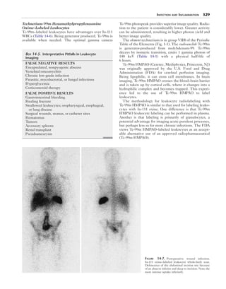
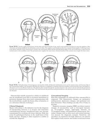
Figure 14-28. Crohn disease on 4-hour In-111 oxine–labeled leukocytes.
Patient with several-year history of regional ileitis and 2 months of recur-
rent and worsening symptoms. Scintigraphy confirms active inflammation
of ileum.
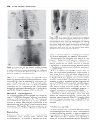
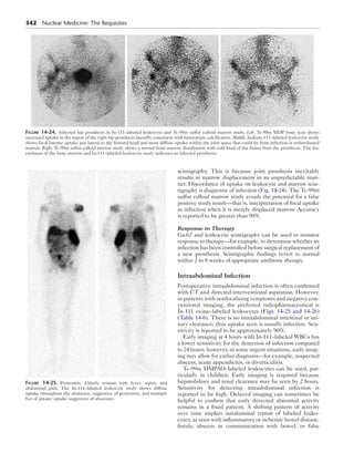
Figure 14-29. Infected polycystic kidneys on In-111–labeled leukocyte scan for renal stones, recurrent urinary tract infections, and recent persistent
fever of uncertain cause. Low-dose CT (above) demonstrates very large multicystic kidneys. Fused SPECT/CT images (below) show multiple areas of
increased uptake (yellow) in the renal cysts diagnostic of infection.](https://image.slidesharecdn.com/14infectioninflammation-141117030350-conversion-gate01/85/14-infection-inflammation-23-320.jpg)


This document discusses radiopharmaceuticals used for infection imaging. It begins by describing gallium-67 citrate, which was the first infection-seeking radiopharmaceutical and is still used today, though more limited. Radiolabeled leukocytes are now usually preferred. FDG is also increasingly used. The document then discusses the pathophysiology of inflammation and infection in more detail. It describes how various radiopharmaceuticals, including gallium-67 citrate and radiolabeled leukocytes, are taken up at sites of infection and their mechanisms of uptake, distributions, and dosimetry.