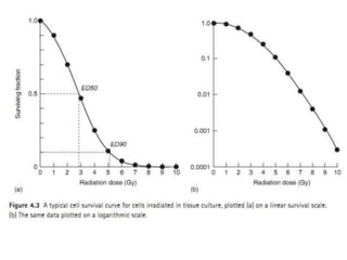
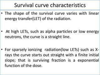
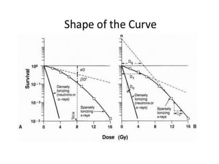
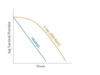
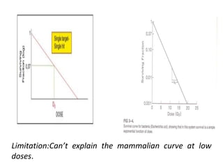
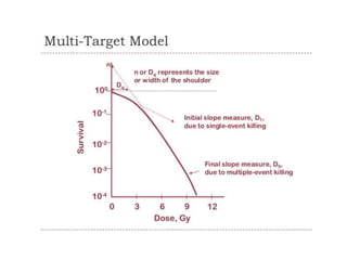
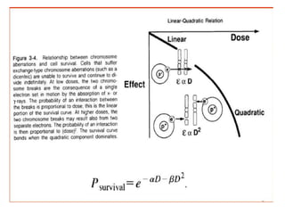
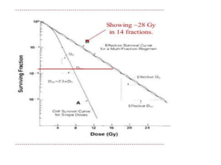
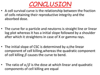
CELL SURVIVAL CURVES
Presenter- Dr Luri Borah
1st Year PGT, Dept. of Radiotherapy
Moderator- Dr. Mouchumee Bhattacharyya
Addl. Prof. , Dept of Radiotherapy
Defined as
“ The relationship between the radiation dose
and the proportion of cell that survive”.
• 100Gy - nonproliferating system
• 2Gy- proliferating system
Cell Death
Reproductive
death
Functional
death
Clonogenic: Survivor that retains reproductive
integrity and proliferate indefinitely.
Radiation and cell death
• Cells generally die in two ways:
1. Mitotic death
2. Apoptosis.
• The shape of cell survival curve tells us the
radiosensitivity, repair and recovering ability of
the cell. The shape of the curve gets altered if
the conditions are changed.
The In Vitro Survival Curve
• To conduct the experiment a known number of cells
are seeded in multiple petridishes and exposed to
varying dose of radiation and incubated.
• They are allowed to grow colonies and the same is
counted. One or two dishes are used as controls
and no radiation given.
• Two parameters are used to quantify the cells:
1. Plating Efficiency(PE)
2. Survival fraction(SF)
Contd…
Survival curve characteristics
• The shape of the survival curve varies with linear
energy transfer(LET) of the radiation.
• At high LETs, such as alpha particles or low energy
neutrons, the curve is a straight line.
• For sparsely ionizing radiation(low LETs) such as X-
rays the curve starts out straight with a finite initial
slope; that is surviving fraction is a exponential
function of the dose.
Models of Description of the curve
• Single target model
• Multiple target model
• Linear Quadratics model
Single Target-single hit theory
• It states that “ the objective of treating a tumour
by radiotherapy is to damage every single
potentially malignant cell to such an extent that it
cannot continue to proliferate”
• Hence, the surviving fraction (S) is described by
poisson statistics, as follows:
S = exp(-P)
Where P is the mean number of probability for not
having hit.If the probability is a linear function of
dose(D) ,then the equation will be:
Contd….
S = exp(- D/D˳)
Where D˳ is the mean lethal dose that will give one
hit per target, if D=D˳, then S= exp(-1) = 0.37
• D˳ is the reciprocal of the slope of the linear portion
of the curve .This dose will bring the surviving
fraction to 37%. Hence D˳ is referred to as D₃₇.
• The typical value for mammalian cell is 1-2Gy for
low LET radiations.
Limitation:Can’t explain the mammalian curve at low
doses.
Multitarget theory
• According to this theory, some cells contain more
than one target and each of this targets should
receive the hit.
• The curve starts with less sensitive region at low
dose and tend to become exponential at larger
doses. So they present a shoulder at small doses.
• Here the SF is given by the equation:
S = 1- [1-exp(-D/nD˳)]ⁿ
Where n is the no of targets in the organisms.
contd…
• . A survival curve is defined with these parameters:
1.Initial slope (D₁), is the reciprocal of initial dose
called mean lethal dose
2. Final slope(D˳), is the reciprocal of final slope and
the required to reduce the survival to 37%
3. Extrapolation number(n),corresponding to the
number of targets. If ‘n’ is larger ,width of the
shoulder is broad,if the ‘n’ is small,width is small.
4. Quasithreshold dose(Dq), is the width of the
horizontal line drawn parallel to X-axis at the survival
level of 1 and represents sublethal damage.
Linear Quadratic model
• It states that inactivation of cell results only when
both strands of DNA molecules or both arms of a
chromosome are damaged which can produced by
passage of single ionizing particle or by
independent interaction of two separate ionizing
particles.
• The SF is given by the formulae:
SF = exp-(⍺d+𝛽d²)
Where d is the dose in Gy
⍺ is the cell kill per Gy of the initial linear
component. It dominates high LET and damage is
irreparable.
Contd….
𝛽 is the cell kill per Gy² of the quadratic component of the
curve. It dominates low LET and the damage is reparable.
• The LQ model explains the cell killing in low dose range( 0-
3Gy).
• The shape of the curve is determined by the ⍺/𝛽 ratio which
the dose at which the linear part of cell killing is equal to
quadratic part of cell killing.
• Higher the ⍺/𝛽 ratio straighter the curve and narrow the
shoulder. It exhibits irreparable damage. It refers high
radiosensitivity of cells.
• Lower ⍺/𝛽 ratio , wide shoulder ,high capability of repair. It
refers to radioresistance of cells.
Contd….
• Tumors tend to have high ⍺/𝛽 ratio whereas it is
much lower for normal tissues
• Typical values for cancer cells range from 8-12 Gy
whereas for late responding normal tissue is 2-4 Gy
and early responding normal tissue is 6-12 Gy.
Mechanism of cell death
• Bystander effect
• Apoptosis
• Mitotic death
• Autophagic death
Bystander effect
• When a population of cell is irradiated,the cells
surrounding the irradiated cell population are also
affected and this effect is called radiation- induced
bystander effect(RIBE).
• This effect is true for ⍺,proton,and soft X-rays.
• Laboratory study with low-LET radiation reveals
that the irradiated cells secrete
molecules/chemicals which are capable of killing
cells.
Apoptosis
• It is a greek word that means programmed cell
death.
• Apoptosis occurs in normal cell and and can be
induced in tumour cell by irradiation.
• In the case of tumor cells it is cell type dependent.
• Apoptosis occurs in particular in certain cell lines
after low dose of radiation, eg. Lymphocyte, serous
salivary glands, certain cells in testis and intestinal
crypts.
Mitotic death
• Reproductive cell death is a result of mitotic
catastrophe,which can occur in the first few cell
divisions after irradiation.
• The mitotic death is the most common radiation
induced cell death and it is due chromosomal
aberrations.
• The irradiated cells may undergo cell cycle and
they may die in the first or second mitotic cycle,
while attempting division.
Autophagic cell death
• Autophagy is a self digestive process that uses
lysosomal degradationof long-lived proteins and
organelles to maintain cellular mechanism of
cells.
• Chemotherapeutic agents and radiotherapy can
induce autophagy abd autophagic cell death.
Radiation damage
Lethal damage
Potentialy lethal
damage
No effect
Incorrect repair
Correct repair
Normal function
restored
Cell death
mutation
lethal
Cell death
viable
Transmit mutation to
next generation
Factors influencing cell survival curve
• LET
• Radiosentivity of the cells
• Fractionation
• Dose rate effect
• Cell aging
• Presence of oxyzen.
LET
• The LET is the avg. energy transferred per unit
length of the track in the medium and its unit is
keV/𝛍m.
• Low LET (X-rays and gamma rays) shoulder of the
curve appears.
• High LET ( neutrons) the curve becomes linear and
surviving fraction becomes a exponential function
of dose.
Fractionation
• If a radiation dose is delivered in a series of equal
fractions, separated by sufficient time for repair of
sublethal damage to occur between doses, the
effective dose- survival curve becomes an
exponential function of dose.
• The shoulder of the curve is repeated many times,
so that the effective survival curve is a straight line
from the original through a point on the single
dose survival curve corresponding to the daily
dose fraction.
Contd...
• The dose required to produce the same effect in SF
increases.
• D˳ is 3Gy,defined to be the dose required to reduce
the SF to 37%.
• For calculation purpose ,it is often useful to use the
D₁₀, the dose required to kill 90% of the population.
For example: D₁₀= 2.3xD˳
Dose Rate effect
• Reduction in dose causes reduced cell killing, due to
repair of sublethal damage.
• Reduction in dose rate generally reduces survival
curve slope.
Cell Aging
Sensitivity of a cell in cell cycle to radiation is as
follows:
M> G1>G2>lateS1>early S2
Oxygen and radiation
• In general, tumour cells contain both aerated and hypoxic
cells.
• During radiation more aerated cells are killed, since they are
radiosensitive, but the percentage of hypoxic cells remain
same
• This implies that after 1st fraction of radiation some of the
hypoxic cells become oxygenated( Reoxygenation) and
become sensitive to radiation which are selectively killed in
subsiquent radiation.
• It has been found that the radiation fraction schedule of
60Gy/30# is sufficient to cure majority of tumours.
Contd…
• This suggest that human tumor do have hypoxic
cells and they get reoxygenated during fractionated
radiotherapy.
• However some tumours that donot oxygenate
quickly cause treatment failure.
• Thus time scale of reoxygenation is required for
tumours to achieve effective cure in radiotherapy.
CONCLUSION
• A cell survival curve is the relationship between the fraction
of cells retaining their reproductive integrity and the
absorbed dose.
• The curve for ⍺ particle and neutorns is straight line or linear
log plot whereas it has a initial slope followed by a shoulder
after which it straightens in case of X or gamma rays.
• The initial slope of CSC is determined by ⍺,the linear
component of cell killing,whereas the quadratic component
of cell killing 𝛽 causes the curve to bend.
• The ratio of ⍺/𝛽 is the dose at which linear and quadratic
components of cell killing are equal
Contd…
• The most common mode of cell death in tumour
cells is mitotic death.
• Different tumors have different radiosensitivity.
• The effective survival curve for a multifraction
regimen is an exponential function of dose.
• The avg value of the effective D₀ for multifraction
survival curve is about 3Gy.
• The D₁₀ is related to D₀ by
D₁₀= 2.3 x D₀.
THANK YOU

Cell survival curve ppt.

  • 1.
    CELL SURVIVAL CURVES Presenter-Dr Luri Borah 1st Year PGT, Dept. of Radiotherapy Moderator- Dr. Mouchumee Bhattacharyya Addl. Prof. , Dept of Radiotherapy
  • 2.
    Defined as “ Therelationship between the radiation dose and the proportion of cell that survive”. • 100Gy - nonproliferating system • 2Gy- proliferating system
  • 3.
    Cell Death Reproductive death Functional death Clonogenic: Survivorthat retains reproductive integrity and proliferate indefinitely.
  • 4.
    Radiation and celldeath • Cells generally die in two ways: 1. Mitotic death 2. Apoptosis. • The shape of cell survival curve tells us the radiosensitivity, repair and recovering ability of the cell. The shape of the curve gets altered if the conditions are changed.
  • 5.
    The In VitroSurvival Curve • To conduct the experiment a known number of cells are seeded in multiple petridishes and exposed to varying dose of radiation and incubated. • They are allowed to grow colonies and the same is counted. One or two dishes are used as controls and no radiation given. • Two parameters are used to quantify the cells: 1. Plating Efficiency(PE) 2. Survival fraction(SF)
  • 6.
  • 9.
    Survival curve characteristics •The shape of the survival curve varies with linear energy transfer(LET) of the radiation. • At high LETs, such as alpha particles or low energy neutrons, the curve is a straight line. • For sparsely ionizing radiation(low LETs) such as X- rays the curve starts out straight with a finite initial slope; that is surviving fraction is a exponential function of the dose.
  • 12.
    Models of Descriptionof the curve • Single target model • Multiple target model • Linear Quadratics model
  • 13.
    Single Target-single hittheory • It states that “ the objective of treating a tumour by radiotherapy is to damage every single potentially malignant cell to such an extent that it cannot continue to proliferate” • Hence, the surviving fraction (S) is described by poisson statistics, as follows: S = exp(-P) Where P is the mean number of probability for not having hit.If the probability is a linear function of dose(D) ,then the equation will be:
  • 14.
    Contd…. S = exp(-D/D˳) Where D˳ is the mean lethal dose that will give one hit per target, if D=D˳, then S= exp(-1) = 0.37 • D˳ is the reciprocal of the slope of the linear portion of the curve .This dose will bring the surviving fraction to 37%. Hence D˳ is referred to as D₃₇. • The typical value for mammalian cell is 1-2Gy for low LET radiations.
  • 15.
    Limitation:Can’t explain themammalian curve at low doses.
  • 16.
    Multitarget theory • Accordingto this theory, some cells contain more than one target and each of this targets should receive the hit. • The curve starts with less sensitive region at low dose and tend to become exponential at larger doses. So they present a shoulder at small doses. • Here the SF is given by the equation: S = 1- [1-exp(-D/nD˳)]ⁿ Where n is the no of targets in the organisms.
  • 17.
    contd… • . Asurvival curve is defined with these parameters: 1.Initial slope (D₁), is the reciprocal of initial dose called mean lethal dose 2. Final slope(D˳), is the reciprocal of final slope and the required to reduce the survival to 37% 3. Extrapolation number(n),corresponding to the number of targets. If ‘n’ is larger ,width of the shoulder is broad,if the ‘n’ is small,width is small. 4. Quasithreshold dose(Dq), is the width of the horizontal line drawn parallel to X-axis at the survival level of 1 and represents sublethal damage.
  • 19.
    Linear Quadratic model •It states that inactivation of cell results only when both strands of DNA molecules or both arms of a chromosome are damaged which can produced by passage of single ionizing particle or by independent interaction of two separate ionizing particles. • The SF is given by the formulae: SF = exp-(⍺d+𝛽d²) Where d is the dose in Gy ⍺ is the cell kill per Gy of the initial linear component. It dominates high LET and damage is irreparable.
  • 20.
    Contd…. 𝛽 is thecell kill per Gy² of the quadratic component of the curve. It dominates low LET and the damage is reparable. • The LQ model explains the cell killing in low dose range( 0- 3Gy). • The shape of the curve is determined by the ⍺/𝛽 ratio which the dose at which the linear part of cell killing is equal to quadratic part of cell killing. • Higher the ⍺/𝛽 ratio straighter the curve and narrow the shoulder. It exhibits irreparable damage. It refers high radiosensitivity of cells. • Lower ⍺/𝛽 ratio , wide shoulder ,high capability of repair. It refers to radioresistance of cells.
  • 22.
    Contd…. • Tumors tendto have high ⍺/𝛽 ratio whereas it is much lower for normal tissues • Typical values for cancer cells range from 8-12 Gy whereas for late responding normal tissue is 2-4 Gy and early responding normal tissue is 6-12 Gy.
  • 23.
    Mechanism of celldeath • Bystander effect • Apoptosis • Mitotic death • Autophagic death
  • 24.
    Bystander effect • Whena population of cell is irradiated,the cells surrounding the irradiated cell population are also affected and this effect is called radiation- induced bystander effect(RIBE). • This effect is true for ⍺,proton,and soft X-rays. • Laboratory study with low-LET radiation reveals that the irradiated cells secrete molecules/chemicals which are capable of killing cells.
  • 25.
    Apoptosis • It isa greek word that means programmed cell death. • Apoptosis occurs in normal cell and and can be induced in tumour cell by irradiation. • In the case of tumor cells it is cell type dependent. • Apoptosis occurs in particular in certain cell lines after low dose of radiation, eg. Lymphocyte, serous salivary glands, certain cells in testis and intestinal crypts.
  • 26.
    Mitotic death • Reproductivecell death is a result of mitotic catastrophe,which can occur in the first few cell divisions after irradiation. • The mitotic death is the most common radiation induced cell death and it is due chromosomal aberrations. • The irradiated cells may undergo cell cycle and they may die in the first or second mitotic cycle, while attempting division.
  • 27.
    Autophagic cell death •Autophagy is a self digestive process that uses lysosomal degradationof long-lived proteins and organelles to maintain cellular mechanism of cells. • Chemotherapeutic agents and radiotherapy can induce autophagy abd autophagic cell death.
  • 28.
    Radiation damage Lethal damage Potentialylethal damage No effect Incorrect repair Correct repair Normal function restored Cell death mutation lethal Cell death viable Transmit mutation to next generation
  • 29.
    Factors influencing cellsurvival curve • LET • Radiosentivity of the cells • Fractionation • Dose rate effect • Cell aging • Presence of oxyzen.
  • 30.
    LET • The LETis the avg. energy transferred per unit length of the track in the medium and its unit is keV/𝛍m. • Low LET (X-rays and gamma rays) shoulder of the curve appears. • High LET ( neutrons) the curve becomes linear and surviving fraction becomes a exponential function of dose.
  • 31.
    Fractionation • If aradiation dose is delivered in a series of equal fractions, separated by sufficient time for repair of sublethal damage to occur between doses, the effective dose- survival curve becomes an exponential function of dose. • The shoulder of the curve is repeated many times, so that the effective survival curve is a straight line from the original through a point on the single dose survival curve corresponding to the daily dose fraction.
  • 32.
    Contd... • The doserequired to produce the same effect in SF increases. • D˳ is 3Gy,defined to be the dose required to reduce the SF to 37%. • For calculation purpose ,it is often useful to use the D₁₀, the dose required to kill 90% of the population. For example: D₁₀= 2.3xD˳
  • 34.
    Dose Rate effect •Reduction in dose causes reduced cell killing, due to repair of sublethal damage. • Reduction in dose rate generally reduces survival curve slope. Cell Aging Sensitivity of a cell in cell cycle to radiation is as follows: M> G1>G2>lateS1>early S2
  • 36.
    Oxygen and radiation •In general, tumour cells contain both aerated and hypoxic cells. • During radiation more aerated cells are killed, since they are radiosensitive, but the percentage of hypoxic cells remain same • This implies that after 1st fraction of radiation some of the hypoxic cells become oxygenated( Reoxygenation) and become sensitive to radiation which are selectively killed in subsiquent radiation. • It has been found that the radiation fraction schedule of 60Gy/30# is sufficient to cure majority of tumours.
  • 37.
    Contd… • This suggestthat human tumor do have hypoxic cells and they get reoxygenated during fractionated radiotherapy. • However some tumours that donot oxygenate quickly cause treatment failure. • Thus time scale of reoxygenation is required for tumours to achieve effective cure in radiotherapy.
  • 38.
    CONCLUSION • A cellsurvival curve is the relationship between the fraction of cells retaining their reproductive integrity and the absorbed dose. • The curve for ⍺ particle and neutorns is straight line or linear log plot whereas it has a initial slope followed by a shoulder after which it straightens in case of X or gamma rays. • The initial slope of CSC is determined by ⍺,the linear component of cell killing,whereas the quadratic component of cell killing 𝛽 causes the curve to bend. • The ratio of ⍺/𝛽 is the dose at which linear and quadratic components of cell killing are equal
  • 39.
    Contd… • The mostcommon mode of cell death in tumour cells is mitotic death. • Different tumors have different radiosensitivity. • The effective survival curve for a multifraction regimen is an exponential function of dose. • The avg value of the effective D₀ for multifraction survival curve is about 3Gy. • The D₁₀ is related to D₀ by D₁₀= 2.3 x D₀.
  • 40.