Downloaded 198 times


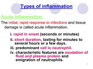


Acute inflammation is characterized by rapid onset, short duration, and neutrophil infiltration. It serves to destroy, dilute, or wall off injurious agents. The key steps are vascular changes that increase blood flow and permeability, allowing plasma proteins and leukocytes to exit circulation and reach injured tissues. Leukocytes are recruited through chemotaxis and remove pathogens via phagocytosis. Mediators like histamine and prostaglandins regulate vascular changes and leukocyte behavior. Common patterns include purulent inflammation seen in abscesses and serous inflammation seen in effusions. Acute inflammation typically resolves completely or leads to scarring, and can progress to chronic inflammation if the cause persists.


