This document discusses non-PET imaging techniques for oncology applications. It focuses on peptide receptor imaging using radiolabeled somatostatin analogs such as In-111 pentetreotide, which binds to somatostatin receptors overexpressed on many neuroendocrine tumors. In-111 pentetreotide scintigraphy has high sensitivity for detecting carcinoid tumors and other neuroendocrine tumors but lower sensitivity for insulinomas. It provides whole body imaging of tumor lesions and localization of disease when combined with SPECT or SPECT/CT. While F-18 FDG PET is useful for more aggressive tumors, In-111 pentetreotide remains an important functional imaging tool for somatostatin receptor-expressing neuro
Positron emission tomography (PET) using fluorine-18 fluorodeoxyglucose (F-18 FDG) has expanded for clinical oncology applications. PET provides metabolic tumor imaging, which can complement anatomical imaging from computed tomography (CT). F-18 FDG uptake is increased in cancer cells due to higher glucose metabolism. Combined PET/CT imaging provides precise anatomical localization of metabolic abnormalities seen on PET. PET can help characterize tumors, detect recurrence, and assess treatment response in ways that CT cannot by directly measuring cellular activity levels. Standard protocols involve patient preparation, intravenous F-18 FDG injection, and imaging after uptake and clearance periods.
Nuclear medicine application in neuroendocrine tumors (net)Ramin Sadeghi
This document discusses the use of nuclear medicine techniques for staging and treatment monitoring of neuroendocrine tumors. Positron emission tomography using radiolabeled somatostatin analogues like Ga-68 DOTATATE is recommended for staging most well-differentiated neuroendocrine tumors. In-111 or Tc-99m octreotide scintigraphy is also used but has lower sensitivity than PET. F-18 FDG PET is used for poorly differentiated and extrapulmonary neuroendocrine tumors. I-123 MIBG, somatostatin receptor imaging, or FDG PET are used for pheochromocytoma/paraganglioma staging if metastasis is suspected. Lu-177 DOTATATE
Multiple endocrine neoplasia (MEN) is characterized by tumors involving two or more endocrine glands. There are four main types of MEN: MEN type 1 is associated with tumors of the parathyroid, pancreas, and pituitary glands. MEN type 2 is associated with medullary thyroid carcinoma, pheochromocytomas, and parathyroid tumors. MEN type 3, also called MEN 2B, involves the same tumors as MEN 2A but also includes other features. MEN type 4 involves tumors associated with MEN 1 and CDNK1B gene mutations. The tumors associated with each MEN type are inherited in an autosomal dominant pattern and can be diagnosed clinically
Multiple endocrine neoplasia (MEN) type 1 is characterized by tumors of the parathyroid glands, pancreas, and pituitary gland. It is caused by mutations in the MEN1 gene which codes for the menin protein. Patients with MEN1 often present with hyperparathyroidism, pancreatic neuroendocrine tumors, or prolactin-secreting pituitary adenomas. Diagnosis involves blood tests and imaging to identify affected organs. Treatment depends on the specific tumors but may include parathyroidectomy, surgical removal of pancreatic tumors, and medication for functional tumors. Ongoing screening is important due to the risk of additional tumors developing over time.
This document discusses multiple endocrine neoplasia (MEN) syndromes, which are rare genetic disorders where multiple endocrine tumors occur in multiple endocrine glands. It describes the six known MEN syndromes and their associated genetic mutations. Specific syndromes discussed in detail include MEN type 1 and type 2A, which are associated with tumors of the parathyroid, pancreas, and pituitary glands. Timely surgical removal of affected glands can prevent or cure malignancies associated with these syndromes.
Multiple endocrine neoplasia type 1 (MEN1) is an autosomal dominant genetic condition characterized by tumors of the parathyroid glands, endocrine pancreas, and pituitary gland. It is caused by mutations in the MEN1 tumor suppressor gene, resulting in DNA damage. Affected individuals require lifelong screening for primary hyperparathyroidism, pancreatic neuroendocrine tumors, and prolactin- or growth hormone-secreting pituitary adenomas. Surgical management focuses on resection of symptomatic tumors.
This document summarizes multiple endocrine neoplasia type 1 (MEN1), a rare hereditary cancer syndrome characterized by tumors of the parathyroid glands, pancreas, and pituitary gland. MEN1 is caused by mutations in the MEN1 gene and is inherited in an autosomal dominant pattern. Diagnosis involves identifying biochemical abnormalities and tumors associated with MEN1. Management focuses on surgery to remove tumors, with the goal of preventing complications from hormone hypersecretion. Lifelong screening is required due to the high risk of recurrent and multiple tumors developing over time.
Multiple endocrine neoplasia syndromes are inherited conditions where two or more endocrine glands develop non-cancerous or cancerous tumors due to mutations in genes regulating cell growth. There are two main types: type 1 affects the parathyroid, pituitary, pancreas and thyroid glands. Type 2 affects the parathyroid, thyroid and adrenal glands and is divided into 2A and 2B. Symptoms vary depending on affected glands but include high blood calcium and kidney problems. Diagnosis involves family history, hormone testing, imaging and genetic testing. Treatment focuses on surgically removing tumors and managing hormone levels with medication.
Poster presentation at the 2016 WORLD GASTROINTESTINAL SYMPOSIUM on tepotinib a selective inhibitor of c-MET by S. Faivre, J.-F. Blanc, P. Merle, A. Fasolo, A. Iacobellis, V. Grando, T. Decaens, J. Trojan, E. Villa, U. Stammberger, R. Bruns, E. Raymond
1Oncology Unit, Beaujon University Hospital, Clichy, France; 2Service d’hépato-gastroentérologie et d’oncologie digestive, Groupe Hospitalier Saint André, Bordeaux, France; 3Service d'Hépato-Gastro-Entérologie, Hôpital de la Croix Rousse, Lyon, France; 4Dipartimento di Oncologia Medica, Ospedale San Raffaele IRCSS, Milan, Italy; 5Servizio di Endoscopia Digestiva, Ospedale Casa Sollievo della Sofferenza IRCCS, San Giovanni Rotondo, Italy; 6Service Hépatologie, Hôpital Jean-Verdier, Bondy, France; 7Service d'hepato-gastro-enterologie, CHU de Grenoble - Hôpital Nord, Grenoble, France; 8Gastrointestinal Oncology, Goethe University Hospital, Frankfurt, Germany; 9Policlinico di Modena, Modena, Italy; 10Merck KGaA, Darmstadt, Germany
This study examined placental samples from 60 women with preeclampsia and 60 healthy women at 29-35 weeks of gestation. It investigated the expression levels of ADAMTS-1 and ADAMTS-4, which are involved in extracellular matrix remodeling, between the two groups. The study found significantly lower expression levels of ADAMTS-1 and ADAMTS-4 in placental samples from women with preeclampsia compared to healthy women. It also observed morphological changes in the placentas of women with preeclampsia. The study concluded that decreased expression of ADAMTS-1 and ADAMTS-4 may affect placental morphology, trophoblast invasion, and angiogenesis in preeclampsia
1. The study explored the role of Sema4D in bone metastasis of breast cancer. Sema4D expression was downregulated in MCF-7 breast cancer cells using siRNA transfection. Proliferation, invasion, and osteoblast differentiation were then measured.
2. Proliferation and invasion were reduced in the Sema4D siRNA group compared to control groups. Bone nodule area and AKT phosphorylation were increased in the Sema4D siRNA group.
3. Downregulating Sema4D expression attenuated inhibition of osteoblast differentiation, likely by enhancing AKT phosphorylation. Sema4D may play a role in regulating bone metastasis of breast cancer.
This document describes a case study of a rare case of cardiac metastases from solid papillary carcinoma (SPC) of the breast. A 67-year-old woman with a history of breast cancer underwent surgery to remove a tumor in her right atrium. Pathological examination of the tumor found that it was an invasive SPC of the breast that had metastasized to the heart. Immunohistochemical staining confirmed the diagnosis. Analysis of this case improves understanding of the diagnosis and treatment of metastatic SPC of the breast to rare sites like the heart.
Thanks to Prof. Chavdar Pavlov, MD, PhD, MScD - Department Head of Therapy, Head of the Centre for Evidence-Based Medicine, I.M. Sechenov First Moscow State Medical University (Sechenov University), Moscow, Russia for helping organizing this event in Moscow.
The document discusses the use of diagnostic algorithms and antibody panels to identify tumor types through immunohistochemistry (IHC). It provides examples of algorithms that use sequential testing of antibodies to differentiate between common tumor types like carcinoma vs sarcoma, or to identify the primary origin of a metastatic tumor. The algorithms aim to increase diagnostic accuracy and guide treatment by narrowing down the possible tumor types. Key details about specific antibodies, their targets, and staining patterns are also presented.
Multiple endocrine neoplasia and neuroendocrine tumour of pancreasePrince Lathiya
Multiple endocrine neoplasia (MEN) is characterized by tumors in multiple endocrine tissues. The document discusses MEN types 1 and 2, which are caused by mutations in the MEN1 and RET genes, respectively. MEN type 1 is associated with tumors of the parathyroid glands, pancreas, and pituitary gland. Parathyroid tumors are the most common manifestation. MEN type 2A is associated with medullary thyroid cancer, pheochromocytomas, and parathyroid tumors, while type 2B additionally involves mucosal neuromas. Surgical removal of affected tissues is the main treatment approach.
This document discusses tumor markers, which are biochemical indicators of the presence of tumors. Tumor markers can help diagnose or predict the prognosis of cancers, and can be used to monitor the effectiveness of cancer treatment or detect relapse. The document provides examples of several common tumor markers including CEA, AFP, HCG, PSA, and various isoenzymes and glycoproteins; and describes the specific cancers each marker may indicate.
Gastroenteropancreatic neuroendocrine tumors arise from neuroendocrine cells throughout the gastrointestinal tract and pancreas. They can be classified based on location and grade. Common types include carcinoid tumors, insulinomas, gastrinomas, glucagonomas, and VIPomas. Diagnosis involves biopsy for immunohistochemistry and laboratory/imaging evaluation. Treatment depends on the type and stage of tumor, and may include surgery, somatostatin analogs, interferon alpha, or chemotherapy. Complete resection of localized disease can often result in cure, while metastatic tumors generally require palliative approaches.
Immunohistochemistry is an important complimentary tool for diagnosis of cancer"Lawrence Richards
IHC is an important diagnostic tool that can provide valuable information to pathologists. When used along with H&E staining and other special stains, IHC helps identify cell types and origins that can aid in cancer diagnosis, classification, and determining appropriate treatment. Different tumor types express specific protein markers detectable by IHC. While IHC results must be interpreted carefully, they can help diagnose poorly differentiated tumors, identify primary sites of metastatic cancers, and guide personalized cancer treatment decisions. Large antibody panels are used for different cancer types to provide diagnostic, prognostic and predictive information through protein expression profiling.
This document summarizes a presentation on new targets and agents for hepatocellular carcinoma. It discusses several new targets including VEGFR, PDGFR, c-MET, FGFR4, TGF-β, and PD-1/PDL1 that are being investigated with new agents. Regorafenib and nivolumab are highlighted as agents that have shown survival benefits in late stage trials for HCC. Other agents discussed include tepotinib as a c-MET inhibitor and galunisertib as a TGF-β inhibitor. Combination approaches are also a focus, including using PD-L1 inhibitors with other agents and combining galunisertib with sorafenib.
This document discusses multiple endocrine neoplasia type 2 (MEN2), which includes MEN2A, MEN2B, and familial medullary thyroid cancer (FMTC). The key features are medullary thyroid cancer, pheochromocytoma, and parathyroid abnormalities. MEN2 is caused by mutations in the RET proto-oncogene. Treatment involves screening and prophylactic surgery to prevent primary tumors from developing. Lifelong biochemical screening is recommended for affected individuals and their relatives.
This document discusses various hereditary cancer syndromes known as multiple endocrine neoplasia (MEN) syndromes. It describes MEN type 1 (MEN1), MEN type 2A, MEN type 2B, and mixed syndromes. MEN1 is characterized by tumors of the parathyroid glands, pancreas, and pituitary gland. MEN2A causes medullary thyroid carcinoma (MTC), pheochromocytoma, and parathyroid tumors. MEN2B results in MTC, pheochromocytoma, and other neural tumors. Genetic testing and screening are important for early detection and treatment of tumors associated with these syndromes.
The document discusses medullary carcinoma of the thyroid gland. It begins with an overview of thyroid anatomy and the characteristics of medullary thyroid carcinoma, which originates from C cells that produce calcitonin hormone. The document then examines the causes, types, risk factors, signs and symptoms, diagnostic tests, treatment options including surgery, radiotherapy and chemotherapy, and prognosis for medullary thyroid carcinoma. It also references several medical studies and articles on topics such as variants of medullary thyroid carcinoma and detection of metastases.
This document discusses the laboratory diagnosis of cancer through various methods such as histological examination of tissue samples, cytological examination of cell samples, immunohistochemistry, serum tumor markers, and electron microscopy. It provides examples of commonly used tumor markers like CEA, AFP, HCG, PSA and CA-125 that can aid in the diagnosis, monitoring, and detection of cancer recurrence of certain cancer types like colon, liver, testicular, prostate and ovarian cancers. The prognosis of cancer depends on factors like the growth rate and characteristics of the tumor as well as the patient's overall health status.
The document discusses neuroendocrine tumors of the gastrointestinal tract. It covers the histological classification of NETs based on mitotic count and Ki-67 index. Diagnosis involves clinical presentation, biochemical evaluation, radiological imaging including CT, MRI, somatostatin receptor scintigraphy, and Ga-68 dotatate PET/CT. Management depends on primary tumor site and includes endoscopic or surgical resection with or without lymphadenectomy. Advanced metastatic disease may be treated with molecularly targeted therapies like everolimus or antiangiogenic agents. Peptide receptor radionuclide therapy is also an option for somatostatin receptor positive tumors.
NEOPLASIA: Clinical Features of Tumors, Grading and Staging & Laboratory Diag...Dr. Roopam Jain
This document discusses neoplasia, including the clinical features and effects of tumors on the host. It describes local effects like compression and obstruction, as well as systemic manifestations such as cancer cachexia, fever, and paraneoplastic syndromes. Grading and staging of tumors is covered, examining differentiation and the prognostic value of staging. Methods for laboratory diagnosis of cancer are summarized, including histological analysis and the use of tumor markers.
This document discusses several anti-metabolite chemotherapy drugs, including their mechanisms of action, indications, trade names, dosages, toxicities and administration. It covers fluorouracil, capecitabine, gemcitabine, cytarabine, methotrexate, and leucovorin. For each drug, it provides details on its structural classification, mechanisms, cancer types it is used for, common toxicities and dose-limiting toxicities, trade names and formulations, storage requirements, reconstitution and administration instructions.
Use of Tumor Markers in Liver, Bladder, Cervical, and Gastric CancersLAB IDEA
This chapter discusses tumor markers for liver cancer. Liver cancer, or hepatocellular carcinoma, is a major cause of cancer death worldwide. Early detection is important for effective treatment but many cases are asymptomatic and detected late. The guidelines evaluate tumor markers like alpha-fetoprotein for surveillance of high-risk patients and diagnosis of liver cancer, noting their limitations. Recommendations are provided on the appropriate use of tumor markers in conjunction with imaging for managing liver cancer.
This document discusses neuroendocrine tumors (NETs) of the gastroenteropancreatic system, focusing on gastric and duodenal NETs. It covers the molecular pathogenesis, classification, biomarkers, imaging techniques, and treatment approaches for gastroenteropancreatic NETs. Key points include that gastric NETs are classified into three types, duodenal NETs can originate from gastrinomas or somatostatinomas, and biomarkers like chromogranin A, 5-HIAA, and pancreastatin can help in diagnosis along with localization techniques like endoscopic ultrasound, CT, MRI, and somatostatin receptor imaging.
This document discusses neuroblastoma and nephroblastoma (Wilms tumor). Neuroblastoma is the most common extracranial solid tumor in children, arising from the sympathetic nervous system. It presents heterogeneously from spontaneous regression to aggressive forms. The causes are largely unknown. Nephroblastoma (Wilms tumor) is the most common malignant renal tumor of childhood. Both tumors are diagnosed through imaging and urine/blood tests. Prognosis and treatment varies depending on tumor histology and staging. Aggressive forms of neuroblastoma and anaplastic Wilms tumor remain difficult to treat.
This document provides information on multiple endocrine neoplasia (MEN) syndromes. It describes MEN type 1 and type 2, including their characteristic manifestations and genetic causes. MEN type 1 is associated with tumors of the parathyroid, pancreas and pituitary glands, and is caused by mutations in the MEN1 gene. MEN type 2 involves medullary thyroid cancer and pheochromocytoma, and is caused by RET gene mutations. The document outlines the clinical features, diagnostic evaluation, management approaches, and treatment strategies for the various tumors involved in each type of MEN.
Thyroid nodules are common and their clinical importance relates to excluding malignancy. The document reviews advances in thyroid nodule evaluation and management according to current guidelines. Key points include: thyroid ultrasound and fine needle aspiration biopsy are recommended for initial evaluation, with FNA indicated for nodules ≥1 cm or those with suspicious ultrasound features; cytology results are categorized as diagnostic (satisfactory) or nondiagnostic (unsatisfactory); and molecular testing of thyroid nodules is mentioned but not described in detail.
The document summarizes current diagnosis and treatment strategies for neuroendocrine tumors. It discusses the classification and grading of neuroendocrine tumors based on primary tumor site and biomarkers like Ki67. Imaging techniques like octreoscan, MIBG scintigraphy, and PET using tracers like 18F-DOPA and 68Ga-DOTA-octreotide are described. Treatment options discussed include surgery, medical therapies like somatostatin analogs, chemotherapy, targeted radionuclide therapy using 177Lu-DOTA-octreotate and peptide receptor radionuclide therapy.
Pancreatic neuroendocrine tumors (PNETs) are rare tumors comprising 2-4% of pancreatic tumors. They originate from endocrine cells in the gastrointestinal tract. The most common functional PNETs are insulinomas, gastrinomas, glucagonomas, VIPomas, and somatostatinomas. PNETs are classified based on their malignant potential. Treatment involves surgical resection of localized tumors along with management of hormone hypersecretion. PNETs may also be associated with genetic syndromes like MEN1.
The document discusses medullary carcinoma of the thyroid gland. It begins with an overview of thyroid anatomy and the characteristics of medullary thyroid carcinoma, which originates from C cells that produce calcitonin hormone. The document then examines the causes, types, risk factors, signs and symptoms, diagnostic tests, treatment options including surgery, radiotherapy and chemotherapy, and prognosis for medullary thyroid carcinoma. It also references several medical studies and articles on topics such as variants of medullary thyroid carcinoma and using PET scans to detect breast metastases.
This document discusses pharmacological and analytical interferences that can occur in hormone assays used to diagnose adrenal incidentalomas. It describes several issues that can lead to false hormone concentrations and misinterpretation of results, including circadian rhythms, medical treatments, analytical method limitations, and preanalytical factors like posture and diet. The review focuses on specific interferences that may occur in testing for pheochromocytoma, primary aldosteronism, and other adrenal tumor types. Addressing preanalytical variables, using appropriate reference ranges, and interpreting results in light of comorbidities and medications can help reduce interference effects.
This document discusses testicular cancer, including:
- Risk factors include history of undescended testes, contralateral testicular tumor, or Klinefelter syndrome.
- Tumors are classified as germ cell tumors (most common), interstitial cell tumors, lymphoma, or other rare tumors.
- Seminoma and non-seminomatous germ cell tumors (NSGCT) are the main types of germ cell tumors.
- Diagnostic workup includes scrotal ultrasound, serum tumor markers, chest imaging and lymph node assessment to determine clinical stage according to the TNM system.
This document provides an overview of thyroid cancer, including classification, incidence, risk factors, diagnostic evaluation, surgical treatment, use of radioiodine, staging, follow-up recommendations, and management of recurrent or metastatic disease. Key points discussed include the increasing incidence of differentiated thyroid cancer, diagnostic evaluation using ultrasound and fine needle aspiration, total thyroidectomy as the standard initial surgery, use of radioiodine ablation for selected patients, lifetime follow-up including physical exam and serum thyroglobulin testing, and treatment of recurrence or metastasis with surgery, radioiodine, external beam radiation, or chemotherapy depending on the specific clinical situation.
This document provides an overview of thyroid cancer, including classification, incidence, risk factors, diagnostic evaluation, surgical treatment, use of radioiodine, staging, follow-up recommendations, and management of recurrent or metastatic disease. Key points discussed include the increasing incidence of differentiated thyroid cancer, diagnostic evaluation using ultrasound and fine needle aspiration, total thyroidectomy as the standard initial surgery, use of radioiodine ablation for selected patients, lifetime follow-up including serum Tg testing and neck ultrasound, and treatment of recurrent disease with surgery, radioiodine, external beam radiation, or chemotherapy depending on the presentation.
This document summarizes recent advances in understanding and treating primary brain tumors in adults. It focuses on gliomas and primary CNS lymphomas, which are the most common types. For gliomas, important progress includes improved imaging techniques for diagnosis and assessing tumor grade. Large clinical trials have established standard treatments and confirmed the prognostic value of specific molecular alterations. Genome-wide studies have improved understanding of tumor biology, which could lead to better classification and targeted therapies. For primary CNS lymphomas, high-dose methotrexate regimens increase survival but standards of care and the role of whole-brain radiation remain unclear and depend on patient age. Current focus is on new polychemotherapy regimens to reduce or delay whole-
Pituitary adenomas account for approximately 15% of intracranial tumors. They can produce different hormones depending on the cell of origin, resulting in varying endocrine syndromes and management approaches. Treatment involves transsphenoidal surgery, radiation therapy, and medical therapy depending on the type and characteristics of the adenoma. Aggressive adenomas that are invasive or recurrent may require multiple treatment approaches.
This document discusses paraneoplastic syndromes, which are rare disorders triggered by an altered immune response to cancer. Symptoms result from substances produced by tumors that cause endocrine, neurological, skin or other issues. Diagnosis involves testing for autoantibodies, imaging to find tumors, and ruling out other potential causes. While treatment focuses on the underlying cancer, paraneoplastic symptoms may persist even if the cancer improves.
Stereotactic Radiosurgery and Radiotherapy of Pituitary Adenomas Clinical Whi...Brainlab
Learn more: https://www.brainlab.com/iplan-rt
Pituitary adenomas (PAs) are the third most common intracranial tumors in surgical practice, accounting for approximately 10 to 25% of all intracranial neoplasms. Radiological series suggest that unsuspected PAs may be present in one out of six people and autopsy specimens reveal a prevalence of 14%. Histopathologically, PAs are mostly benign lesions located on the anterior lobe of the pituitary gland. Because of their invasive growth tendency, these adenomas may cause significant morbidity in affected patients, expressed by visual, endocrinologic and neurologic symptoms.
This document discusses the classification, clinical features, diagnosis and management of thyroid malignancies. It describes the main types as differentiated (papillary and follicular), medullary, and undifferentiated (anaplastic). Papillary carcinoma is the most common and has a good prognosis. Follicular carcinoma has a greater risk of metastases. Medullary carcinoma arises from C-cells and may be part of MEN syndrome. Anaplastic carcinoma has a very poor prognosis. Surgical resection with lymph node dissection is the main treatment, along with radioiodine ablation for differentiated types. Targeted therapies are used for advanced or metastatic disease.
The guidelines recommend FNA for:
(A) Nodules >1cm in greatest dimension with high suspicion sonographic pattern.
(B) Nodules >1cm in greatest dimension with intermediate suspicion sonographic pattern.
II. Thyroid nodule diagnostic FNA may be considered for:
(A) Nodules 0.5-1cm with high suspicion sonographic pattern
(B) Nodules 1-1.5cm with low suspicion sonographic pattern
(C) Nodules showing significant growth over time (by ultrasound) even if none of the above criteria are met.
Classification of pitutary tumor & their managementanadjharims
The document discusses classification and management of pituitary tumors. It notes that pituitary tumors constitute 12-15% of all brain tumors and are commonly adenomas. It classifies pituitary tumors based on hormone secretion, size, and histological staining. Treatment approaches include medication, surgery such as trans-sphenoidal resection, and radiation therapy. Specific treatments are discussed for types of tumors such as prolactinomas, acromegaly, and Cushing's disease. Surgical techniques, risks, and postoperative management are also outlined.
Neuroblastoma is the most common extracranial solid tumor in children, arising from neural crest cells. It often presents with metastases at diagnosis. Treatment involves surgery, chemotherapy, radiation, stem cell transplant, and immunotherapy depending on risk factors like age, stage, genetics. Prognosis depends on stage, MYCN amplification, DNA ploidy, with stage 4 and amplified MYCN indicating poorer outcomes. Multimodal therapy has improved survival rates.
The document discusses the anatomy, physiology, and pathologies of the pituitary gland. It begins by describing the normal anatomy of the pituitary, including its location and that it is divided into anterior and posterior lobes. It then discusses the hormones produced by the anterior pituitary and the epidemiology of pituitary adenomas. Various types of pituitary tumors and other sellar masses are outlined. The natural history, clinical presentations, evaluation, and treatment options for pituitary tumors are summarized, including pharmacotherapy, surgery, and radiation therapy. Specific details are provided about prolactinomas, acromegaly, and Cushing's disease.
This document summarizes the first case of brain perfusion SPECT with acetazolamide challenge in Bangladesh. A 30-year old female with left middle cerebral artery infarct underwent brain perfusion SPECT before revascularization surgery to evaluate cerebrovascular reserve. Baseline imaging showed reduced perfusion in the left hemisphere infarct area. Post-acetazolamide imaging showed diminished perfusion deficit, indicating moderate peri-infarct ischemia and reserve. The patient was managed medically with improvement seen after four months. This demonstrated the first use of cerebrovascular reserve evaluation and easy Z-score imaging in Bangladesh.
Normal organ biological effective dose azmal su jamilaazmal sarker
This document discusses the concept of biological effective dose (BED) which is a radiobiological model used to predict clinical outcomes when radiation treatment parameters are altered. It defines BED and provides the formula. It also discusses α and β values, dose limiting organs for various radiopharmaceutical therapies, and safety measures and dose limits reported in literature to protect organs like the kidney, liver, lung, and bone marrow.
The preface summarizes updates to the 4th edition of the textbook "Nuclear Medicine: The Requisites". Several advances in radiopharmaceuticals and imaging instrumentation are described, including the increasing clinical use of hybrid PET/CT and SPECT/CT systems. New FDA-approved radiotracers like DaTscan and Amyvid are mentioned. Part I includes a new chapter on molecular imaging and revisions to physics, instrumentation, and imaging chapters. Part II's oncology and pearls chapters were extensively updated. The book continues to provide an organized overview of nuclear medicine principles and clinical applications for medical residents and practitioners.
This document provides an index for a medical textbook on nuclear medicine. It lists topics covered in the textbook alphabetically from A to Z. For each topic, it provides the relevant page numbers for figures, tables, and boxes pertaining to that topic within the textbook. The index allows readers to quickly look up and locate information on specific topics covered in the textbook.
The foreword discusses the updates made in the fourth edition of the textbook "Nuclear Medicine: The Requisites". It notes that the fields of nuclear medicine have continued advancing, with SPECT evolving into SPECT/CT and the increased use of PET and PET/CT. New radiopharmaceuticals have also been developed. These updates have injected new vitality into the specialty of nuclear medicine. The foreword hopes that this edition of the textbook will serve as a concise introduction and review text for residents, fellows, and specialists in nuclear medicine and radiology.
This document is the fourth edition of the textbook "Nuclear Medicine: The Requisites" by Harvey A. Ziessman, Janis P. O'Malley, and James H. Thrall, with Frederic H. Fahey as Associate Editor. The book provides an overview of nuclear medicine and is authored by experts in the field, including professors of radiology and directors of nuclear medicine programs at major hospitals and universities.
This 3 sentence summary provides high-level information about the document:
The document includes copyright information for the book "Nuclear Medicine: The Requisites" published in 2014 by Elsevier, covering topics such as radiopharmaceuticals, emission computed tomography, and nuclear medicine methods. It also contains standard legal disclaimers about changes in medical knowledge and treatment over time, and limitations of liability for any injuries resulting from use of content in the book. The book is part of the "Requisites" series on radiology.
Single-photon emission computed tomography (SPECT) uses gamma-ray emitting radiotracers and gamma cameras to produce 3D images of organ function. Positron emission tomography (PET) uses radiotracers that emit positrons and detects coincident gamma rays to produce 3D images of metabolic processes. Hybrid imaging combines anatomical and functional imaging by performing PET/CT or SPECT/CT scans, allowing correlation of metabolic and functional data with anatomical structures.
This chapter provides pearls, pitfalls, and frequently asked questions about radiopharmaceuticals, nuclear medicine physics, radiation detection and instrumentation. It contains brief explanations and definitions in response to multiple choice questions on these topics. The questions and answers reinforce key concepts and provide practical knowledge for day-to-day practice.
Myocardial perfusion scintigraphy uses radiopharmaceuticals like Tc-99m sestamibi, Tc-99m tetrofosmin, and Tl-201 chloride to evaluate regional myocardial blood flow and viability. These agents are extracted by viable myocardial cells in proportion to blood flow. Defects seen on scintigraphy indicate reduced perfusion due to ischemia or infarction. Over time, Tl-201 redistributes within the myocardium and blood, allowing assessment of both flow and volume. Newer Tc-99m agents provide better image quality than Tl-201 and allow imaging at earlier timepoints.
This document summarizes imaging techniques for the central nervous system. It describes how radionuclide brain scans were an early method for diagnosing brain diseases before CT. Technetium-99m agents are now commonly used as they can cross the blood-brain barrier. Positron emission tomography using F-18 fluorodeoxyglucose allows assessment of regional cerebral glucose metabolism and can identify tumor viability. Single photon emission computed tomography with agents like Tc-99m HMPAO and Tc-99m ECD provide regional cerebral blood flow information. Anatomy, blood flow, imaging protocols, and dosimetry are also discussed.
This document discusses radiopharmaceuticals used for infection imaging. It begins by describing gallium-67 citrate, which was the first infection-seeking radiopharmaceutical and is still used today, though more limited. Radiolabeled leukocytes are now usually preferred. FDG is also increasingly used. The document then discusses the pathophysiology of inflammation and infection in more detail. It describes how various radiopharmaceuticals, including gallium-67 citrate and radiolabeled leukocytes, are taken up at sites of infection and their mechanisms of uptake, distributions, and dosimetry.
Radionuclide imaging can detect gastrointestinal motility disorders by visualizing the movement of radiolabeled liquids and foods through the esophagus and stomach. Esophageal transit scintigraphy involves having the patient swallow small amounts of radiotracer every 30 seconds to assess esophageal emptying, which is slowed in conditions like achalasia. Gastroesophageal reflux can also be identified and quantified by detecting spikes of radiotracer moving from the stomach into the esophagus. These nuclear medicine studies provide a physiological evaluation of gastrointestinal motility that is helpful for diagnosing disorders noninvasively.
This document summarizes pulmonary embolism (PE) diagnosis. It states that over 650,000 PE cases are diagnosed annually in the US, with a mortality rate of 30% that can be reduced to 3-10% with treatment. Computed tomography angiography (CTA) has become widely used as the primary diagnostic method, though some patients cannot undergo CTA due to contrast allergy or renal failure. Ventilation/perfusion (V/Q) scanning remains important and has significantly lower radiation exposure than CTA, making it preferable for some patients. Both CTA and V/Q scanning have limitations and neither has proven clearly superior to the other for PE diagnosis.
This document summarizes renal anatomy and function and provides an overview of nuclear medicine techniques used to evaluate the kidneys. It describes how radiopharmaceuticals like Tc-99m DTPA and Tc-99m MAG3 are used to measure glomerular filtration rate and effective renal plasma flow. Tc-99m MAG3 has largely replaced previous agents due to its high extraction efficiency and ability to provide both functional and anatomic renal imaging. The document outlines the clearance pathways and kinetics of various renal radiotracers and provides examples of their ability to detect abnormalities.
This document discusses hepatobiliary scintigraphy and cholescintigraphy. It provides details on:
- The radiopharmaceuticals used, including technetium-99m labeled hepatobiliary agents.
- The normal physiology and pharmacokinetics of radiotracer uptake, distribution in the liver and biliary system, and clearance from the body.
- The standard protocol for performing cholescintigraphy and interpreting normal versus abnormal findings to diagnose hepatobiliary diseases.
The document summarizes skeletal scintigraphy (bone scan) imaging. It describes how bone scan works by using radiotracers that localize to areas of bone formation/turnover. Technetium-99m methylene diphosphonate (Tc-99m MDP) is commonly used as it rapidly distributes to bone and is cleared from soft tissues. Imaging involves whole body scans 2-4 hours later to visualize bone abnormalities indicative of tumors, fractures or infections. Single photon emission computed tomography (SPECT) provides three-dimensional localization of lesions and improved specificity when fused to CT images. The distribution of uptake in normal bone changes with age and osteoarthritic changes are common benign findings.
This document discusses thyroid scintigraphy and uptake studies using various radiopharmaceuticals. It begins by describing the early use of radioactive iodine in nuclear medicine and its continued importance in thyroid diagnosis and therapy. It then provides details on the anatomy and physiology of the thyroid gland and iodine metabolism. The remainder of the document focuses on the radiopharmaceuticals used in thyroid scintigraphy, including their physical properties, dosimetry considerations, and imaging characteristics. In particular, it compares the properties and use of radioactive iodine isotopes I-123 and I-131 as well as technetium-99m pertechnetate.
This document discusses tomographic imaging techniques used in nuclear medicine, specifically single-photon emission computed tomography (SPECT) and positron emission tomography (PET). It describes how these modalities acquire projection data at different angles around the patient, which is then reconstructed into cross-sectional images. Filtered backprojection is the most common reconstruction method, though iterative techniques are increasingly used due to producing images with less noise and artifacts. The document focuses on the geometry of data acquisition, representation of projection data as sinograms, and image reconstruction methods.
This document discusses different types of radiation detectors used in nuclear medicine, including gas detectors, scintillators, and semiconductors. It describes key properties and operating principles of each type. Gas detectors like ionization chambers and Geiger-Müller counters are used to measure radiation exposure and detect contamination. Scintillation detectors like sodium iodide crystals coupled with photomultiplier tubes are common for clinical imaging due to their higher sensitivity compared to gas detectors. Semiconductor detectors provide the best energy resolution but require cryogenic cooling, while newer room-temperature semiconductors are being developed for nuclear medicine applications.
Histololgy of Female Reproductive System.pptxAyeshaZaid1
Dive into an in-depth exploration of the histological structure of female reproductive system with this comprehensive lecture. Presented by Dr. Ayesha Irfan, Assistant Professor of Anatomy, this presentation covers the Gross anatomy and functional histology of the female reproductive organs. Ideal for students, educators, and anyone interested in medical science, this lecture provides clear explanations, detailed diagrams, and valuable insights into female reproductive system. Enhance your knowledge and understanding of this essential aspect of human biology.
Travel Clinic Cardiff: Health Advice for International TravelersNX Healthcare
Travel Clinic Cardiff offers comprehensive travel health services, including vaccinations, travel advice, and preventive care for international travelers. Our expert team ensures you are well-prepared and protected for your journey, providing personalized consultations tailored to your destination. Conveniently located in Cardiff, we help you travel with confidence and peace of mind. Visit us: www.nxhealthcare.co.uk
These lecture slides, by Dr Sidra Arshad, offer a simplified look into the mechanisms involved in the regulation of respiration:
Learning objectives:
1. Describe the organisation of respiratory center
2. Describe the nervous control of inspiration and respiratory rhythm
3. Describe the functions of the dorsal and respiratory groups of neurons
4. Describe the influences of the Pneumotaxic and Apneustic centers
5. Explain the role of Hering-Breur inflation reflex in regulation of inspiration
6. Explain the role of central chemoreceptors in regulation of respiration
7. Explain the role of peripheral chemoreceptors in regulation of respiration
8. Explain the regulation of respiration during exercise
9. Integrate the respiratory regulatory mechanisms
10. Describe the Cheyne-Stokes breathing
Study Resources:
1. Chapter 42, Guyton and Hall Textbook of Medical Physiology, 14th edition
2. Chapter 36, Ganong’s Review of Medical Physiology, 26th edition
3. Chapter 13, Human Physiology by Lauralee Sherwood, 9th edition
Promoting Wellbeing - Applied Social Psychology - Psychology SuperNotesPsychoTech Services
A proprietary approach developed by bringing together the best of learning theories from Psychology, design principles from the world of visualization, and pedagogical methods from over a decade of training experience, that enables you to: Learn better, faster!
Osteoporosis - Definition , Evaluation and Management .pdfJim Jacob Roy
Osteoporosis is an increasing cause of morbidity among the elderly.
In this document , a brief outline of osteoporosis is given , including the risk factors of osteoporosis fractures , the indications for testing bone mineral density and the management of osteoporosis
Travel vaccination in Manchester offers comprehensive immunization services for individuals planning international trips. Expert healthcare providers administer vaccines tailored to your destination, ensuring you stay protected against various diseases. Conveniently located clinics and flexible appointment options make it easy to get the necessary shots before your journey. Stay healthy and travel with confidence by getting vaccinated in Manchester. Visit us: www.nxhealthcare.co.uk
Does Over-Masturbation Contribute to Chronic Prostatitis.pptxwalterHu5
In some case, your chronic prostatitis may be related to over-masturbation. Generally, natural medicine Diuretic and Anti-inflammatory Pill can help mee get a cure.
Kosmoderma Academy, a leading institution in the field of dermatology and aesthetics, offers comprehensive courses in cosmetology and trichology. Our specialized courses on PRP (Hair), DR+Growth Factor, GFC, and Qr678 are designed to equip practitioners with advanced skills and knowledge to excel in hair restoration and growth treatments.
Cosmetology and Trichology Courses at Kosmoderma Academy PRP (Hair), DR Growt...
12 oncol non pet
1. 265
Chapter 12
Oncology: Non–Positron
Emission Tomography
Many tumor imaging topics are described elsewhere,
including thyroid cancer in Chapter 6, “Endocrinology”;
bone tumors in Chapter 7, “Skeletal Scintigraphy”; brain
neoplasms in Chapter 15, “Central Nervous System”; and
a variety of uses of whole-body and brain fluorine-18 fluo-rodeoxyglucose
(F-18 FDG) in Chapter 11, “Oncology:
Positron Emission Tomography.” This chapter covers a
range of additional topics, including agents that accumu-late
in tumors without specific binding and molecules
designed to target a characteristic attribute of the tumor
cell for imaging or therapy. An overview of some imaging
and therapeutic radiopharmaceuticals used in oncology is
provided in Box 12-1. Although F-18 FDG positron emis-sion
tomography (PET) has replaced many agents clini-cally,
the use of non-PET radioisotopes for oncology
applications is also showing a surge in clinical growth.
PEPTIDE RECEPTOR IMAGING
Numerous endogenous peptides that modulate tumor cell
growth and metabolism have been identified, including
several hormones and growth factors that interact with
receptors on the tumor cell membrane. Among these are
somatostatin, vasoactive intestinal peptide (VIP), tumor
necrosis factor, and angiogenesis factor. This section dis-cusses
somatostatin derivatives that are available for neu-roendocrine
tumor imaging and therapy.
Neuroendocrine Tumors
Neuroendocrine tumors can arise in most organs. Although
the histiologic grade is important, they are most often classi-fied
by their site of origin, as unique pathologic and clinical
features of the tumors are often dependent on where the
tumors arise. However, other shared characteristics are seen,
including the presence of membrane-bound secretory gran-ules
and an ability to produce specific peptides and hor-mones.
Examples of these neoplasms include carcinoid,
several gastropancreatic tumors (e.g., gastrinoma, insulin-oma),
and small and large cell neuroendocrine cancers.
Tumors arising from the neural crest, including those deriv-ing
from chromaffin cells such as pheochromocytoma and
paraganglioma and those of neuroectodermal origin such as
neuroblastoma and ganglioneuroblastoma, are often included
in these discussions. Neuroendocrine tumors have long been
associated with the inheritable multiple endocrine neoplasia
(MEN) syndromes and other tumors (Table 12-1).
Somatostatin receptors are found on many different cells
and tumors of neuroendocrine origin (Fig. 12-1). Soma-tostatin,
a peptide 14 amino acids in length, is produced in
the hypothalamus, pituitary gland, brainstem, gastrointes-tinal
tract, and pancreas. In the central nervous system, it
acts as a neurotransmitter. Outside of the brain, it func-tions
as a hormone inhibiting release of growth hormone,
insulin, glucagon, gastrin, serotonin, and calcitonin. It
appears to play a role in angiogenesis inhibition, is involved
in the immune function of white blood cells, and has an
antiproliferative effect on tumors.
Box 12-1. Radiopharmaceuticals for Oncology
Applications
ORGAN-SPECIFIC
Cold Spot Imaging
Thyroid: I-123, Tc-99m pertechnetate
Liver: Tc-99m sulfur colloid
Hot Spot Imaging
Bone: Tc-99m MDP, Tc-99m HDP
NONSPECIFIC
Ga-67: Lymphoma, others
Tl-201 chloride: Bone sarcomas, brain tumors
Tc-99m sestamibi: Breast cancer, parathyroid adenomas,
and Tl-201 applications
Tc-99m tetrofosmin: Similar to sestamibi
F-18 fluorodeoxyglucose: Multiple tumors, especially
aggressive disease
TUMOR-TYPE SPECIFIC
I-131: Papillary and follicular thyroid cancer
I-131 MIBG: Neural crest tumors (adrenal medulla
imaging)
I-131 iodomethylnorcholesterol (NP-59)*: Adrenal
cortical tumor imaging
Tc-99m HIDA: tumors of hepatocyte origin
Radiolabeled monoclonal antibodies:
Tumor antigens
In-111 satumomab*: Ovarian and colorectal cancer
Tc-99m arcitumomab (CEA-Scan)*: Colorectal cancer
In-111 capromab pendetide (ProstaScint): Prostate
cancer
Tc-99m Veraluma*: Small cell lung cancer
In-111 ibritumomab tiuxetan (Zevalin): Lymphoma
I-131 tositumomab (Bexxar): Lymphoma
Radiolabeled peptides: Somatostatin
receptors
In-111 pentreotide (OctreoScan): Neuroendocrine
tumors
Tc-99m depreotide*: Lung cancer
*No longer clinically available in the United States.
CEA, Carcinoembryonic antigen; HDP, hydroxyphosphonate; HIDA, hepatobiliary
iminodiacetic acid; MDP, methylene diphosphonate.
2. 266 Nuclear Medicine: The Requisites
Several agents have been developed that readily bind to
somatostatin receptors (Fig. 12-2). Octreotide is an
8-amino-acid peptide that maintains the ability to bind to
native hormone receptors but is resistant to enzymatic
degradation with a 1.5- to 2-hour half-life, as opposed to
the 2- to 3-minute half-life of endogenous somatostatin.
Nonradiolabeled octreotide (Sandostatin) has been
approved by the U.S. Food and Drug Administration
(FDA) as a therapeutic agent, suppressing growth in acro-megaly
and controlling symptoms in carcinoid syndrome.
Indium-111 Pentetreotide
(Indium-111 OctreoScan)
Pharmacokinetics and Dosimetry
Radiolabeling indium-111 pentetreotide In-111-DTPA-pentetreotide
(In-111 pentetreotide [OctreoScan; Mallinck-rodt,
Hazelwood, MO]) involves complexing octreotide
with diethylenetriaminepentaacetic acid (DTPA) to bind
In-111. In-111 pentetreotide is rapidly cleared by the kid-neys,
with 50% of the dose excreted by 6 hours and 85% by
Table 12-1 Multiple Endocrine Neoplasia (MEN) Syndromes
Lesion MEN-I MEN-IIA MEN-IIB
Pituitary adenoma +
Pancreatic islet cell tumor +
Parathyroid adenoma + +
Pheochromocytoma + +
Medullary thyroid cancer + +
Ganglioneuroma +
Figure 12-1. Somatostatin receptors are found on many tumors, includ-ing
those derived from neuroendocrine cells. GH, Growth hormone; TSH,
thyroid-stimulating; VIPoma, vasoactive intestinal polypeptide-secreting
tumors; Z-E, Zollinger Ellison.
Ala-Gly-Cys-Lys-Asn-Phe-
Phe
D-Trp
Lys
s
s
D-Phe-Cys-
s
s
Thr-OL-Cys-
Phe
Cys-Ser-Thr-Phe-
Thr
Somatostatin Octreotide
D-Phe-Cys-
123I
s
s
Thr-OL-Cys-
Phe
D-Trp
Lys
Thr
D-Trp
Lys
Thr
111In-DTPA-D-Phe-Cys-
s
s
Thr-OL-Cys-
Phe
D-Trp
Lys
Thr
I-123 octreotide In-111 pentetreotide
Figure 12-2. Comparison of somatostatin analogs octreotide, I-123
pentetreotide, and In-111 pentetreotide.
A Ant
Octreoscan 4 hr
Post B Ant
Octreoscan 24 hr
Post
Figure 12-3. Normal In-111 pen-tetreotide
whole-body scans at (A)
4 hours and at (B) 24 hours. Note
increasing bowel activity over time.
Significant renal uptake is classic
but may be less intense in some
patients. Activity in the spleen is
more intense than in the liver in
this case, although this varies. Ant,
Anterior; Post, posterior.
3. Oncology: Non–Positron Emission Tomography 267
24 hours after injection. A low level (2%) of hepatobiliary
excretion also occurs. At 4 hours after injection, 10% of the
dose remains in circulation; at 20 hours, less than 1% is in
circulation. Whereas rapid clearance enhances the target-to-background
ratio, bowel activity increases over time and can
cause problems detecting abdominal lesions (Fig. 12-3).
Estimates of radiation dosimetry are provided in Table 12-2.
At least five different subtypes of human somatostatin
receptors (SSTR) have been identified (SSTR1 to SSTR5).
These receptors are expressed to varying degrees on dif-ferent
tumors. The commercially available radiopharma-ceutical
In-111 pentetreotide binds with high affinity to
the SSTR2 and SSTR5 subtypes, to a lesser extent with
SSTR3, and not at all with SSTR1 or SSTR4. Identifying
the specific receptor subtypes on tumors is also important
as future targeted therapeutic agents.
Methodology
A sample protocol for imaging In-111 pentetreotide is pro-vided
in Box 12-2. Although early imaging allows visualiza-tion
of the abdomen in the absence of significant bowel
activity, 24-hour images are generally most sensitive. Addi-tional
images at 48 hours can be done with further bowel
cleansing if needed. The addition of single-photon emis-sion
computed tomography (SPECT) adds considerably to
specificity and may improve sensitivity, as well. Fusing
SPECT images to a CT or, ideally, acquisition with SPECT/
CT allows the best localization and identification of lesions.
Image Interpretation and Applications
Normal activity can be seen not only in the kidneys, blad-der,
liver, spleen, and colon but also occasionally in the
thyroid and gallbladder. False positive uptake can occur in
benign inflammatory conditions such as granulomatous
disease, inflammatory bowel disease, chest radiation, bleo-mycin
therapy, and sites of recent surgery. Close correla-tion
with history is needed to avoid confusion.
Approximately 80% to 90% of tumors are visible by 4
hours. Because of decreasing background, more lesions are
seen by 24 hours. Many tumors can be diagnosed with pla-nar
imaging (Figs. 12-4 and 12-5). However, SPECT is
essential in the abdomen and can help identify disease in
normal-size nodes in any area (Fig. 12-6). Lesions around
the kidneys can be difficult to visualize because of the
high renal uptake.
Overall, the sensitivity of In-111 pentetreotide is highest
for carcinoid at 85% to 95%. This is particularly the case for
extrahepatic sites of disease because liver lesions may
show only background levels of activity. Neuroendocrine
tumors of the pancreas such as gastrinomas, glucaganomas,
vasoactive intestinal polypeptide-secreting tumors (VIPo-mas),
and nonfunctioning islet cell tumors are also detected
approximately 85% of the time (75%-100%). Pheochromo-cytomas,
neuroblastomas, and paragangliomas can be eval-uated
with iodine-123 meta-iodo-benzyl-guanidine
(MIBG), but In-111 pentetreotide may be preferable for
the detection of extraadrenal lesions. The use of In-111
pentetreotide is more limited in insulinomas, with low
sensitivity. In addition, sensitivity for medullary thyroid
cancer is only moderate (Box 12-3).
False negative examination results are seen in tumors
that do not express sufficient receptor quantities or the
appropriate receptor subtype (e.g., SSTR2). Tumors that
do demonstrate In-111 pentetreotide uptake are good can-didates
for octreotide drug therapy. Sensitivity appears to
be lower in patients on octreotide therapy during imaging;
however, many clinicians do not want to stop treatment for
imaging examinations.
Experimental protocols with beta-emitters such as
iodine-131, yttrium-90, and lutetium-177 labeled to
octreotide have been used to treat poorly controlled neuro-endocrine
tumors. Some sites have used even higher doses
of In-111 with its Auger electron emissions for therapy.
Although the use of F-18 FDG PET has grown in most
areas of tumor imaging, the sensitivity is low for many neu-roendocrine
tumors. F-18 FDG PETCT may be useful in
Table 12-2 Indium-111 Pentetreotide (Indium-111 Octreotide)
Radiation Dosimetry
Administered dose
mCi (MBq)
Organ receiving
highest dose
rad/mCi (mGy/MBq)
Effective dose
rem/mCi
(mSv/MBq)
6 (222) Spleen 2.1 (0.57) 0.20 (0.054)
Data from SNM Procedure Guideline version 2.0. 2011. Modified from ICRP 106(37).
Box 12-2. Indium-111 Pentetreotide (Octreotide):
Protocol Summary
PATIENT PREPARATION
Hydrate patient before injection and at least 1 day after
Consider:
Oral laxative (e.g., bisacodyl) for abdominal lesions
(when no active diarrhea present)
Discontinuing octreotide therapy 24 hours before
injection
RADIOPHARMACEUTICAL
Children: 0.14 mCi/kg (5 MBq/kg)
Adults: 6 mCi (222 MBq) In-111 octreotide, intrave-nously
INSTRUMENTATION
Gamma camera: Large field of view,
Collimator: Medium energy
Windows: 20% centered at 173 keV and 247 keV
ACQUISITION
Imaging at 24 hours preferable but 4-hour images
may be helpful; 48-hour images may be used if gut
activity is initially limiting.
Planar Spot Views
10 to 15 minutes per view
512 × 512 or 256 × 256 word matrix
Whole-Body Images
Dual-head camera 3 cm/min (approximately 30
minutes head to below hips)
1024 × 512 or 1024 × 256 word matrix
SPECT Critical Areas
128 × 128 matrix, 3-degree angular sampling,
360-degree rotation, 20 to 30 sec/stop
Fusion to CT or SPECT/CT preferable
4. 268 Nuclear Medicine: The Requisites
Ant Post
Figure 12-4. Whole-body In-111 pentetreotide images reveal numerous
metastases from carcinoid. The primary tumor was not seen with lesions in
the abdomen corresponding to adenopathy. Ant, Anterior; Post, posterior.
Figure 12-5. Metastatic gastrinoma with diffuse hepatic lesions on
In-111 pentetreotide. Note: Normal renal activity is variable and less
intense in this example.
A
B
Figure 12-6. In-111 pentetreotide SPECT/CT images (A) localize a small focus of activity to a peripancreatic retroperitoneal lymph node, difficult to
see on planar images and (B) show marked uptake in a precarinal nodal metastasis, which would be falsely called normal by CT size criteria.
5. Oncology: Non–Positron Emission Tomography 269
more aggressive, poorly differentiated cases. In addition,
new gallium-68 labeled DOTA-TOC agents show supe-rior
sensitivity to In-111 pentetreotide.
ADRENAL SCINTIGRAPHY
In the previous edition of this text, adrenocortical imaging
with I-131-6β-iodomethyl-19-norcholesterol (NP-59) was
described. NP-59, most commonly used to localize tumors
causing Cushing disease or hyperaldosteronism, is pres-ently
no longer available for clinical use in the United
States and thus is not discussed here. However, I-123
MIBG is now FDA approved and widely available.
I-123 and I-131 Meta-iodo-benzyl-guanidine
Use of adrenal medullary scintigraphy has been increasing at
many centers, because it has proved valuable for the diagno-sis,
staging, and management of adrenergic neuroectodermal
tumors, particularly pheochromocytomas, extraadrenal para-gangliomas,
and neuroblastomas. MIBG is a norepinephrine
analog that accumulates in adrenergic and neuroblastic
tumors. Although it has been labeled for clinical use with
both I-131 and I-123, the I-123 radiolabel is generally pre-ferred.
The major advantage is improved image quality, but
dosimetry is a consideration as well, especially in children.
Radiopharmaceutical Localization and Dosimetry
Adrenal uptake occurs via a type I, energy-dependent,
active amine transport mechanism. MIBG localizes in cyto-plasmic
storage vesicles in presynaptic adrenergic nerves.
In addition to normal uptake in the adrenal medulla, the
tracer localizes avidly in organs with rich adrenergic inner-vation,
including the heart, salivary glands, and spleen (Fig.
12-7). Unlike I-131 MIBG, the normal adrenal medulla is
frequently visualized with I-123 MIBG, particularly with
SPECT. Normal uptake is seen in the liver, spleen, kid-neys,
liver, heart, and salivary glands. Genitourinary and
colon clearance is often seen. Variable activity has been
reported in lungs, gallbladder, salivary glands, and nasal
mucosa. The bones are not normally seen. Dosimetry for
I-123 and I-131 MIBG is outlined in Table 12-3.
Methodology
Numerous drugs interfere with MIBG uptake, and a careful
drug history should be obtained before imaging. Interfering
drugs include tricyclic antidepressants, reserpine, guanethi-dine,
certain antipsychotics, cocaine, and the alpha- and
beta-blocker labetalol (Table 12-4).
A sample imaging protocol is supplied in Box 12-4. The
patient is required to take potassium iodide (SSKI) or
other thyroid-blocking medications starting at least 1 day
Box 12-3. Indium-111 Pentetreotide Sensitivity
in Various Applications
HIGH
Carcinoid (86%-95%)
Islet cell tumors (75%-100%)
Gastrinoma, glucaganoma, VIPoma
Adrenal medullary tumors (>85%)
Pheochromocytoma, neuroblastoma, paragangliomas
Small cell lung cancer (80%-100%)
MODERATE
Medullary thyroid (50%-75%)
Insulinoma (25%-70%)
Medulloblastoma (61%-93%)
Meningioma (50% and 100% reported)
LOW
Pituitary adenoma
Astrocytoma grade IV (higher in grades I and II)
Breast cancer
Melanoma
Renal cell carcinoma
Data from SNM Procedure Guidelines, version 2.0, 2011.
Figure 12-7. Normal I-123 MIBG study. Prominent uptake in salivary
glands, heart, liver, both adrenals (faint in posterior view, on right), and
colon and bladder clearance.
Table 12-3 Radiation Dosimetry of Iodine-123 and Iodine-131 Meta-iodo-benzyl-guanidine
Organ with highest absorbed dose: bladder (mGy/MBq) Effective dose(mSv/MBq)
Agent 5 yr old 15 yr old Adult 5 yr old 15 yr old Adult
I-123 0.084 0.061 0.048 0.037 0.017 0.013
I-131 1.70 0.73 0.59 0.43 0.19 0.14
Data from the Society of Nuclear Medicine Procedure Guidelines.
6. 270 Nuclear Medicine: The Requisites
before injection to prevent uptake of free radioiodine by
the thyroid and continuing for 3 to 6 days to prevent free
radioiodine uptake in thyroid (Table 12-5). Although I-123
can be imaged with a low-energy collimator, a small frac-tion
of the photons (<3%) may be high energy (440-625
keV [2.4%] and 625-784 keV [0.15%]), reducing image
quality. Therefore a medium-energy collimator may be
preferable. Images are acquired 24 hours after injection.
For pheochromocytoma, posterior and anterior views of
the abdomen are most important. Additional images from
the pelvis to the base of the skull are indicated to detect
extraadrenal pheochromocytoma and neuroblastomas.
Whole-body imaging is indicated for patients with neuro-blastoma.
SPECT and or SPECT/CT are routine at many
centers, improving sensitivity and accuracy.
Clinical Applications
Pheochromocytoma
Pheochromocytoma is an uncommon catecholamine-secret-ing
tumor derived from chromaffin cells. When these tumors
arise outside of the adrenal gland, they are termed extraadre-nal
pheochromocytomas, or paragangliomas. Because of
excessive catecholamine secretion, they can precipitate life-threatening
hypertension or cardiac arrhythmias. Ten percent
of pheochromocytomas are bilateral, 10% are extraadrenal
(paragangliomas), and 10% are malignant. Paragangliomas
may be found from the bladder up to the base of the skull.
Box 12-4. Iodine-123 and Iodine-131 MIBG:
Summary Protocol
PATIENT PREPARATION
Hydration
Thyroid blockade (Table 12-5) with Lugol solution or
potassium iodide. Begin day before injection and
continue 1 to 2 days for I-123 and 3 to 6 days for
I-131 MIBG
Discontinue interfering medications (Table 12-4)
RADIOPHARMACEUTICAL
IV dose injections must be done slowly, over at least
5 minutes
I-123 MIBG
Children: 0.14 mCi/kg (5.2 MBq/kg); minimum 1.0 mCi
(20 MBq) and maximum 10.8 mCi (400 MBq)*
Adults: 10.8 mCi (400 MBq)
I-131 MIBG: Adults 1.2-2.2 mCi (40-80 MBq)
INSTRUMENTATION
Gamma camera: Large field of view for planar images
Modern SPECT/CT hybrid systems recommended
for SPECT
Collimator:
I-123: Medium energy, parallel hole
I-131: High energy, parallel hole
ACQUISITION
I-123: Image at 20 to 24 hours. Delayed images less
than 2 days for equivocal cases.
I-131: Image 1 to 2 days after injection. Repeat
images at day 3 if needed.
Spot views: 75,000 to 100,000 counts for I-123
preferred, 256 × 256 matrix or 128 × 128 with zoom.
Whole body planar images (5 cm/sec) and limited
spot views (500,000 counts or 10 minutes) can be
done in adults.
SPECT: 3-degree steps, 25 to 35 sec/step, 120
projections, 128 × 128 matrix.
Uncooperative patients: consider 6-degree steps, or
64 × 64 matrix with shorter time per frame.
*Data from Gelfand MJ, Parisi MT, Treves ST. North American consensus guide-lines
for administered radiopharmaceutical activities in children and adolescents.
J Nucl Med. 2011;52(2):318-322.
MIBG, Meta-iodo-benzyl-guanidine.
Table 12-5 Daily Doses of Thyroid Blockade Compounds
Drug Adults
Child
(15-50 kg)
Child
(5-15 kg)
Child
(<5 kg)
CAPSULES*
Potassium iodate 170 80 40 20
Potassium iodide 130 65 32 16
Potassium
400 300 200 100
perchlorate
SOLUTION
Lugol solution 1% 1 drop/kg to max 40 (20 drops twice daily)
*Dose in milligram per day. Data from Giammarile F, Chiti A, Lassmann M, et al.
EANM procedure guidelines for I-131 MIBG therapy. Eur J Nucl Med Mol Imaging.
2008;35(5):1039-1047.
Table 12-4 Medications Discontinue Before MIBG Imaging
Drug Related drugs Mechanism No. days to stop
Antihypertensive/cardiac
agents
Bretylium, guanethidine, reserpine
Calcium channel blockers (amlodipine, nifedipine, nicardipine)
Labetalol
Deplete granules
Deplete granules
Deplete granlues and
inhibit uptake
7-14
Antipsychotics Butyrophenones (droperidol, haloperidol)
Loxapine
Phenothiazines (chlorpromazine, fluphenazine, promethazine, others)
Inhibit uptake
Inhibit uptake
Inhibit uptake
21-28
7-21
21-28
Cocaine/opioids Inhibit uptake 7-14
Sympathicomimetics Amphetamine, dopamine, ephedrine, isoproterenol, phenoterol,
phenylephrine, phenylpropanolamine, pseudoephedrine,
salbutamol, terbutaline, xylometazoline
Deplete granules 7-14
Tramadol Inhibit uptake 7-14
Tricyclic antidepressants Amitriptyline (and derivatives), amoxapine, doxepine, others Inhibit uptake 7-21
MIBG, Meta-iodobenzylguanidine.
7. Oncology: Non–Positron Emission Tomography 271
Paragangliomas are often associated with multiple endo-crine
neoplasia (MEN) types IIA and IIB, von Hippel-
Lindau disease, neurofibromatosis, tuberous sclerosis, and
Carney syndrome. Adrenomedullary hyperplasia develops
in patients with MEN type IIA and can be difficult to diag-nose
with CT or magnetic resonance imaging (MRI).
MIBG scintigraphy is uniquely suited to detect medullary
hyperplasia and has been used to assist decision making
for timing of surgery.
The diagnosis of pheochromocytoma is suggested by
detection of elevated blood or urinary catecholamines,
although there are many other causes for these laboratory
findings. CT or MRI is often the first imaging modality
used. If an adrenal mass is demonstrated in this setting,
the diagnosis is inferred and further workup before surgery
is often unnecessary. I-131 or I-123 MIBG can sometimes
be helpful to confirm the cause of a detected mass on ana-tomical
imaging and detect extraadrenal tumors and less
commonly, metastatic disease.
The characteristic scintigraphic appearance of a pheo-chromocytoma
or extraadrenal paraganglioma is focal
intense MIBG uptake (Figs. 12-8 and 12-9). The tumor-to-
background ratio is quite high. The sensitivity for
detection is approximately 90% and specificity greater
than 95%.
Neuroblastoma
Neuroblastoma is an embryonal malignancy of the sympa-thetic
nervous system, typically occurring in children
younger than 4 years of age. Seventy percent of tumors
originate in the retroperitoneal region, from the adrenal or
the abdominal sympathetic chain, and 20% occur in the
chest, derived from the thoracic sympathetic chain (Fig.
12-10). Patients with localized tumors have good prognosis
and outcome; however, those with metastatic disease fare
poorly. At the time of diagnosis, more than 50% of patients
present with metastatic disease, 25% have localized dis-ease,
and 15% have regional extension. Metastatic disease
typically involves lymph nodes, liver, bone, and bone mar-row.
Most patients present with signs and symptoms
related to tumor growth.
Bone scans are used to detect osseous metastases. How-ever,
a common location for metastases is in the metaphy-seal
areas of long bones, which can be hard to detect due
to high normal uptake in growth plates. MIBG has supe-rior
sensitivity for detection of metastases compared to
the bone scan, partly because these tumors initially
involve the bone marrow. The combination of the two
studies results in the highest sensitivity for metastatic
detection.
MIBG sensitivity for detection of neuroblastoma is
greater than 90%, with specificity of 95%. The study is
used for staging, detecting metastatic disease, restaging,
and determining patient response to therapy. Whole-body
scanning is routine for imaging patients with this disease
(Fig. 12-11). SPECT and SPECT/CT for specific areas
can be quite useful (Fig. 12-12).
Other tumors that take up MIBG include carcinoid and
medullary carcinoma of the thyroid. However, the sensi-tivity
for tumor detection is considerably lower than for
neuroblastoma or pheochromocytoma.
The high uptake of MIBG in neuroectodermal tumors
has led to investigations of the use of high-dose I-131
MIBG therapy to treat metastatic neuroblastoma in
patients who have failed prior conventional therapies.
Investigations are ongoing.
MONOCLONAL ANTIBODY IMAGING
AND THERAPY
In recent years, important radiolabeled antibodies have
been developed and approved by the FDA for tumor
imaging and therapy. Although some are no longer clini-cally
available, agents for treating lymphoma and imaging
prostate cancer remain valuable problem-solving tools.
Background
Antibodies are proteins produced by lymphocyte plasma
cells in response to exposure to foreign antigens. An IgG
antibody consists of two identical heavy (H) and two light
(L) chains linked by a disulfide bridge (Fig. 12-13). Each
chain is made up of two regions. The variable region (Fab′)
is responsible for specifically binding to a cell surface anti-gen.
The constant region (Fc) is involved with cell destruc-tion
through complement fixation and antibody-dependent
cell cytotoxicity.
A desired monoclonal antibody can be produced by fus-ing
myeloma cancer cells with lymphocytes from the
spleen of a mouse immunized with a particular antigen.
These “hybridoma” cells retain both the specific antibody
production capacity of the lymphocytes and the immortal-ity
of cancer cells. Immunoassay screening identifies the
murine (or mouse) monoclonal antibody clone desired.
A monoclonal antibody with high affinity and specificity
for the antigen of interest is then harvested.
However, the human immune system recognizes these
murine monoclonal antibodies as foreign and mounts an
Figure 12-8. Pheochromocytoma on I-123 MIBG scan. Patient with
poorly controlled hypertension and very elevated serum and urinary cat-echolamines.
Focal increased uptake is noted in the region of the left
adrenal consistent with a pheochromocytoma.
8. 272 Nuclear Medicine: The Requisites
A
B
Figure 12-9. Metastatic pheochromocytoma on I-123 MIBG SPECT/CT scan. A 70-year-old man with extensive metastases. CT above and fused
images below, transverse (left), sagittal (middle), and coronal (right) selected slices. A, Metastases noted in the ischium, sacrum, and lumbar spine. B,
Conglomeration of retroperitoneal nodes with metastases.
9. Oncology: Non–Positron Emission Tomography 273
Figure 12-10. Imaging findings of neuroblastoma. Coronal CT image
(top) reveal a large left abdominal mass (arrows) that had arisen from the
retroperitoneum in an adult. I-131 MIBG images (bottom left) show
increased uptake in the mass as well as throughout the bones from diffuse
metastases. Tc-99m MDP bone scan (bottom right) shows abnormal soft tis-sue
uptake in the mass, which is not uncommon. The bone lesions are seen
but were harder to detect in some areas.
Figure 12-11. Metastatic neuroblastoma on I-123 MIGB whole-body
scan. A 7-year-old boy with stage IV tumor after two bone marrow trans-plants.
Extensive metastases are seen throughout the bones.
A B
Figure 12-12. Primary neuroblastoma on I-123 MIBG scan. A 9-year-old girl with posterior mediastinal mass. A, Planar anterior and posterior whole body
images. The posterior planar image shows uptake in the chest just above the liver. B, Low-resolution SPECT/CT clearly localizes the paraspinal mass.
10. 274 Nuclear Medicine: The Requisites
immunological response known as a human antimouse anti-body
(HAMA) response. This can range from mild, with
fever and hives, to severe, with shortness of breath and
hypotension, or even fatal anaphylaxis. The HAMA response
is related to the amount of antibody and the antibody size.
Antibody fragments formed from the smaller active regions
contribute less to the HAMA response. However, the poten-tial
for serious reactions remains a concern even with frag-ments.
Chimera monoclonal antibodies replacing the murine
Fc portion of the antibody with a human component and
fully human monoclonal antibodies created through phage
display recombinant DNA technology are being explored.
Fragment antibodies not only have fewer immunogenic
side effects but are also better suited for imaging, clearing
more rapidly from the background. Because of this rapid
clearance, same-day imaging can be performed, and these
agents can be labeled with technetium-99m. Several Tc-
99m–labeled antibody fragments have been used clini-cally
in the past, such as Tc-99m carcinoembryonic antigen
(CEA) scan for colon cancer, but are no longer available.
The monoclonal antibody imaging agents currently in
clinical use are derived from whole murine antibodies.
Because these whole antibodies clear more slowly, they are
labeled with radioisotopes with longer half-lives, such as
In-111. Normal distribution on images also differs.
Whereas antibody fragments demonstrate significant renal
activity because of their clearance, whole antibodies tend
to be seen in the liver.
Whatever the antibody type, it is designed for specific
targets associated with the tumor. Examples include the
surface antigens, such as CEA on colon cancer cells. Other
tumors express increased numbers of normal antigens or
receptors on their surfaces, such as the CD20 receptor on
certain lymphomas.
Lymphoma
Non-Hodgkin lymphoma is the most common hematologi-cal
cancer and the sixth most common cause of death.
Whereas high-grade non-Hodgkin lymphoma can be cured,
the low-grade form is generally considered incurable.
Patients ultimately relapse after initial positive responses,
tumors become refractory, and transformations to high-grade
tumors are common. The mean survival is 8 to
10 years with conventional chemotherapy and radiation
therapy.
Of the several immunological targets available, the CD20
antigen is present on 90% of the B-cell lymphomas but is
not highly expressed on the membranes of normal hemato-poietic
cells, antibody-producing mature plasma cells, early
B-cell precursors, or other lymphoid tissues. The nonradio-labeled
antibody agent rituximab (Rituxan) has been suc-cessful
in treating patients by recruiting the immune
system to aid in killing the cell. Similar antibodies labeled
with beta-emitters such as Y-90 and I-131 can directly kill
the tumor cell (Table 12-6). Because beta radiation travels
only a short distance, only nearby cells are irradiated. This
limits damage to normal cells but causes a “cross-fire”
effect, killing adjacent tumor cells not bound by the anti-body.
Therefore radiolabeled monoclonal antibody thera-pies
show better tumor response than treatment with a
nonradiolabeled monoclonal antibody.
Yttrium-90 Ibritumomab Tiuxetan (Zevalin)
Ibritumomab tiuxetan (Zevalin) was the first FDA-approved
radiolabeled antibody therapy agent (Fig.
12-14). The murine immunoglobulin IgG1 kappa mono-clonal
CD20 antibody uses the chelator molecule tiuxetan
to form a stable link to either In-111 or Y-90. The In-111
label (In-111 ibritumomab tiuxetan or In-111 Zevalin)
can be used for an initial biodistribution scan. However,
no imaging can be done with Y-90 because it is a pure
beta-emitter. It has a high-energy (2.29 MeV) beta-particle
that travels only 5 mm, depositing an effective dose of
radiation very close to the binding site.
Indications and Contraindications
Y-90 ibritumomab tiuxetan was originally indicated for the
treatment of relapsed, refractory, or transformed CD20+
non-Hodgkin lymphoma. However, it is now approved as
a first-line therapy. It is absolutely contraindicated in
patients with a known hypersensitivity reaction to murine
Figure 12-13. IgG antibody. The molecule can be digested enzy-matically
by papain, resulting in three parts, two Fab′ fragments and
one Fc fragment, or by pepsin to produce one F(ab′)2 and one Fc sub-fragment.
Fab′ may be produced by splitting the disulfide bond of
F(ab′)2. SS, disulfide bond.
Table 12-6 Comparison of Monoclonal Antibody
Lymphoma Therapy Agents
Factors compared
Y-90 rituximab
(Zevalin)
I-131 tositumomab
(Bexxar)
Radionuclide half-life 64 hr 8 days
Beta particle 2.293 MeV 5-mm
path
0.606 MeV 8-mm
path
Gamma emission No Yes, 364 keV
Pretreatment
No Yes
dosimetry
Pretreatment
unlabeled antibody
Rituximab
chimeric
Tositumomab murine
HAMA 1%-2% 60%
Outpatient therapy + +/−
HAMA, Human antimouse antibody.
11. Oncology: Non–Positron Emission Tomography 275
proteins (HAMA), greater than 25% tumor involvement of
marrow, and impaired marrow reserves. Patients should
not have had myelotoxic therapies with autologous bone
marrow transplant or stem cell rescue. External beam radi-ation
should not have involved more than 25% of the mar-row.
The neutrophil count must be over 1500 cells/mm3,
and the platelet count must be over 100,000.
Dosimetry and Radiation Safety
Y-90 ibritumomab tiuxetan has an average half-life in the
blood of 27 hours (14-44 hours) and a biological half-life of
46 to 48 hours, resulting in a typical dose to the tumor of
15 to 17 Gy. Elimination is primarily through the urine
(7%), although most of the agent remains in the body. The
radiation dosimetry is listed in Table 12-7.
No special shielding is needed with this pure beta-emitter,
and treatment can be done as an outpatient. In
fact, few special radiation safety precautions are needed.
This situation contrasts with I-131 tositumomab (Bexxar)
therapy, which is stored and administered through heavily
shielded equipment.
Patients should remain well hydrated and void fre-quently.
Even though radiation exposure is limited,
patients should limit prolonged close contact, sleep apart
from others, and restrict time in public places for the first 4
to 7 days. They should be encouraged to prevent exchange
of bodily fluids in the first week and use careful hygiene in
the bathroom because of the urinary clearance.
Methodology
The full protocol originally consisted of three parts
(Table 12-8), although biodistribution scans with In-111
are no longer mandatory in the United States. Many
times the tumor was difficult to see on these images, but
therapy was still indicated and effective. Unlabeled
rituximab (Rituxan) is given to block CD20 antigens on
cells circulating in the blood and spleen before therapy
and before any biodistribution scan. The patient must be
closely monitored during this infusion because serious,
potentially fatal reactions can occur.
If biodistribution scans are to be done, 5 mCi (185 MBq)
of In-111–labeled ibritumomab tiuxetan is administered
over 10 minutes within 4 hours of the rituximab. Whole-body
planar images are done within 2 to 24 hours and
between 48 and 72 hours later to assess distribution of the
radiopharmaceutical. An optional third image can be
obtained over the next 90 to 120 hours. Normally, urinary
tract and bowel activity are low, but fairly high liver and
spleen uptake are present. The blood pool activity should
markedly decrease over the studies. Altered blood pool
A B
Figure 12-14. Before therapeutic infusion of the pure beta-emitter,
Y-90 ibritumomab tiuxetan, an initial biodistribution scan with In-111
ibritumomab tiuxetan was required in the past. Images at 24 hours (A)
and 48 hours (B) show normal activity in the kidneys, liver, and blood
pool. Expected uptake is present in the lymph nodes involved with
tumor in the midabdomen, left groin, and left pelvis
Table 12-7 Radiation Dosimetry of Indium-111
Ibritumomab Tiuxetan (In-111 Zevalin)
Administered dose
Organ receiving
highest dose
rad/mCi
(mGy/MBq)
Effective dose
rem/mCi (mSv/MBq)
5 mCi (185 MBq)
Spleen
2.11 (0.57) 0.19 (0.05)
Table 12-8 Yttrium-90 Ibritumomab Tiuxetan: Summary
Protocol
I Day 1
II Biodistribution
scans*
III Therapy
(day 7-9)
Pretreatment:
Acetaminophen 650 mg
Diphenhydramine
50 mg
Unlabeled rituximab
(Rituxan) 250 mg/m2
At initial test rate
50 mg/hr
After 4 hr:
In-111 Zevalin 5 mCi
(185 MBq)
1. First scan 2-24 hr
2. Second scan 48-72 hr
3. Optional third scan
48-72 hr
Expect:
• Blood pool activity
first scan to decrease
• Moderate liver or
spleen uptake
• Low uptake in
kidneys
Unacceptable lung
uptake
Pretreatment:
Unlabeled
rituximab
250 mg/m2
After 4 hr:
Y-90 Zevalin
0.4 mCi/kg
platelets >150k
0.3 mCi/kg
platelet
100-150k
*No longer mandatory in the United States.
12. 276 Nuclear Medicine: The Requisites
distribution would include activity increasing rather than
decreasing over time in lung, liver, heart, urinary tract, or
bowel uptake. Any of these changes could lead to unac-ceptable
radiation to the organ in question, such as the
kidneys.
Toxicity
Significant side effects may occur from this therapy. Usu-ally
within 7 to 9 weeks, blood counts reach a nadir with a
30% to 70% reduction in platelets and neutrophils (median
neutrophil count 800, platelet count 40,000, hemoglobin
10.3). The cytopenia may last from 7 to 35 days. Roughly
7% of neutropenic patients are prone to febrile neutrope-nia
and infections. Thrombocytopenia may result in hem-orrhage.
Up to one third of patients will subsequently
experience disease transformation to a more aggressive
lymphoma. It is unclear whether this is a side effect of
therapy or the natural course of the disease. A small num-ber
(1.4%) of patients will develop myelodysplasia or acute
myelogenous leukemia.
Patients treated with Y-90 ibritumomab tiuxetan experi-ence
a HAMA response only 1% to 2% of the time. This
low level could be due to the protective effects of the pre-dosing
administration of rituximab or the immunocompro-mised
status of the patient.
Results
This is a very effective therapy. Overall, 75% (67%-83%)
of patients experience some response, with 15% to 37% of
patients showing complete remission. These values are
significantly better than the results of nonlabeled rituxan
monoclonal antibody therapy alone. The duration of
response ranges from 0.5 to 24.9 months.
Iodine-131 Tositumomab (Iodine-131 Bexxar)
I-131 tositumomab (I-131 Bexxar) is a murine IgG2a mono-clonal
antibody developed to target CD20, which is the same
target for Y-90 ibritumomab tiuxetan. Y-90 ibritumomab
tiuxetan and I-131 tositumomab have several similarities
and differences. I-131 tositumomab is recommended for
CD20+ follicular non-Hodgkin lymphoma (with or without
transformation) refractory to rituximab but is not considered
a first-line therapy.
Method
A protocol outline is provided in Table 12-9. Unlike Y-90
ibritumomab tiuxetan, the I-131 tositumomab therapy
dose can be imaged using the 364-keV gamma emissions.
The tositumomab regimen uses the same tositumomab
monoclonal antibody for pretreatment (in the nonradiolabeled
form) as for dosimetry and therapy (in the I-131–
labeled form). The I-131 label means that, unlike a pure
beta-emitter, the dose requires shielding. Also, it must be
determined before discharging the patient that the expo-sure
to others will not be greater than 500 mrem from the
patient. I-131 tositumomab protocols require additional
scans to determine dosimetry, which adds to inconve-nience
compared to Y-90 ibritumomab tiuxetan.
Because the I-131 radiolabel can disassociate from the
monoclonal antibody and result in unwanted thyroid expo-sure,
the patient must receive thyroid-blocking medica-tion,
such as with SSKI beginning at least 1 day before the
studies and continuing for 2 weeks.
The patient is first treated with nonlabeled tositumomab
to block excess CD20 sites. This helps decrease nonspe-cific
antibody targeting. Following this, the dosimetry stud-ies
are done with a low dose of I-131 tositumomab. Serial
scans allow calculation of how fast activity clears from the
body (residence time) before calculating the therapy dose,
which is also based on the patient’s platelet levels.
Toxicity
As with Y-90 ibritumomab tiuxetan, similar significant
hematological side effects can occur. Up to 15% of patients
may require supportive care such as transfusions and colony-stimulating
factor. Long-term side effects are possible, such
as the myelodysplastic syndrome and secondary leukemia.
Hypothyroidism may occur if proper premedication is not
given to block the thyroid from taking up I-131.
HAMA titers are commonly elevated initially, although
reports of symptomatic reactions are rare. The patients
who had extensive previous chemotherapy became sero-positive
approximately 10% of the time, whereas patients
who received I-131 tositumomab as a first-line therapy had
initially elevated titers up to 70% of the time.
Results
Overall, 63% (54%-71%) of patients refractory to rituximab
showed response. Of these, 29% of the patients experi-enced
complete response. The median duration of
response was 26 months, significantly longer than with
Y-90 ibritumomab tiuxetan.
Prostate Cancer
Background
Prostate cancer staging is based on the combination of
physical examination, histopathological Gleason score,
and serum prostate-specific antigen (PSA). Therapy with
radical prostatectomy is not undertaken when evidence
shows nodal involvement or distant spread. Lymph nodes
are the most common site of metastatic involvement,
usually occurring in a stepwise fashion from periprostatic
or obturator nodes to internal or external iliac nodes, and
Table 12-9 Therapy Protocol for I-131 Tositumomab
Day 0 Day 1 Day 2, 3, or 4 Day 6 or 7 Day 7 up to 14
Thyroid blockade
(continue 2 wk)
1. Premedicate acetaminophen 650 mg,
tositumomab 450 mg over 60 min
2. I-131 tositumomab 5 mCi over
20 min
3. Prevoid whole-body dosimetry scan
Dosimetry scan Dosimetry scan
Calculate drug dose:
>150k, 75 cGy
100-150k, 65 cGy
Therapy administration:
1. Pretreat with tositumomab
450 mg over 60 min
2. Therapy dose I-131 tositumomab
over 20 min
13. Oncology: Non–Positron Emission Tomography 277
then to common iliac and periaortic nodes. Frequent
sites of distant metastases are the skeleton, liver, and
lungs.
If the PSA fails to decline after prostatectomy or begins
to rise, residual or recurrent tumor is likely. Identification
of patients with isolated disease in the prostate bed is
important. If disease is localized to the prostate fossa or
pelvis, radiation therapy offers the potential for effective
treatment. However, if recurrence involves periaortic
lymph nodes or other distant sites outside the therapy
field, radiation therapy exposes the patient to significant
morbidity with no potential for cure.
Staging is often difficult. Bone scans are indicated with a
serum PSA greater than 10 to 20 ng/mL or a high Gleason
score. CT and MRI have limited value because of their
low sensitivity for detecting nodal involvement. In addi-tion,
F-18 FDG PET has low sensitivity for prostate can-cer
and plays no role in staging or detecting early
recurrence.
Indium-111 Capromab Pendetide (ProstaScint)
Attempts to target PSA with radiolabeled antibodies
proved insensitive and resulted in significant HAMA
reactions. In-111 capromab pendetide (In-111 ProstaScint)
is a murine monoclonal antibody that targets prostate-spe-cific
membrane antigen (PMSA), a glycoprotein expressed
by more than 95% of prostate adenocarcinomas (Fig.
12-15). PMSA expression is higher in malignant than non-malignant
cells and is higher in metastatic lesions than in
primary tumors. Unlike PSA, it is not affected by hormone
levels. In-111 capromab pendetide has been found useful
for detection of soft tissue metastases in two situations:
patients with newly diagnosed cancer at increased risk for
advanced disease and patients with rising PSA after pros-tatectomy
or radiation.
In-111 capromab pendetide has a biological half-life of
72 hours, with 10% of the dose excreted in the urine
within 72 hours and a smaller amount through the bowel.
Normal distribution includes the liver, spleen, bone mar-row,
and blood pool structures. Clearance occurs into the
bowel and bladder. Nonspecific binding in blood pool
structures is especially problematic because the lymph
nodes being evaluated lie in close approximation to the
vessels.
The highest radiation dose is received by the liver, fol-lowed
by the spleen and kidneys. HAMA response has
been reported in 8% of patients after one dose and in 19%
of those receiving multiple injections. These levels will
usually return to normal after a few months. Overall
adverse effects were noted in 4% of patients, mostly mild
and reversible. Issues included elevated bilirubin, hyper-tension,
hypotension, and injection site reaction.
Acquisition
Planar whole-body images or spot views from skull to mid-femur
are useful to assess biodistribution and identify dis-tant
metastases. SPECT should be performed from the
lower edge of the liver down through the bottom of the
pelvis, with attenuation correction if possible. Registering
images retrospectively to CT using manual or automated
software techniques is time consuming and often limited
by differences in positioning. Acquisition with SPECT/
CT is preferred where available (Fig. 12-16). Some sites
still perform a dual acquisition of a Tc-99m red blood cell
SPECT along with the In-111 examination to help map
the blood pool background.
Interpretation
The learning curve for interpretation of In-111 capromab
pendetide studies is steep. The FDA approved this radio-pharmaceutical
for clinical use and interpretation only by
physicians who have undergone specific training in the
acquisition and interpretation of these studies. There are
several reasons for the difficulty in addition to the high
levels of nonspecific blood pool activity. With SPECT,
images are low count and low resolution. Bowel and blad-der
clearance can complicate interpretation. Increased
uptake may be seen in the prostate bed after radiation
therapy, although uptake in the prostate bed after surgery
or radiation must be regarded with suspicion for residual or
recurrent tumor. False positive examination findings have
been reported at surgical sites, from arthritis, tortuous ves-sels,
and bowel (Fig. 12-17).
In-111 capromab pendetide has been shown more accu-rate
than CT or MRI for detection of lymph node
A B
Figure 12-15. Prostate cancer lymph node metastases. A, In-111 capro-mab
pendetide whole-body scans 96 hours after dosing reveal multiple
abnormal foci in the midabdomen from lymph node metastases. B,
Images from a different patient show a slightly altered biodistribution.
Even though uptake in the liver is more intense, blood pool activity is
limited and the study remains diagnostic with visible nodal involvement
in the abdomen.
14. 278 Nuclear Medicine: The Requisites
Figure 12-16. Left, A patient with high clinical suspicion for recurrent prostate cancer but no definite metastases on enhanced CT. Marked uptake was
seen in the small retroperitoneal lymph nodes on In-111 capromab pendetide SPECT/CT. Middle and right, From metastases. (Courtesy Dr. Leonie
Gordon.)
metastases. The sensitivity for nodal involvement ranges
from 49% to 62%; this compares to sensitivities of 4% for
CT and 15% for MRI in one of the same reports. The
specificity ranges from 72% to 97%, but negative predic-tive
values are all quite high (94%).
Assessment of the prostate bed varies. While sensitivity
maybe as high as 89.6%, specificity can be as low as 18.2%.
In addition, the study does not discriminate between pros-tate
and seminal vesicle involvement.
Evaluation of the bone is limited, perhaps because of
the low PMSA expression and significant background mar-row
activity. In general, bone scan is used for identification
of osseous metastases.
NONSPECIFIC BINDING TUMOR AGENTS
Technetium-99m Sestamibi
Potential uses of Tc-99m sestamibi (Tc-99m MIBI) gen-erally
parallel those of thallium-201 but with superior
dosimetry and imaging characteristics (Table 12-10).
Known as Cardiolite for cardiac perfusion studies and
commonly used to identify parathyroid adenomas, it is
also marketed as Miraluma for breast imaging. Cellular
uptake occurs as a result of passive diffusion, and it is
concentrated in the mitochondria. Like with Tl-201,
tumor activity is greater in more active cells, including
many tumors (Fig. 12-18).
Breast Imaging Background
A great need exists for noninvasive methods of evaluating
the breast for early tumor diagnosis and follow-up. Mam-mography
remains the primary breast cancer screening
method, with reported sensitivities ranging from 75% to
95%. Specificity is a known problem with mammography,
with many patients undergoing biopsy for benign lesions.
In addition, the sensitivity of mammography is much lower
in younger women, especially those with dense breasts.
Ultrasound is an extremely valuable problem-solving tool,
particularly in the breast. However, it is highly user depen-dent,
and low positive predictive values have been
reported, on the order of only 10%.
Contrast-enhanced MRI with gadolinium has been
shown sensitive for breast cancer detection and is strongly
advocated for screening women in high-risk groups such as
those with BRCA1 and BRCA2 genetic mutations. However,
patterns of gadolinium enhancement and clearance are
often difficult to interpret, and the specificity of breast MRI
is somewhat limited. In addition, many patients cannot
A B
Figure 12-17. False positive on In-111 capromab pendetide scan. A,
Abnormal activity lateral to the right hip on coronal images (upper) local-izes
to stranding along the muscles from prior procedure on fused axial
(middle) and coronal (lower) images. Additional uptake in the femoral neck
was also related to the surgery. The bone scan (B) done just 2 weeks earlier
should show mild nonspecific uptake in the femur and arthritic changes in
the hips. No areas suggestive of bone metastasis are seen elsewhere.
15. Oncology: Non–Positron Emission Tomography 279
Table 12-10 Physical Characteristics of Nonspecific Tumor Binding Agents
undergo the examination because of renal failure, claustro-phobia,
large body habitus, or arthritis.
Fluorine-18 Fluorodeoxyglucose Positron Emission
Mammography
F-18 FDG PET/CT is useful for the staging and restag-ing
of locally advanced breast carcinoma. However, the
sensitivity of a whole-body scanner is more limited in
breast cancer than in many other tumors, particularly in
low-grade breast tumors. A dedicated breast positron
emission mammography, or PEM, camera has recently
entered clinical use. These cameras have very high
intrinsic resolution, on the order of 1 to 2 mm and are
superior to whole-body scanners for the detection of pri-mary
tumors. However, PEM imaging still has many of
the disadvantages of whole-body FDG PET imaging,
including the same preparation (fasting, controlled
serum glucose levels), a 50- to 60-minute delay after
injection, and a need for significant dosing room shield-ing.
The 10 mCi (370 MBq) recommended F-18 FDG
dose results in a substantial breast radiation dose, over
10 times the effective dose of digital mammography
(Table 12-11). Current PEM research appears promising
when F-18 FDG doses half the amount originally recom-mended
are used. F-18 FDG PEM seems well suited to
follow the effects of therapy, identify tumor recurrences,
and visualize multifocal or synchronous lesions in the
ipsilateral and contralateral breast.
Molecular Breast Imaging With Technetium-99m
Sestamibi
The ability of Tc-99m sestamibi to detect breast cancers
with traditional gamma camera imaging has been well doc-umented
over the years. However, imaging the breast with
a routine gamma camera is technically challenging, requir-ing
special efforts to position the prone patient with the
breast hanging off the table or through a special holder cut-out.
The recent clinical introduction of dedicated single-head
and dual-head dedicated breast imaging cameras
that provide molecular breast imaging or breast-specific
gamma imaging has resulted in tremendous growth. Single-head,
high-resolution gamma cameras with a small field of
view first allowed reliable detection of tumors less than
1 cm. Newer commercially available solid-state detector
Radiotracer
Physical
half-life
(hr) Decay
Photopeaks
Injected dose
mCi (MBq)
Organ receiving
highest dose rad/mCi
(mGy/MBq)
Effective dose
rem/mCi
keV Abundance (%) (mSv/MBq)
Ga-67 78 EC 93
185
300
394
41
23
18
4
10 (370) Colon
0.74 (0.2)
0.44 (0.12)
Tl-201 73 EC 69-83 94 3 (111) Kidneys
1.7 (0.46)
0.85 (0.23)
Tc-99m
sestamibi
6 IT 140 88 Varies Gallbladder
0.14 (0.039)
0.033 (0.009)
Data from Dosimetry: International Commission on Radiological Protection. Radiation dose to patients from radiopharmaceuticals. International Commission on Radiological
Protection. Publ. No. 53, 1998.
EC, Electron capture; IT, isomeric transition.
A B
Figure 12-18. Scintimammography. A palpable right breast mass shows obvious accumulation of Tc-99m sestamibi in the upper outer breast on lateral
(A) and anterior (B) images on a routine gamma camera scan.
16. 280 Nuclear Medicine: The Requisites
systems, such as cadmium zinc telluride (CZT), show even
higher intrinsic spatial resolution and the potential for even
greater sensitivity at lower administered doses. In addition,
newer dual-head detector cameras may better image tumors
at low doses, helping minimize the impact of distance from
the detector.
Uptake and Dosimetry
The methoxy-isobutyl-isonitrile lipophilic cation passively
diffuses into the cell, and retention results from attraction
between the positively charged lipophilic molecule
and the negatively charged mitochondria. Up to 90% of
Tc-99m sestamibi is concentrated in the mitochondria.
Clearance from the cells is slow, allowing more than ade-quate
time for imaging. P-glycoprotein, increased in cases
expressing a multidrug resistance gene, pumps cations and
lipophilic substances out of cells and may have an impact
on the use of Tc-99m sestamibi in following therapy
response.
The breast is highly radiation sensitive, and the risk for
radiation-induced cancers from imaging studies such as mam-mography
have been calculated. Efforts to bring the effective
dose down to at least the level of a mammogram (2-4 mCi
[75-150 MBq]) are highly desirable. However, in some
patient populations, the risk-to-benefit ratios suggest that
even the higher doses of Tc-99m sestamibi may be worth-while
if scans can identify cancers that cannot otherwise be
found, such as in high-risk patients with dense breasts.
Methodology
A sample protocol is listed in Box 12-5. However, the dose
of Tc-99m sestamibi recommended was based on recom-mendations
from standard cameras with a large field of
view. Significantly lower doses are being investigated for
molecular breast imaging in which dedicated breast cam-eras
put the breast in close approximation with the detec-tors.
Many sites use 8 mCi (296 MBq), and early studies
report success with much lower doses—as low as 2 to 4
mCi (74-148 MBq).
To help prevent false positive examination results, imag-ing
should be done after a delay of 3 to 4 weeks after core
biopsy or 2 weeks after fine-needle aspiration. Imaging early
in the menstrual cycle (days 2-12) is also recommended.
Interpretation
Normal breast parenchyma shows mild, usually symmet-ric
activity. Small focal areas of uptake are most suggestive
of malignancy, and patchy uptake is likely benign.
Box 12-5. Technetium-99m Sestamibi
Scintimammography and Breast-Specific Gamma
Imaging: Summary Protocol
PATIENT PREPARATION
None
RADIOPHARMACEUTICAL
8 mCi (296 MBq) Tc-99m sestamibi intravenously
*Consider lower doses (2-4 mCi [74-148 MBq]) for
dual head cadmium zinc telluride detector small
field of view dedicated breast cameras.
INSTRUMENTATION AND ACQUISITION
Small field of view dedicated single-head or dual-head
breast camera
Begin imaging 5 to 10 minutes after injection
Immobilize breast with light compression
Image 7 to 10 min/view (craniocaudad [CC] and
mediolateral oblique [MLO])
Image injection site
Additional views optional: True (90-degrees)
lateral, axillary tail, cleavage view, exaggerated
CC, implant displacement
Routine gamma camera (not preferred)
Begin imaging 5 to 10 minutes after injection
Place patient prone on table with breasts hanging
dependent, preferably in holder through cutouts
10 minutes/view for prone lateral and supine
anteroposterior chest, including axilla
Image injection site
Obtain marker view of any palpable nodule
However, the intensity of the uptake may not parallel the
aggressiveness of the lesion. Uptake in the axilla may rep-resent
nodal metastasis, particularly if no dose infiltration
is seen.
The sensitivity of breast-specific gamma imaging is high,
on the order of 91% to 95%. Lesions larger than 1 cm are
generally easily seen. For subcentimeter lesions, sensitiv-ity
varies, with reports of 3 to 7 mm routinely visualized,
although sensitivity decreases with size. Limited data have
shown superior detection of tumors over mammography in
patients with dense breasts and higher sensitivity for lobu-lar
carcinomas than other breast imaging modalities,
including MRI. In addition, early studies show specificity
at least similar to that of MRI, although higher (65%-90%)
Table 12-11 Comparison of Radiation Doses in Breast Imaging*
Modality Breast dose mGy/mCi (mGy/MBq) Effective dose mSv/mCi (mSv/MBq)
Tc-99m sestamibi 0.141 (0.0038) 25 mCi = 3.5 mGy 0.333 (0.009) 25 mCi = 8.3 mSv
8 mCi = 2.66 mSv
4 mCi = 1.33 mSv
2 mCi = 0.67 mSv
F-18 FDG PET 0.318 (0.0086) 10 mCi = 3.2 mGy 0.703 (0.019) 10 mCi = 7.03 mSv
Digital mammography 1-1.2 mGy/view (4-5 mGy/4 views) 0.48-0.6 mSv/4 views
*Based on International Commission on Radiological Protection weighting.
FDG, Fluorodeoxyglucose; PET, positron emission tomography.
17. Oncology: Non–Positron Emission Tomography 281
in some cases. A 2011 multicenter trial, Weigert et al
reported sensitivity of 91%, specificity of 77%, positive
predictive value of 57%, and negative predictive value of
96%. The high negative predictive value suggests that Tc-
99m sestamibi could be used to evaluate questionable
lesions on mammogram. However, with a false negative
rate of 6% for breast-specific gamma imaging, biopsy
should be performed when mammogram indicates a need
but scintigraphic imaging is normal.
Thallium-201 Tumor Imaging
Although the potassium analog Tl-201 chloride is com-monly
known as a cardiac perfusion radiopharmaceutical,
it has long been known to accumulate in many tumors,
with some listed in Table 12-12. Multiple factors influence
cellular uptake, including blood flow delivery and the
membrane sodium-potassium adenosine triphosphatase
(ATPase) pumping. Overall, distribution is proportional to
blood flow, although activity in tumor relates to cellular
activity and viability. Biological clearance is primarily via
the kidneys. Given the suboptimal imaging characteristics
and poor dosimetry compared to technetium-based agents
(Table 12-10), it is not surprising that applications are lim-ited
to occasional use, such as in the brain to assess possi-ble
recurrent glioma or help differentiate toxoplasmosis
from intracranial lymphoma.
Gallium-67 Tumor Imaging
Gallium-67 (Ga-67) was initially used as a bone-imaging
agent and is still sometimes used for the assessment of
infection. Ga-67 is sensitive for many cancers, including
hepatocellular cancer, sarcomas, and lung cancer (Table
12-12). For years, Ga-67 was primarily used in the
assessment of lymphoma (Fig. 12-19). Eventually, CT
became the primary imaging modality in lymphoma,
with Ga-67 used as a problem-solving tool. Recently,
Ga-67 has become relatively obsolete for tumor imag-ing,
with the dissemination of F-18 FDG and PET/CT
cameras.
Although its biodistribution is complex, Ga-67 has a
biological behavior similar to that of iron. It is carried in
the blood by iron transport proteins, such as transferrin.
Excretion of 15% to 25% of the dose occurs through the
kidneys in the first 24 hours, with the bowel becoming the
major route of excretion after that. Radiation dosimetry
and imaging characteristics are not optimal (Table 12-10),
with high-energy photons and significant background
activity. However, sensitivity is high, improved by using a
higher injected dose, delaying imaging for 3 to 7 days for
background clearance, and performing the routine acqui-sition
of SPECT images of the chest, abdomen, and
pelvis.
LYMPHOSCINTIGRAPHY: THE SENTINEL
LYMPH NODE
Background
Cancers such as melanoma and breast carcinoma tend to
metastasize first to regional lymph nodes. Mapping the
lymphatic drainage around a tumor has improved staging
in patients with early cancer (no clinically evident nodal
involvement) by identifying the lymph node (or nodes)
most likely to reveal occult metastases. This node, the
sentinel lymph node (SLN), directly drains the region of
the tumor. Once identified, it can be excised for close his-tological
scrutiny. This process has not only proved more
accurate for staging than dissecting larger numbers of
nodes in the perceived drainage basin but also has mark-edly
decreased morbidity, such as lymphedema, from
lymphadenectomy.
SLN biopsy is indicated for patients at risk for node
metastases, especially early stage, with no clinically evi-dent
metastatic disease. Although the procedure has been
explored in a variety of tumors, such as cancers of the head
and neck, cervix, and colon, it has been widely accepted
for melanoma and breast cancer (Fig. 12-20).
Radiopharmaceutical and Dosimetry
No particular radiopharmaceutical is approved for lympho-scintigraphy
in the United States, and many agents have
been used, such as Tc-99m human serum albumin (HSA),
Tc-99m dextran, Tc-99m DTPA-mannosyl-dextran, and
Tc-99m ultrafiltered sulfur colloid. However, the most
commonly used agents are Tc-99m sulfur colloid in the
United States, Tc-99m nanocolloid in Europe, and Tc-99m
antimony trisulfide in Canada and Australia. Small parti-cles
migrate quickly, but show shorter lymph node resi-dence
times, and larger particles may not migrate. Because
particles sizes for sulfur colloid tend to be large, recom-mended
preparation includes slowly passing the dose
through a 0.22-μ millipore filter.
Methodology
SLN biopsy is preferably done before wide local excision of
the primary lesion to avoid problems from surgery altering
lymph flow. The injection can be performed on the day of
surgery, or within 18 hours of surgery on the day before. Vari-ous
techniques have been used. An intradermal injection,
raising a tight wheal, generally results in good dose migration
through the lymphatic vessels. For melanoma, the dose is
injected in 4 to 6 divided doses within 1 cm around the lesion
or scar. Different injection sites have been used for breast
Table 12-12 Tumor Imaging and Other Applications
of Non–Positron Emission Tomography Agents
Agent Historical Uses Current Applications
Ga-67 Lymphoma
Hepatocellular cancer
(Lung cancer)
Pneumocystis pneumonia
Osteomyelitis spine
(Infections)
Tl-201 Bone and soft tissue sarcoma
Kaposi sarcoma
Recurrent glioma
Intracranial lymphoma
Tc-99m
sestamibi
Routine gamma camera breast
imaging parathyroid adenoma
Parathyroid adenoma
BSGI (MBI)
Intracranial uses like
Tl-201
BSGI, Breast-specific gamma imaging; MBI, molecular breast imaging.
18. 282 Nuclear Medicine: The Requisites
A B
Figure 12-19. Ga-67 images from a patient with Hodgkin disease (A) revealing tumor in the mediastinum and right perihilar region as well as an
injection granuloma in the left buttock (black arrowhead). B, Follow-up scan after therapy shows resolution of tumor in the thorax but development of a
new area of activity in the left upper quadrant (white arrowhead). SPECT images (not shown) determined this activity was in the region of the stomach
and correlated with gastritis reported clinically. Ant, Anterior; Lt, left; Post, posterior; Rt, right.
cancer, including intradermal, subdermal, subcutaneous, per-itumoral,
periareolar, and subareolar. Although peritumoral
injection, using ultrasound guidance if needed, is recom-mended
for deep tumors, subdermal or intradermal injec-tions
are sufficient in most cases, resulting in rapid dose
migration and very rare visualization of internal mammary
nodes. Periareolar injections can be used if the tumor is in the
upper quadrant near the axilla, to avoid confusion or crosstalk
between the injection site and any nodes. Vigorous massage
of the injection site helps promote dose movement.
Imaging should begin immediately, with the patient
injected on the camera table, because the sentinel nodes
may be visualized in minutes. Sequential images are per-formed
at 5-minute intervals until the node is detected,
usually within 60 to 90 minutes. Transmission images
using a cobalt-57 sheet source placed between the patient
and the camera for 10 to 15 seconds can help provide ana-tomical
landmarks.
Any visualized nodes are marked on the skin. An intra-operative
gamma probe is used for detection, with the sen-tinel
node at least 2 times above background, but usually
quite a bit more. Nodes removed at surgery are rechecked
with the probe to make sure they show increased activity.
Findings
The first “hot spot” seen is considered the sentinel node.
If additional sites of increased activity are found, these are
R L
Injection site LLAT RLAT
Figure 12-20. Lymphoscintigraphy in a patient with melanoma illustrates the value of imaging in detecting unpredictable patterns of drainage. Activ-ity
moves from the region of the lesion on the trunk to sentinel lymph nodes in both axilla. L, Left; LLAT, left lateral; R, right; RLAT, right lateral.
19. Oncology: Non–Positron Emission Tomography 283
also removed. Although intraoperative vital blue dye injec-tion
and preoperative radiolabeled colloids have been used
independently, together they appear to result in the great-est
sensitivity for the SLN. There is no consensus on a
procedure if the node cannot be visualized.
Melanoma
Of the many factors influencing prognosis in melanoma,
the primary lesion’s thickness (depth of tumor invasion
measured in millimeters), lesion ulceration, and mitotic
rate are the most useful in staging early tumors and pre-dicting
occult lymph node involvement. Lymph node
involvement is the most important independent predictor
of survival.
Currently, the American Joint Committee on Cancer
uses lesion thickness cutoffs of 1.0, 2.0, and 4.0 mm, simi-lar
to the old Breslow classification, to set the T-stage
parameters. Patients with intermediate thickness tumors,
greater than 1.0 mm but less than 4.0 mm, may benefit
most from SLN biopsy. Of patients with tumors less than
1.0 mm (stage I), the procedure can be considered if there
are other high-risk factors such as the presence of a high
mitotic rate in the biopsy (≥1 mitosis/mm2) or lesion ulcer-ation
(stage IB).
SLN biopsy accuracy is much higher than clinical assess-ment.
False negative rates are reported at 5%, with recur-rences
of 3% to 6% seen among those who had been found
negative for disease initially on SLN assessment. Also,
complication rates are much lower than for complete lymph
node dissection: 10.1% versus 37.2% in Multicenter Selec-tive
Lymphadenectomy Trial I (MSLTI) and 4.6 versus
23.2% in the Sunbelt Melanoma Trial.
Limited data have been reported on survival benefit
from SLN biopsy. Preliminary results from the large
MSLTI showed no difference in survival between patients
who were in an observation group and those who under-went
SLN biopsy (86.6% vs. 87.1% p = 0.58). However,
5-year survival among patients in the observation group
who did develop lymph node metastases was much lower
than in patients in the SLN biopsy group who had positive
lymph nodes initially (52.4% vs. 72.4%).
Breast Cancer
Axillary node status is a major prognostic factor in early-stage
breast cancer. Even in small, T1 tumors (≤2 cm),
axillary nodes are involved at initial staging 10% to 30% of
the time, and this number increases to 45% for T2 lesions
(2.1-3.0 cm). Some centers limit SLN biopsy to those with
unifocal tumors smaller than 2 to 3 cm; others offer the
procedure to patients with large T2 or T3 lesions (>5 cm)
and multifocal or multicentric lesions. In breast cancer, use
of the sentinel node biopsy has largely replaced axillary
lymph node dissection.
Most of the time, the SLN is identified in the axillary
region. The presence of internal mammary nodes may be
demonstrated with greater frequency with periareolar
injections. The significance of internal mammary lymph
node visualization is often unclear. Some disagreement
exists on surgical management of these nodes, with many
not including the findings in their assessment at staging.
Clearly, the internal mammary nodes are frequently
involved in breast cancer and attention to this region is
warranted, particularly in medially located primary tumors,
when restaging, such as with PET/CT.
Lymphoscintigraphy detects the SLN in 90% to 98% of
cases. False negative rates of 7% to 8.5% are typical,
although some significantly higher levels have been
reported. Although this is above the 5% target felt to be
acceptable, many surgeons do not perform confirmatory
axillary dissection if the SLN is free of tumor. The risk for
lymphedema is considerable lower after SLN biopsy than
from axillary dissection (5% vs. 13%).
Some recent trials have shown low recurrence rates
(0.12%-0.6%) in patients initially free of spread on SLN
biopsy. This is well within the expected range. However, a
clear impact on survival has not been demonstrated. The
preliminary results in the National Surgical Adjuvant
Breast and Bowel Project trial, for example, showed an
8-year survival rate of 90.3% in SLN biopsy cases com-pared
to 91.8% in the axillary lymph node dissection group.
INTRAARTERIAL RADIOACTIVE
MICROSPHERES
Background
Tumors, both primary and metastatic, commonly involve
the liver. In the case of hepatocellular cancer, surgical resec-tion
and liver transplantation are the only methods for cure,
but the majority of patients present with unresectable dis-ease.
In the setting of liver metastases, additional palliative
or adjuvant therapy is frequently needed in addition to che-motherapy
to reduce tumor burden or symptoms.
Many new treatments have grown over the past several
years, including direct ablation, hepatic arterial chemo-therapy
pumps, chemoembolization, and most recently,
drug-eluting and radioactive microspheres. Procedures
such as thermal ablation (microwave and radiofrequency),
cryoablation, and percutaneous injections have proved
effective but are not suitable for patients with large or mul-tiple
lesions.
Transarterial chemoembolization (TACE) has been rec-ommended
as a front-line therapy for patients with large or
multifocal hepatocellular tumors. TACE involves a combi-nation
of a chemotherapy agent with an embolic agent
(steel coils, microspheres, particles, sponges) that induces
ischemic necrosis and locally delivers chemotherapy.
Newer drug-eluting microspheres provide an advantageous
sustained chemotherapy agent release in comparison with
TACE. Radiolabeled microspheres also can be delivered
via the hepatic artery, thereby offering the advantage of
delivering a large dose of radiation directly to the region of
the tumor, so-called selective internal radiation therapy.
These directed intraarterial therapy techniques take
advantage of the fact that 80% of the blood supply to hepatic
tumors originates from the hepatic artery and three quarters
of the blood perfusing normal liver parenchyma is from the
portal venous system. Therefore lesions preferentially take
up the dose and do not need to be ablated individually.
Two radioembolization microsphere agents are available
clinically: Y-90 SIR-Sphere and Y-90 Therasphere. Y-90
SIR-Sphere has been approved for use with adjuvant che-motherapy
in hepatic metastases from colon cancer and Y-90
Therasphere for unresectable hepatocellular carcinoma.
20. 284 Nuclear Medicine: The Requisites
Radiopharmaceutical
Physical characteristics of the two Y-90 microsphere agents
are outlined in Table 12-13. The beta emissions from the
Y-90 label have a mean penetration length of 2.5 mm and
energy of 0.94 MeV, resulting in 100 to 150 Gy intratu-moral
dose. However, nearby tumor cells are relatively
spared. With the physical half-life of Y-90 at 2.68 days,
approximately 94% of the dose is delivered by 11 days.
Dose calculation is based on many factors, including the
tumor burden with in the liver (Table 12-14) and the
amount of shunting present from the lungs into the liver.
The typical doses given are in the range of 40 to 70 mCi
(1.5-2.5 GBq). The microspheres will remain in the liver
and do not degrade physically.
Methodology
Potential patients must be carefully screened before
undergoing radiolabeled microsphere therapy. The func-tional
status of the patient, liver function, and estimated
tumor burden are all examined. Patency of the portal vein
must be established because portal vein thrombosis has
been a contraindication, although some studies suggest it
need not be absolute. Arteriography of hepatic vasculature
is performed, and anomalous vessels that could result in
accidental delivery into stomach, bowel, or other struc-tures
may be embolized. With the catheter in the expected
position for therapy administration, arteriovenous shunt-ing
is assessed with a Tc-99m macroaggregated albumin
(MAA) scan (Fig. 12-21). The Tc-99m MAA is adminis-tered
into the liver. The catheter is removed and the groin
stabilized. The patient is then scanned anteriorly and pos-teriorly.
The shunt fraction is calculated using a geometric
mean calculation based on counts obtained from regions of
interest over the lungs and over the liver. For Y-90 SIR-spheres,
the dose is adjusted according to Table 12-15 to
help prevent radiation pneumonitis. When using Y-90
Theraspheres, the activity is higher on the glass beads, so
a lower level of shunting (<10%) is accepted.
After these procedures, the patient returns another day
for the therapy itself. The catheter is placed in the same
position under fluoroscopic guidance. The dose is admin-istered
with a very slow push to prevent refluxing the dose
into the systemic circulation. The patient can then be
taken to the nuclear medicine department and imaged
using the bremsstrahlung radiation emitted from the Y-90
to confirm proper localization (Fig. 12-22). SPECT and
SPECT allow the best visualization and comparison to the
CT findings.
Patients can be discharged to home after the procedure.
Some radiation safety precautions are needed concerning
urine because a small amount of activity is excreted
through the kidneys. The patient should be instructed to
flush the toilet twice and use careful hand hygiene in the
bathroom for the first 24 hours after the procedure.
Findings
Y-90 microsphere therapy has been shown to be relatively
safe and effective. Side effects potentially include gastric
ulcers, radiation hepatitis or cholecystitis, and radiation
pneumonitis. Myelosuppression is not expected with cur-rent
agents.
After Y-90 microsphere therapy, significant response can
be seen in tumor appearance on CT with a decrease in size
Table 12-13 Physical Characteristics of Radiolabeled
Microspheres
Agent Radiolabel
Particle size
(microns)
Particle
material
Activity
(Bq/particle)
SIR-Sphere Y-90 35
20-60
Resin 50
Therasphere Y-90 25
20-30
Glass 2500
Table 12-14 Yttrium-90 Microsphere Therapy Calculations
Liver involvement by tumor (%)
Recommended Y-90 dose
(GBq)
>50 3
25-50 2.5
<25 2
Figure 12-21. Intraarterial administration of Tc-99m MAA into the
liver allows quantification of any shunt through the liver. Heterogeneous
uptake seen above in the liver is not unusual, but the high levels of lung
activity indicate a problem, despite all visible accessory vessels having
been embolized. The liver-lung shunt was calculated to be 40%, and
therapy with the Y-90–labeled microspheres was canceled.
Table 12-15 Yttrium-90 SIR-SPHERE Dose Correction Based
on Lung Shunting*
Hepatopulmonary shunting (%) Dose reduction (%)
<10 0
10-15 20
15-20 40
>20 100
*Maximum allowable shunting for Y-90 Therasphere = 10%
21. Oncology: Non–Positron Emission Tomography 285
and development of necrosis within the lesion. F-18 FDG
PET/CT has been done at 1 month and may help monitor
response (Fig. 12-23). The majority of patients demon-strate
at least partial response. Studies have shown
responses in patients who were not responding to chemo-therapy,
with hepatocellular cancer in some patients
becoming resectable. Limited data suggest some improve-ment
in median survival, particularly in those receiving
higher doses.
BONE PAIN PALLIATION
Metastatic disease to the bone is a common problem causing
significant pain and disability in patients with cancer.
Numerous methods are available for the treatment of bone
pain. These include analgesics, chemotherapy drugs, hor-monal
therapy, bisphosphonates, external beam radiation,
and even surgery. Radiopharmaceuticals are an important
addition to this list of treatments. Radiopharmaceuticals
available for treatment of bone pain are listed in Table 12-16.
Bone-seeking radiopharmaceuticals have been used to
treat bone pain from cancer for decades. These agents local-ize
to bone, in areas of bone repair and turnover. Therefore
they deposit in areas of metastasis. The therapeutic effects
depend on the emission of beta particles. Beta particles are
high energy but travel only millimeters from the site of
deposition. This ensures the effects are limited to the
abnormal bone and normal tissue is spared. These agents
are extremely useful because they can be given in addition
to other therapies such as external beam radiation or even
after external beam therapy has reached maximal limits.
Phosphorus-32
Phosphorus-32 is one of the earliest known bone-seeking
radioisotopes. It has been used in intraperitoneal infusion
for treatment of tumors such as ovarian cancer and in the
treatment of polycythemia vera. It is available for intrave-nous
administration for bone pain palliation. A range of
skeletal absorbed doses have been calculated (25-63 rad/
mCi [0.68-1.733 cGy/MBq]). However, it appears that the
normal marrow receives a high dose relative to the tumor as
a result of distribution of P-32 in the inorganic matrix and
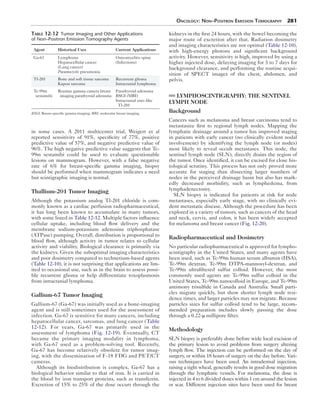
Figure 12-22. Planar Y-90 microspheres image after intrahepatic artery
injection for therapy using the bremsstrahlung radiation. Although planar
images can determine whether the distribution is adequate, SPECT
fused with CT provides better correlation.
A B
Figure 12-23. Monitoring therapy. F-18 FDG PET/CT images of the liver in a patient with unresectable hepatocellular carcinoma (A) before and
(B) 1 month after Y-90 Therasphere administration show marked improvement.
Table 12-16 Radiopharmaceuticals for Bone Pain Palliation
Radionuclide Pharmaceutical
Physical half-life
(days)
β Max
(MeV) Beta mean (MeV)
Maximal tissue
distance (mm)
Gamma photon
(keV)
P-32 Orthophosphate 14.3 1.71 0.695 8 —
Sr-89 Chloride 50.5 1.46 0.583 6.7 —
Sm-153 EDTMP 1.95 0.8 0.224 3.4 103
Re-186 HEDP 3.8 1.07 0.349 4.7 137
EDTMP, Ethylenediamine tetra; HEDP, hydroxyethylidene-1, 1-diphosphonic acid.
22. 286 Nuclear Medicine: The Requisites
A B
Figure 12-24. Sm-153 palliation of metastatic disease bone pain. A, The whole-body Tc-99m MDP bone scan before therapy confirms the presence
of osseous metastasis that will accumulate the therapy agent. B, The whole-body scan done with the Sm-153 therapy dose shows close correlation with
the lesions seen on bone scan. Ant, Anterior; Post, posterior.
cellular regions. Also, the lack of a gamma emission means
no external imaging can be done to assess distribution.
Strontium-89
Strontium-89 is a pure beta-emitter approved by the FDA
for management of metastatic bone pain under the trade
name Metastron. It selectively localizes in areas of bony
turnover, predominantly in blastic lesions. It is retained lon-ger
in regions of metastasis than normal bone. The pathway
of excretion is predominantly through urine, with about one
third bowel excretion. Dosimetry is outlined in Table 9-18.
A 4-mCi (148 MBq) dose is administered intravenously
slowly over 1 to 2 minutes. An alternative dose of 55 μCi/
kg (2.04 MBq/kg) may be used. Repeat dosing is possible,
but factors such as initial response, hematological status,
and current status must be considered in each case. In gen-eral,
a repeat administration is not recommended before
90 days have elapsed.
Toxicity
Because toxicity occurs to marrow components, platelets,
and white blood cells, the hematological status of each
patient must be evaluated before therapy. After obtaining a
baseline platelet count, platelets should be measured at
least every other week. Typically, platelets will decrease
30% from baseline and reach the nadir 12 to 16 weeks after
therapy. Toxicity is generally mild; however, Sr-89 must be
used with caution in those with white blood cell counts less
than 2400 and platelets less than 60,000. A small number of
patients experience transient worsening of symptoms.
Approximately 20% of patients will become pain free.
Around 75% of patients will experience some significant
decrease in pain, although some series have reported up to
90% of patients experience some symptom relief. Pain
relief begins approximately 7 to 20 days after injection and
generally lasts 3 to 6 months.
Samarium-153
Samarium-153 (Quadramet) is a beta-emitting radiophar-maceutical
that has the added advantage of a gamma emis-sion
that can be detected for external imaging. It has been
approved for use in patients with osteoblastic metastases
that can be visualized on a nuclear medicine bone scan
(Fig. 12-24).
Sm-153 is administered in a 1.0-mCi/kg (37 MBq/kg)
dose intravenously over the course of 1 minute. Approxi-mately
50% of the dose is localized to bone. It accumulates
in metastatic lesions in a 5:1 ratio compared with normal
bone. Patients should be well hydrated and void fre-quently
because the primary route of clearance is through
the urine. Approximately 35% of the dose is excreted in
the first 6 hours.
23. Oncology: Non–Positron Emission Tomography 287
As in Sr-89, the bone marrow toxicity is a limiting fac-tor.
Toxicity is usually mild, although serious side effects
and even fatalities have been reported. Platelets
decreased on the order of 25% from baseline and white
blood cells by 20%.
The short range of the Sm-153 beta particle should be
advantageous when considering the dose to normal mar-row.
A response rate on the order of 83% has been reported.
Pain relief is generally noted within 2 weeks, with a dura-tion
of 4 to 40 weeks.
Rhenium-186 Hydroxyethylidene
Diphosphonate
Rhenium-186 hydroxyethylidene diphosphonate (HEDP)
is formed by combining a diphosphonate useful for bone
pain therapy, etidronate, with a beta-emitter. Re-186
HEDP is another agent that may be useful for the pallia-tion
of bone pain. It emits a gamma ray useful for imaging
and lesion identification. It rapidly localizes to bone, with
approximately 14% retained in bone. The remainder is
rapidly cleared, with approximately 70% of the dose
excreted in the urine 6 hours after injection.
Suggested Reading
Breast
Berg WA, Zheng Z, Lehrer D, et al. Detection of breast cancer with the addition
of annual screening ultrasound or single-screening MRI to mammography in
women with elevated breast cancer risk. JAMA. 2012;307:1775-1877.
Berg WA, Madsen KS, Schilling K, et al. Comparative effectiveness of positron
emission mammography and MRI in the contralateral breast of women with
newly diagnosed breast cancer. AJR Am J Roentgenol. 2012;198:219-232.
Conners AL, Hruska CB, Tortorelli CL, et al. Lexicon for standardized
interpretation of gamma camera molecular breast imaging: observer agreement
and diagnostic accuracy. Eur J Nucl Med Mol Imaging. 2012:Jan 31 [Epub ahead
of print.]
Goldsmith SJ, Parsons W, Guiberteau MJ, et al. SNM practice guideline for breast
scintigraphy with breast-specific γ-cameras. 2010. Available at: http://snm.org/
guidelines.
Hendrick RE. Radiation doses and cancer risks from breast imaging studies.
Radiology. 2010;257(1):246-253.
O’Connor MK, Hua L, Rhodes DJ, et al. Comparison of radiation exposure and
associated radiation-induced cancer risks from mammography and molecular
imaging of the breast. Med Phys. 2010;37(12):6187-6198.
Rhodes DJ, Hruska CB, Phillips SW, Whaley DH, O’Connor MK. Dedicated
dual-head gamma imaging for breast cancer in women with dense breasts.
Radiology. 2011;258(1):106-118.
Tadwalkar RV, Rapelyea JA, Torrente J, et al. Breast-specific gamma imaging as an
adjunct modality for the diagnosis of invasive breast cancer with correlation to
tumor size and grade. Br J Radiol. 2011;85(1014):e212-e216.
Wahner-Roedler DL, Boughey JC, Hruska CB, et al. The use of molecular breast
imaging to assess response in women undergoing neoadjuvant therapy for breast
cancer: a pilot study. Clin Nucl Med. 2012;37(4):344-350.
Wang CL, MacDonald LR, Rogers JV. Positron emission mammography:
correlation of estrogen receptor, progesterone receptor, and human epidermal
growth factor receptor 2 status and 18F-FDG. AJR Am J Roentgenol.
2011;197(2):W247-W255.
Weigert JM, Bertrand ML, Lankowsky L, Stern LH, Kieper DA. Results of a
multicenter registry to determine the clinical impact of breast-specific gamma
imaging, a molecular breast imaging technique. AJR Am J Roentgenol.
2011;198(1):W69-W75.
Somatostatin Receptor Imaging
Balon HR, Goldsmith SJ, Siegel BA, et al. Society of nuclear medicine
procedure guideline for somatostatin receptor scintigraphy with In-111
pentetreotide, version 2.0. 2002. Available at: http://interactive.snm.org/docs/
SRS_Final_v2_0.pdf.
Kabasakal L, Demirci E, Ocak M. Comparison of (68)Ga-DOTATAE and (68)
Ga-DOTANOC PET/CT imaging in the same patient group with neuroendo-crine
tumors. Eur J Nucl Med Mol Imaging. 2012;39(8):1271-1277.
Kwekkeboom DJ, Krenning EP. Somatostatin receptor imaging. Semin Nucl Med.
2002;32(2):84-91.
Adrenal Imaging
Bombardieri E, Giammarile F, Aktolum C, et al. 131I/123I-Metaiodobenzylguanidine
(MIBG) scintigraphy-procedure guidelines for tumor imaging. 2010. Available
at: http://interactive.snm.org/docs/EANM_guideline_for_I131_I123_MIBG_
Scintigraphy.pdf.
Antibody Imaging
Hagenbeek A. Radioimmunotherapy for NHL: experience of 90Y-ibritumomab
tiuxetan in clinical practice. Leuk Lymphoma. 2003;44(suppl 4):S37-S47.
Rieter WJ, Keane TE, Ahlman MA, et al. Diagnostic performance of In-111
capromab pendetide SPECT-CT in localized and metastatic prostate cancer.
Clin Nucl Med. 2011;36:872-878.
Taneja SS. ProstaScint® scan: contemporary use in clinical practice. Rev Urol.
2004;6(suppl 10):S19-S28.
Wong TZ, Turkinton TG, Polascik TJ, Coleman RE. ProstaScint (capromab
pendetide) imaging using hybrid gamma camera-CT technology. AJR Am J
Roentgenol. 2005;184(2):676-680.
Wyngaert JK, Noz ME, Ellerin B, et al. Procedure for unmasking localization
information from ProstaScint scans for prostate radiation therapy treatment
planning. Int J Radiat Oncol Biol Phys. 2004;60(2):654-662.
Lymphoscintigraphy
Buscombe J, Paganelli G, Burak ZE, et al. Sentinel node in breast cancer
procedural guidelines. Eur J Nucl Med Mol Imaging. 2007;34(12):2154-2159.
Gershenwald JE, Ross MI. Sentinel-lymph-node biopsy for cutaneous melanoma.
N Engl J Med. 2011;364(18):1738-1745.
Hindie E, Groheux D, Brenot-Rossi I. The sentinel node procedure in breast
cancer: nuclear medicine as a starting point. J Nucl Med. 2011;52(3):405-414.
Lyman GH, Giuliano AE, Somerfield MR, et al. American society of clinical
oncology guideline recommendations for sentinel lymph node biopsy in early
stage breast cancer. J Clin Oncol. 2005;23(30):7703-7720.
Intraarterial Microsphere Therapy
Ahmadzadehfar H, Biersack HJ, Ezzidin S. Radioembolization of liver tumors with
yttrium-90 microspheres. Semin Nucl Med. 2010;40(2):105-121.
Kalva SP, Thabet A, Wicky S. Recent advances in transarterial therapy of primary
and secondary liver malignancies. Radiographics. 2008;28(1):101-117.