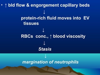
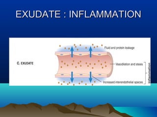
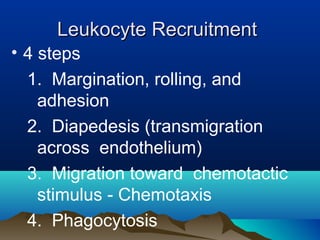
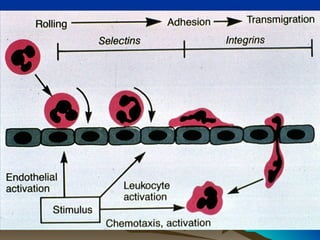
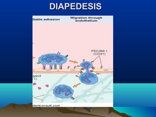
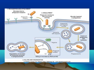
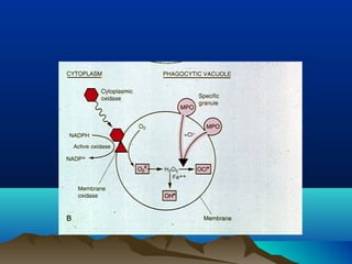
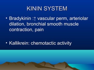
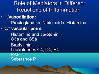
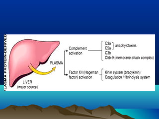
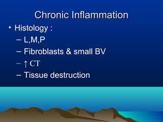
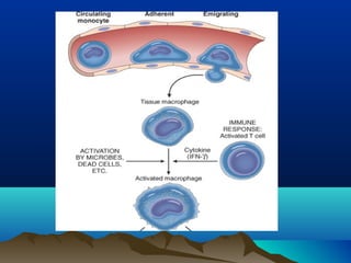
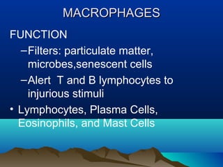
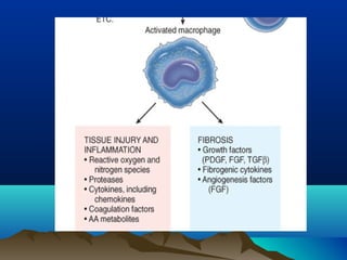
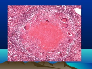
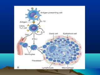
INFLAMMATIONINFLAMMATION
Dr Sapna MDr Sapna M
Dept pathologyDept pathology
SNIMS,KERALASNIMS,KERALA
INFLAMMATIONINFLAMMATION
• Protective response intended
to eliminate initial cause of cell
injury , necrotic cells and
tissues resulting from original
insult
Introduction:Introduction:
• “Inflame” – to set fire
• Inflammation is “dynamic
response of vascularised
tissue to injury.”
-itis-itis
AppendicitisAppendicitis
CellulitisCellulitis
MeningitisMeningitis
PneumonitisPneumonitis
NephritisNephritis
MyocarditisMyocarditis
TYPES OF INFLAMMATIONTYPES OF INFLAMMATION
• ACUTE
• CHRONIC
Acute Inflammation-FeaturesAcute Inflammation-Features
• Rapid onset
• Short duration( mts. to few days)
• Fluid , plasma protein exudation
• Neutrophil accumulation
ChronicChronic Inflammation-FeaturesInflammation-Features
• Onset- insidious
• Longer duration (days to years)
• Lymphocytes ,macrophages ,plasma
cells
• Vascular proliferation & fibrosis
(scarring)
HEAT REDNESS SWELLING PAIN LOSS OF
5 Cardinal Signs of Inflammation5 Cardinal Signs of Inflammation
4 cardinal clinical signs of inflammation4 cardinal clinical signs of inflammation
described by Celsus, 1 A.D.:described by Celsus, 1 A.D.:
• Rubor - redness
• Tumor - swelling
• Calor - heat
• Dolor - pain
• Virchow added a fifth--loss of
function(functio laesa)
Steps Of Inflammatory ResponseSteps Of Inflammatory Response
• Remembered as five Rs:
• (1) Recognition of injurious agent
• (2) Recruitment of leukocytes
• (3) Removal of agent
• (4) Regulation of response
• (5) Resolution (repair).
•ACUTE INFLAMMATION
Stimuli for Acute InflammationStimuli for Acute Inflammation
• Infections (b, v, f, p)
• Trauma
• Physical & chemical agents
• Tissue necrosis (MI)
• Foreign bodies
• Immune reactions
Acute Inflammation-componentsAcute Inflammation-components
ACUTE INFLAMMATIONACUTE INFLAMMATION
Vascular changes Cellular events
•Vasodilation
Increased vascular permeability.
Acute Inflammation-componentsAcute Inflammation-components
• 1.Vascular changes:
• Vasodilation
• ↑ vascular perm.
• 2.Cellular events
• Cellular recruitment & activation
VASCULAR CHANGESVASCULAR CHANGES
• 1.Changes In Vascular Caliber And Flow
• Transient vasoconstriction (sec.)
• Arteriolar vasodilation( redness
warmth)
• ↑ bld flow & engorgement capillary beds
↓
protein-rich fluid moves into EV
tissues
↓
RBCs conc., ↑ blood viscosity
↓
Stasis
↓
margination of neutrophils
ACUTE INFLAMMATIONACUTE INFLAMMATION
• 2. ↑ Vascular Perm.{ mechanisms }
• 1.Endothelial cell contraction
–immediate transient response
:reversible process, by histamine,
bradykinin, leukotrienes ,15-30 min.
2 .Retraction of EC
: TNF and IL-1, 24 hours or more
↑↑ Vascular Permeability{ mechanisms }Vascular Permeability{ mechanisms }
• 3.Endothelial injury
-immediate sustained response , delayed
prolonged leakage
• 4.Leukocyte-mediated endothelial injury
• 5.Increased transcytosis
• 6.Leakage from new blood vessels.
EXUDATE : INFLAMMATIONEXUDATE : INFLAMMATION
• 1.Vascular changes:
• Vasodilation
• Increased vascular permeability.
Cellular Events: LeukocyteCellular Events: Leukocyte
Recruitment and ActivationRecruitment and Activation
Leukocyte RecruitmentLeukocyte Recruitment
• 4 steps
1. Margination, rolling, and
adhesion
2. Diapedesis (transmigration
across endothelium)
3. Migration toward chemotactic
stimulus - Chemotaxis
4. Phagocytosis
Neutrophil MarginationNeutrophil Margination
ROLLINGROLLING::
Leukocytes tumble on endothelial
surface, transiently sticking along
the way
• Mediated by SELECTIN family of
adhesion molecules
• 3 members of this family
SELECTINSSELECTINS
• E-selectin (CD62E) on EC
• P-selectin (CD62P) EC & platelets
• L-selectin (CD62L) leukocytes
• Selectins bind sialylated
oligosaccharides ( sialyl-Lewis X
on leukocytes)
ROLLINGROLLING
ADHESION & TRANSMIGRATIONADHESION & TRANSMIGRATION
• Adhesion mediated by integrins
• Activated by chemokines
• TNF and IL-1 activate EC to ↑
expression of ligands for integrins
ADHESIONADHESION
Ligands For IntegrinsLigands For Integrins
• ICAM-1(intercellular adhesion molecule1)
- binds integrins LFA-1 (CD11a/CD18)
and Mac-1 (CD11b/CD18),
• VCAM-1 (vascular cell adhesion
molecule1)
- bind integrin VLA-4
DIAPEDESISDIAPEDESIS
• Leukocytes migrate thr’u vessel wall by
squeezing between cells at IC junctions
• Mediated by PECAM-1 (platelet
endothelial cell adhesion molecule 1
(CD31) on leukocytes & EC
CHEMOTAXISCHEMOTAXIS
• Leukocytes migrate toward sites of
infection or injury along a chemical
gradient
• Chemotactic substances
(1) bacterial products (2) cytokines
(3) C5a (4) LTB4.
6 to 24 hours 24 to 48 hours
LEUKOCYTE ACTIVATIONLEUKOCYTE ACTIVATION
• Leukocytes receptors sensing p/o
microbes:
–Toll-like receptors (TLRs) - LPS
–Seven-transmembrane G-protein-
coupled receptors - bacterial
peptides and mediators
PHAGOCYTOSISPHAGOCYTOSIS
• 3 steps
• (1) recognition and attachment of
particle to ingesting leukocyte
• (2) engulfment & formation of
phagocytic vacuole
• (3) killing and degradation of ingested
material.
(1)(1) Recognition & attachment ofRecognition & attachment of
particle to ingesting leukocyteparticle to ingesting leukocyte
• Opsonins : host proteins that coat
microbes and target them for
phagocytosis (opsonization).
• Leukocytes express receptors for
opsonins
OPSONISATION RECEPTORS
OPSONINS RECEPTORS FOR
OPSONINS
IgG FcγRI
Breakdown
products of C3
CR1 and 3
Collectins C1q
2.ENGULFMENT2.ENGULFMENT & FORMATION& FORMATION
OF PHAGOCYTIC VACUOLEOF PHAGOCYTIC VACUOLE
3.Killing and Degradation of Microbes3.Killing and Degradation of Microbes
• Important microbicidal substances :
reactive oxygen species (ROS) and
lysosomal enzymes
• ROS
• HOCl• (hypochlorous radical)
•Superoxide radicals
(superoxide,H2O2, and OH•).
• Reactive nitrogen species( NO)
Lysosomal enzymesLysosomal enzymes
• Lysosomal acid hydrolases-degrade
dead microorganisms
• Elastase - kill bacteria
Defects in Leukocyte FunctionDefects in Leukocyte Function
• Common causes
• BM suppression ( tumors CT,RT)- ↓
leukocyte no.
• Diabetes - abnormal leukocyte
function
Defects in leukocyte function:Defects in leukocyte function:
• Margination and adhesion
–Etoh, steroids, AR leukocyte adhesion
deficiency
• Emigration toward a chemotactic stimulus
–drugs
–chemotaxis inhibitors
• Phagocytosis
–Chronic granulomatous disease
(CGD) :phagocyte oxidase generating ROS
Defects in leukocyte function:Defects in leukocyte function:
• Myeloperoxidase (MPO) deficiency:
Absent MPO-H2O2 system
• Chédiak-Higashi syndrome :impair
fusion of lysosomes with phagosomes
ACUTE INFLAMMATION - THREEACUTE INFLAMMATION - THREE
OUTCOMESOUTCOMES
• Resolution
• Progression to chronic inflammation
• Abscess
• Scarring or fibrosis
Acute inflammation
Chronic inflammation
RepairResolution
Abscess
Injury
MORPHOLOGIC PATTERNS OFMORPHOLOGIC PATTERNS OF
ACUTE INFLAMMATIONACUTE INFLAMMATION
• SEROUS INFLAMMATION
• Watery, protein-poor fluid .
Serum/secretions of mesothelial cells
• Skin blister
• Fluid in a serous cavity - effusion.
SEROUS INFLAMMATIONSEROUS INFLAMMATION
FIBRINOUS INFLAMMATIONFIBRINOUS INFLAMMATION
• Severe injuries, ↑ vascular perm. &
fibrinogen pass
• Histology: eosinophilic meshwork
of threads or amorphous coagulum
• Site : Lining of body cavities
• Resolution
• Organization & scarring
SUPPURATIVE (PURULENT)SUPPURATIVE (PURULENT)
INFLAMMATIONINFLAMMATION
• Purulent exudate (pus) -
neutrophils, necrotic cells, and
edema fluid
• Staphylococci – pyogenic
• Completely walled off ,replaced by
connective tissue.
ABSCESSESABSCESSES
• Focal collections of pus
• Central, large necrotic region
rimmed by neutrophils
(surrounded by vessels and
fibroblastic proliferation )
ABSCESSESABSCESSES
ABSCESSESABSCESSES
ULCERULCER
• Local defect / excavation, surface of an
organ/ tissue prod. by necrosis of cells
& sloughing of inflammatory necrotic
tissue
• Sites (1)mucosa of GIT( Peptic ulcer),
GUT
• (2) Lower extremities, older persons
with circulatory distur.
CHEMICAL MEDIATORS OFCHEMICAL MEDIATORS OF
INFLAMMATIONINFLAMMATION
CELL DERIVED
CHEMICAL MEDIATORS OFCHEMICAL MEDIATORS OF
INFLAMMATIONINFLAMMATION
PLASMA PROTEIN DERIVED
CHEMICAL MEDIATORS OFCHEMICAL MEDIATORS OF
INFLAMMATIONINFLAMMATION
• CELL-DERIVED MEDIATORS
• VASOACTIVE AMINES
• 1.HISTAMINE released by
• (1) physical injury
• (2) immune reactions
• (3) Anaphylatoxins:C3a and C5a
• (4) leukocyte-derived histamine-releasing
proteins
• (5) neuropeptides
• (6) IL-1 and IL-8
HISTAMINEHISTAMINE
• Action: arteriolar dilation , ↑ vascular
perm.
• Inactivated by histaminase
• SEROTONIN (5-hydroxytryptamine)
effects similar to histamine
CELL DERIVED –NEWLYCELL DERIVED –NEWLY
SYNTHESISEDSYNTHESISED
Arachidonic Acid Metabolites:Arachidonic Acid Metabolites:
((eicosanoidseicosanoids))
• Sources: Leukocytes, mast cells, EC,
platelets
• 2 major enzymatic pathways:
• Cyclooxygenase
• Lipoxygenase
• Cyclooxygenase : prostaglandins and
thromboxanes
• Lipoxygenase: leukotrienes and lipoxins
Arachidonic Acid (AA) MetabolitesArachidonic Acid (AA) Metabolites
• CYCLOOXYGENASE PATHWAY
• Products :PGE2, PGD2, PGF2α,
PGI2 (prostacyclin), TXA2
• TXA2: Platelets - thromboxane
synthase ,platelet-aggregating
agent and vasoconstrictor
CYCLOOXYGENASE PATHWAYCYCLOOXYGENASE PATHWAY
• PGI2 : EC - prostacyclin synthase ,
vasodilator and inhibitor of platelet
aggregation
• PGD2: mast cells; + PGE2 & PGF2α
causes vasodilation & edema
• PGE2 : pain sensitivity, fever.
LIPOXYGENASE PATHWAYLIPOXYGENASE PATHWAY
• 5-Lipoxygenase :AA-metabolizing
enzyme in neutrophils
• 5-HPETE (5-
hydroperoxyeicosatetraenoic acid)
• Reduced to 5-HETE (5-hydroxyeicosa
tetraenoic acid) - chemotactic for
neutrophils
• or converted into leukotrienes
Lipoxygenase Pathway- LeukotrieneLipoxygenase Pathway- Leukotriene
• LTA4 gives rise to LTB4 or LTC4
• LTB4: chemotactic agent for
neutrophils
• LTC4 , LTD4 & LTE4: mast cells ,
cause vasoconstriction,
bronchospasm, ↑ vascular perm.
LIPOXINSLIPOXINS
• Inhibitors of inflammation
• Endogenous antagonists of
leukotrienes.
Principal Inflammatory Actions ofPrincipal Inflammatory Actions of
AA MetabolitesAA Metabolites
• Vasodilation:PGI2 (prostacyclin),
PGE1, PGE2, PGD2
• Vasoconstriction:Thromboxane A2,
leukotrienes C4, D4, E4
• ↑ vascular perm. :Leukotrienes C4,
D4, E4 & PGE2, PGD2
• Chemotaxis, leukocyte adhesion
:Leukotriene B4, 5-HETE
Cyclooxygenase enzyme -COXCyclooxygenase enzyme -COX
• Two forms : COX-1 and COX-2
• COX-1 : gastric mucosa, PGs
protective against acid-induced
damage.
• COX inhibitors :Aspirin ,NSAIDs: treat
pain and fever
Platelet-Activating FactorPlatelet-Activating Factor
• Stimulate platelets
• Vasoconstriction & bronchoconstriction
• Vasodilation & ↑vascular perm.
• Leukocyte adhesion, chemotaxis,
degranulation, oxidative burst
• Synthesis of mediators , (eicosanoids).
CytokinesCytokines
• Polypeptide products .Mediators of
inflammation and immune responses
• Molecularly characterized cytokines -
interleukins
• A/c inflam : TNF, IL-1, chemokines
• C/C inflam: IFN-γ , IL-12.
CHEMOKINESCHEMOKINES
• 2 functions : leukocyte recruitment in
inflam. & N anatomic organization of cells
in lymphoid and other tissues
• Four groups
• Two major groups
• CXC :IL-8
• CC chemokines:MCP-1 , MIP-1α,
RANTES (regulated on activation normal
T expressed and secreted) and eotaxin .
Reactive Oxygen Species (ROSReactive Oxygen Species (ROS
• Synthesized via NADPH oxidase
(phagocyte oxidase) pathway
• Action (1) endothelial damage, with
thrombosis & ↑ perm.
• (2) protease activation and
antiprotease inactivation
• (3) direct injury to other cells
• Catalase, SOD, glutathione -↓ toxicity
NONO
• Synthesized from l-arginine, molecular
O2, NADPH by enzyme NOS.
• 3 isoforms of NOS
• Type I (nNOS)
• Type II (iNOS) present in macrophages
& EC induced by IL-1, TNF,IFN-γ,
bacterial endotoxin
• Type III (eNOS)
Nitric OxideNitric Oxide
NO -NO -FunctionsFunctions
• (1) vasodilation (endothelium-derived
relaxation factor)
• (2) antagonism all stages of platelet
activation(a,a,d)
• (3) reduction of leukocyte recruitment
at inflammatory sites
• (4)Microbicidal agent
Lysosomal Enzymes of LeukocytesLysosomal Enzymes of Leukocytes
• Acid proteases:acidic pH optima ,
active within phagolysosomes
• Neutral proteases: elastase,
collagenase , cathepsin, active in
ECM
• Cleave C3 & C5 to C3a & C5a
• Generate bradykinin-like peptides
from kininogen.
AntiproteasesAntiproteases
• α1-antitrypsin-major inhibitor of
neutrophil elastase
• α2-macroglobulin
• α1-antitrypsin deficiency - lung ,
panacinar emphysema
NeuropeptidesNeuropeptides
• Small proteins, such as substance P
• Transmit pain signals
• Regulate vessel tone
• Modulate vascular permeability
• Lung and GIT- Nerve fibers secrete NP
PLASMA PROTEIN-DERIVEDPLASMA PROTEIN-DERIVED
MEDIATORSMEDIATORS
• 3 systems
– Complement
– Kinin
– Coagulation
ComplementComplement
Immunity and inflammation
• Opsonize particles: phagocytosis and
destruction
• ↑ vascular perm. & vasodilatation
• Leuko. chemotaxis
• Generates MAC (C5b-9)
ComplementComplement
Critical steps : activation of C3Critical steps : activation of C3
• (1) Classical pathway : fixation of C1
to antig-antib complexes
• (2) Alternative pathway: bacterial
polysaccharides / microbial cell-wall
components, involve properdin and
factors B , D
• (3) Lectin pathway: plasma lectin
binds mannose residues on microbes
, activates early component of CP
ComplementComplement
• 3 pathways form C3 convertase :cleaves
C3 to C3a and C3b
• C3b deposits on cell or microbial surface
• Binds to C3 convertase complex to form
C5 convertase
• C5 convertase cleaves C5 → C5a & C5b
• Initiate final stages of assembly of C6 to
C9
• Thrombin : cleave C5
ComplementComplement
• 1.Anaphylatoxins: C3a and C5a
↑vascular perm. and vasodilation ,induce
mast cells to release histamine.
• 2.C5a: activates lipoxygenase pathway
• 3.Leukocyte activation, adhesion, and
chemotaxis. C5a
• 4.Phagocytosis: C3b & iC3b act as
opsonins
• 5.Lysis of microbes - MAC
Coagulation and Kinin SystemsCoagulation and Kinin Systems
• 4 systems activated by Hageman
factor (factor XIIa)
• (1) Kinin system
• (2) Clotting system
• (3) Fibrinolytic system
• 4) Complement system.
Coagulation and Kinin SystemsCoagulation and Kinin Systems
Clotting SystemClotting System
• Factor Xa: ↑ vascular perm. and leuk.
Emigration
• Thrombin : leuk. Adhesion
• Fibrinopeptides :↑ vascular perm. and
chemotactic for leuk.
FIBRINOLYTIC SYSTEMFIBRINOLYTIC SYSTEM
• FDP : ↑ vascular perm.
• Plasmin :cleaves C3 to C3a causing
vasodilation and ↑ vascular perm.
KININ SYSTEMKININ SYSTEM
• Bradykinin :↑ vascular perm, arteriolar
dilation, bronchial smooth muscle
contraction, pain
• Kallikrein: chemotactic activity
Role of Mediators in DifferentRole of Mediators in Different
Reactions of InflammationReactions of Inflammation
• 1.Vasodilation:
Prostaglandins, Nitric oxide Histamine
• 2.↑ vascular perm:
Histamine and serotonin
C3a and C5a
Bradykinin
Leukotrienes C4, D4, E4
PAF
Substance P
Mediators in InflammationMediators in Inflammation
• 3. Leukocyte recruitment and
activation
TNF, IL-1
Chemokines
C3a, C5a
Leukotriene B4
Bacterial products, e.g., N-formyl
methyl peptides
Mediators in InflammationMediators in Inflammation
• 4. Fever
IL-1, TNF, PG
• 5. Pain
PG, Bradykinin, Neuropeptides
• 6. Tissue damage
Lysosomal enzymes , ROS,
Nitric oxide
CHRONIC INFLAMMATIONCHRONIC INFLAMMATION
• Prolonged duration .Active
inflammation, tissue injury & healing
simultaneously
• Characterized by
• Infiltration with mononuclear cells
• Tissue destruction
• Repair : angiogenesis and fibrosis
Chronic InflammationChronic Inflammation
• Time course:
–> 48 hours (weeks, months, years)
• Cell type
–Mononuclear cells (Macrophages,
Lymphocytes, Plasma cells)
Chronic InflammationChronic Inflammation
• Histology :
– L,M,P
– Fibroblasts & small BV
– ↑ CT
– Tissue destruction
MACROPHAGESMACROPHAGES
• Derived from circulating
blood monocytes
• SITES:
• Connective tissues
• Liver (Kupffer cells)
• Spleen & LN (sinus
histiocytes)
• CNS( microglial cells)
• Lungs (alveolar
macrophages)
•Mono
nuclear
phagocyte
system
(RES)
MACROPHAGESMACROPHAGES
FUNCTION
–Filters: particulate matter,
microbes,senescent cells
–Alert T and B lymphocytes to
injurious stimuli
• Lymphocytes, Plasma Cells,
Eosinophils, and Mast Cells
Settings For Chronic InflammationSettings For Chronic Inflammation
• 1.Microbes difficult to eradicate
Mycobacteria, Treponema pallidum ,
viruses and fungi
• 2.Immune-mediated inflammatory
diseases : RA ,IBD, Asthma
• 3.Toxic agents :Silica, atherosclerosis
GRANULOMATOUS INFLAMMATIONGRANULOMATOUS INFLAMMATION
• Distinctive pattern of chronic
inflammation
• Characterized by aggregates of
activated macrophages that assume
an epithelioid appearance
Diseases with GranulomatousDiseases with Granulomatous
InflammationInflammation
• Tuberculosis :M. tuberculosis
• Noncaseating tubercle : a focus of
epithelioid cells, rimmed by fibroblasts,
lymphocytes, histiocytes, occasional
giant cells
• Caseating tubercle: central amorphous
granular debris, loss of all cellular detail;
acid-fast bacilli
LANGHANS GIANT CELLS - HORSE SHOE SHAPE
LANGHANS GIANT CELL
Granulomatous InflammationGranulomatous Inflammation
• Leprosy
• Mycobacterium leprae
• Acid-fast bacilli in macrophages;
noncaseating granulomas
• Syphilis:Treponema pallidum,
Gumma
Granulomatous InflammationGranulomatous Inflammation
• Cat-scratch disease :Gram-negative
bacillus , Rounded or stellate
granuloma
• Sarcoidosis : Unknown etiology
Noncaseating granulomas
• Crohn disease :Immune reaction
against intestinal bacterial, self-
antigens noncaseating granulomas
Foreign body granulomas.Foreign body granulomas.
EPITHELIOID CELLSEPITHELIOID CELLS
• Elongated, finely granular, pale
eosinophilic cytoplasm
• Nucleus:central, oval or
elongate,small nucleoli ,slipper
shaped
• Indistinct shape , merge to form
aggregates.
• Lymphocytes secrete cytokines →
macrophage activation
• Multinucleated giant cells
• Hypoxia and free-radical injury leads to
caseous necrosis
• Healing of granulomas accompanied
by fibrosis
SYSTEMIC EFFECTS OFSYSTEMIC EFFECTS OF
INFLAMMATIONINFLAMMATION
• ACUTE-PHASE REACTION/ SYSTEMIC
INFLAMMATORY RESPONSE
SYNDROME.
• TNF, IL-1, and IL- 6
Clinical And Pathologic ChangesClinical And Pathologic Changes
-APR-APR
• 1.Fever- pyrogens –stimulate PG
synthesis in hypothalamus
LPS(EX .P)
LUECOCYTES(IL-1 &TNF)
ENDO.P
AA
PG(PGE2)
STIMULATE NEUROTRANSMITTERS
RESET TEMP.
↑ CYCLOOXYGENASE
2.Acute-phase proteins2.Acute-phase proteins
• 2.Synthesized in liver
• Up-regulated by IL-6
• I) CRP II) Fibrinogen III)Serum amyloid A
(SAA) protein.
• Fibrinogen cause rouleaux- ↑ ESR
• CRP: ↑ risk of MI or stroke in pts. with
atherosclerotic vascular disease
3. Leukocytosis
• TNF & IL-1-release cells from BM .
Shift to left
• CSFs - ↑ output of leukocytes from BM
• Neutrophilia , lymphocytosis ,
eosinophilia , leukopenia
Other manifestationsOther manifestations
• ↑ heart rate and BP
• ↓ sweating
• Rigors (shivering)
• Chills
• Anorexia, somnolence & malaise:
cytokines on brain cells
• Chronic inflammation: wasting syndrome
called cachexia, TNF- mediated appetite
suppression & mobilization of fat stores
SEPTIC SHOCKSEPTIC SHOCK
• High levels of TNF cause DIC,
hypoglycemia, and hypotensive
shock.
1.A 65-year-old woman had fever the past day.
On physical examination her temperature is 39 C
and blood pressure 90/50 mm Hg with heart rate
of 106/minute. A blood culture is positive for
Escherichia coli. Her central line pressure falls
markedly. She goes into hypovolemic shock as a
result of the widespread inappropriate release of a
chemical mediator derived from macrophages.
Which of the following mediators is most likely to
produce these findings?
A Nitric oxide
B Bradykinin
C Histamine
D Prostacyclin
E Complement C3a
2.The major difference between a
transudate and an exudate is
(A) transudate has proteins whereas
exudate is essentially devoid of proteins
(B) both these are only seen during acute
inflammatory processes
(C) both are associated with a Suppurative
or Purulent inflammatory process
(D) transudate is associated with serous
inflammation whereas exudate is
associated with fibrinous inflammation
3.Granulomatous Inflammation is
associated with which of the following
a. distinct acute inflammatory
process
b. distinct chronic inflammatory
process
c. persistent B-cell response to
certain microbes
d. IgM mediated response
4.Which of the following is/are considered
an anaphylatoxins(s)
(A) lipoxins
(B) leukotrines
(C) C3a
(D) IL-1 and TNF
5.What is the most common form of
increased vascular permeability
associated with acute inflammation
(A) direct endothelial injury
(B) endothelial cell retraction
(C) endothelial cell contraction
(D) leukocyte-dependent endothelial
damage
6.Endothelial cell contraction &
retraction occur in
a) Postcapillary venules
b) Capillaries
c) Arterioles
d) All of the above

Inflammation

  • 1.
    INFLAMMATIONINFLAMMATION Dr Sapna MDrSapna M Dept pathologyDept pathology SNIMS,KERALASNIMS,KERALA
  • 2.
  • 3.
    INFLAMMATIONINFLAMMATION • Protective responseintended to eliminate initial cause of cell injury , necrotic cells and tissues resulting from original insult
  • 4.
    Introduction:Introduction: • “Inflame” –to set fire • Inflammation is “dynamic response of vascularised tissue to injury.”
  • 5.
  • 6.
    TYPES OF INFLAMMATIONTYPESOF INFLAMMATION • ACUTE • CHRONIC
  • 7.
    Acute Inflammation-FeaturesAcute Inflammation-Features •Rapid onset • Short duration( mts. to few days) • Fluid , plasma protein exudation • Neutrophil accumulation
  • 9.
    ChronicChronic Inflammation-FeaturesInflammation-Features • Onset-insidious • Longer duration (days to years) • Lymphocytes ,macrophages ,plasma cells • Vascular proliferation & fibrosis (scarring)
  • 11.
    HEAT REDNESS SWELLINGPAIN LOSS OF 5 Cardinal Signs of Inflammation5 Cardinal Signs of Inflammation
  • 12.
    4 cardinal clinicalsigns of inflammation4 cardinal clinical signs of inflammation described by Celsus, 1 A.D.:described by Celsus, 1 A.D.: • Rubor - redness • Tumor - swelling • Calor - heat • Dolor - pain • Virchow added a fifth--loss of function(functio laesa)
  • 13.
    Steps Of InflammatoryResponseSteps Of Inflammatory Response • Remembered as five Rs: • (1) Recognition of injurious agent • (2) Recruitment of leukocytes • (3) Removal of agent • (4) Regulation of response • (5) Resolution (repair).
  • 14.
  • 15.
    Stimuli for AcuteInflammationStimuli for Acute Inflammation • Infections (b, v, f, p) • Trauma • Physical & chemical agents • Tissue necrosis (MI) • Foreign bodies • Immune reactions
  • 16.
  • 17.
  • 18.
    ACUTE INFLAMMATIONACUTE INFLAMMATION Vascularchanges Cellular events •Vasodilation Increased vascular permeability.
  • 19.
    Acute Inflammation-componentsAcute Inflammation-components •1.Vascular changes: • Vasodilation • ↑ vascular perm. • 2.Cellular events • Cellular recruitment & activation
  • 20.
    VASCULAR CHANGESVASCULAR CHANGES •1.Changes In Vascular Caliber And Flow • Transient vasoconstriction (sec.) • Arteriolar vasodilation( redness warmth)
  • 21.
    • ↑ bldflow & engorgement capillary beds ↓ protein-rich fluid moves into EV tissues ↓ RBCs conc., ↑ blood viscosity ↓ Stasis ↓ margination of neutrophils
  • 23.
    ACUTE INFLAMMATIONACUTE INFLAMMATION •2. ↑ Vascular Perm.{ mechanisms } • 1.Endothelial cell contraction –immediate transient response :reversible process, by histamine, bradykinin, leukotrienes ,15-30 min. 2 .Retraction of EC : TNF and IL-1, 24 hours or more
  • 24.
    ↑↑ Vascular Permeability{mechanisms }Vascular Permeability{ mechanisms } • 3.Endothelial injury -immediate sustained response , delayed prolonged leakage • 4.Leukocyte-mediated endothelial injury • 5.Increased transcytosis • 6.Leakage from new blood vessels.
  • 26.
  • 27.
    • 1.Vascular changes: •Vasodilation • Increased vascular permeability.
  • 28.
    Cellular Events: LeukocyteCellularEvents: Leukocyte Recruitment and ActivationRecruitment and Activation
  • 29.
    Leukocyte RecruitmentLeukocyte Recruitment •4 steps 1. Margination, rolling, and adhesion 2. Diapedesis (transmigration across endothelium) 3. Migration toward chemotactic stimulus - Chemotaxis 4. Phagocytosis
  • 32.
  • 33.
    ROLLINGROLLING:: Leukocytes tumble onendothelial surface, transiently sticking along the way • Mediated by SELECTIN family of adhesion molecules • 3 members of this family
  • 34.
    SELECTINSSELECTINS • E-selectin (CD62E)on EC • P-selectin (CD62P) EC & platelets • L-selectin (CD62L) leukocytes • Selectins bind sialylated oligosaccharides ( sialyl-Lewis X on leukocytes)
  • 35.
  • 36.
    ADHESION & TRANSMIGRATIONADHESION& TRANSMIGRATION • Adhesion mediated by integrins • Activated by chemokines • TNF and IL-1 activate EC to ↑ expression of ligands for integrins
  • 37.
  • 38.
    Ligands For IntegrinsLigandsFor Integrins • ICAM-1(intercellular adhesion molecule1) - binds integrins LFA-1 (CD11a/CD18) and Mac-1 (CD11b/CD18), • VCAM-1 (vascular cell adhesion molecule1) - bind integrin VLA-4
  • 39.
  • 40.
  • 41.
    DIAPEDESISDIAPEDESIS • Leukocytes migratethr’u vessel wall by squeezing between cells at IC junctions • Mediated by PECAM-1 (platelet endothelial cell adhesion molecule 1 (CD31) on leukocytes & EC
  • 42.
    CHEMOTAXISCHEMOTAXIS • Leukocytes migratetoward sites of infection or injury along a chemical gradient • Chemotactic substances (1) bacterial products (2) cytokines (3) C5a (4) LTB4.
  • 43.
    6 to 24hours 24 to 48 hours
  • 44.
  • 45.
    LEUKOCYTE ACTIVATIONLEUKOCYTE ACTIVATION •Leukocytes receptors sensing p/o microbes: –Toll-like receptors (TLRs) - LPS –Seven-transmembrane G-protein- coupled receptors - bacterial peptides and mediators
  • 46.
    PHAGOCYTOSISPHAGOCYTOSIS • 3 steps •(1) recognition and attachment of particle to ingesting leukocyte • (2) engulfment & formation of phagocytic vacuole • (3) killing and degradation of ingested material.
  • 48.
    (1)(1) Recognition &attachment ofRecognition & attachment of particle to ingesting leukocyteparticle to ingesting leukocyte • Opsonins : host proteins that coat microbes and target them for phagocytosis (opsonization). • Leukocytes express receptors for opsonins
  • 49.
  • 50.
    OPSONINS RECEPTORS FOR OPSONINS IgGFcγRI Breakdown products of C3 CR1 and 3 Collectins C1q
  • 52.
    2.ENGULFMENT2.ENGULFMENT & FORMATION&FORMATION OF PHAGOCYTIC VACUOLEOF PHAGOCYTIC VACUOLE
  • 56.
    3.Killing and Degradationof Microbes3.Killing and Degradation of Microbes • Important microbicidal substances : reactive oxygen species (ROS) and lysosomal enzymes • ROS • HOCl• (hypochlorous radical) •Superoxide radicals (superoxide,H2O2, and OH•). • Reactive nitrogen species( NO)
  • 57.
    Lysosomal enzymesLysosomal enzymes •Lysosomal acid hydrolases-degrade dead microorganisms • Elastase - kill bacteria
  • 58.
    Defects in LeukocyteFunctionDefects in Leukocyte Function • Common causes • BM suppression ( tumors CT,RT)- ↓ leukocyte no. • Diabetes - abnormal leukocyte function
  • 59.
    Defects in leukocytefunction:Defects in leukocyte function: • Margination and adhesion –Etoh, steroids, AR leukocyte adhesion deficiency • Emigration toward a chemotactic stimulus –drugs –chemotaxis inhibitors • Phagocytosis –Chronic granulomatous disease (CGD) :phagocyte oxidase generating ROS
  • 60.
    Defects in leukocytefunction:Defects in leukocyte function: • Myeloperoxidase (MPO) deficiency: Absent MPO-H2O2 system • Chédiak-Higashi syndrome :impair fusion of lysosomes with phagosomes
  • 61.
    ACUTE INFLAMMATION -THREEACUTE INFLAMMATION - THREE OUTCOMESOUTCOMES • Resolution • Progression to chronic inflammation • Abscess • Scarring or fibrosis
  • 62.
  • 64.
    MORPHOLOGIC PATTERNS OFMORPHOLOGICPATTERNS OF ACUTE INFLAMMATIONACUTE INFLAMMATION • SEROUS INFLAMMATION • Watery, protein-poor fluid . Serum/secretions of mesothelial cells • Skin blister • Fluid in a serous cavity - effusion.
  • 65.
  • 66.
    FIBRINOUS INFLAMMATIONFIBRINOUS INFLAMMATION •Severe injuries, ↑ vascular perm. & fibrinogen pass • Histology: eosinophilic meshwork of threads or amorphous coagulum • Site : Lining of body cavities • Resolution • Organization & scarring
  • 68.
    SUPPURATIVE (PURULENT)SUPPURATIVE (PURULENT) INFLAMMATIONINFLAMMATION •Purulent exudate (pus) - neutrophils, necrotic cells, and edema fluid • Staphylococci – pyogenic • Completely walled off ,replaced by connective tissue.
  • 69.
    ABSCESSESABSCESSES • Focal collectionsof pus • Central, large necrotic region rimmed by neutrophils (surrounded by vessels and fibroblastic proliferation )
  • 70.
  • 71.
  • 72.
    ULCERULCER • Local defect/ excavation, surface of an organ/ tissue prod. by necrosis of cells & sloughing of inflammatory necrotic tissue • Sites (1)mucosa of GIT( Peptic ulcer), GUT • (2) Lower extremities, older persons with circulatory distur.
  • 74.
    CHEMICAL MEDIATORS OFCHEMICALMEDIATORS OF INFLAMMATIONINFLAMMATION CELL DERIVED
  • 75.
    CHEMICAL MEDIATORS OFCHEMICALMEDIATORS OF INFLAMMATIONINFLAMMATION PLASMA PROTEIN DERIVED
  • 76.
    CHEMICAL MEDIATORS OFCHEMICALMEDIATORS OF INFLAMMATIONINFLAMMATION • CELL-DERIVED MEDIATORS • VASOACTIVE AMINES • 1.HISTAMINE released by • (1) physical injury • (2) immune reactions • (3) Anaphylatoxins:C3a and C5a • (4) leukocyte-derived histamine-releasing proteins • (5) neuropeptides • (6) IL-1 and IL-8
  • 77.
    HISTAMINEHISTAMINE • Action: arteriolardilation , ↑ vascular perm. • Inactivated by histaminase • SEROTONIN (5-hydroxytryptamine) effects similar to histamine
  • 78.
    CELL DERIVED –NEWLYCELLDERIVED –NEWLY SYNTHESISEDSYNTHESISED
  • 79.
    Arachidonic Acid Metabolites:ArachidonicAcid Metabolites: ((eicosanoidseicosanoids)) • Sources: Leukocytes, mast cells, EC, platelets • 2 major enzymatic pathways: • Cyclooxygenase • Lipoxygenase • Cyclooxygenase : prostaglandins and thromboxanes • Lipoxygenase: leukotrienes and lipoxins
  • 81.
    Arachidonic Acid (AA)MetabolitesArachidonic Acid (AA) Metabolites • CYCLOOXYGENASE PATHWAY • Products :PGE2, PGD2, PGF2α, PGI2 (prostacyclin), TXA2 • TXA2: Platelets - thromboxane synthase ,platelet-aggregating agent and vasoconstrictor
  • 83.
    CYCLOOXYGENASE PATHWAYCYCLOOXYGENASE PATHWAY •PGI2 : EC - prostacyclin synthase , vasodilator and inhibitor of platelet aggregation • PGD2: mast cells; + PGE2 & PGF2α causes vasodilation & edema • PGE2 : pain sensitivity, fever.
  • 85.
    LIPOXYGENASE PATHWAYLIPOXYGENASE PATHWAY •5-Lipoxygenase :AA-metabolizing enzyme in neutrophils • 5-HPETE (5- hydroperoxyeicosatetraenoic acid) • Reduced to 5-HETE (5-hydroxyeicosa tetraenoic acid) - chemotactic for neutrophils • or converted into leukotrienes
  • 86.
    Lipoxygenase Pathway- LeukotrieneLipoxygenasePathway- Leukotriene • LTA4 gives rise to LTB4 or LTC4 • LTB4: chemotactic agent for neutrophils • LTC4 , LTD4 & LTE4: mast cells , cause vasoconstriction, bronchospasm, ↑ vascular perm.
  • 88.
    LIPOXINSLIPOXINS • Inhibitors ofinflammation • Endogenous antagonists of leukotrienes.
  • 89.
    Principal Inflammatory ActionsofPrincipal Inflammatory Actions of AA MetabolitesAA Metabolites • Vasodilation:PGI2 (prostacyclin), PGE1, PGE2, PGD2 • Vasoconstriction:Thromboxane A2, leukotrienes C4, D4, E4 • ↑ vascular perm. :Leukotrienes C4, D4, E4 & PGE2, PGD2 • Chemotaxis, leukocyte adhesion :Leukotriene B4, 5-HETE
  • 91.
    Cyclooxygenase enzyme -COXCyclooxygenaseenzyme -COX • Two forms : COX-1 and COX-2 • COX-1 : gastric mucosa, PGs protective against acid-induced damage. • COX inhibitors :Aspirin ,NSAIDs: treat pain and fever
  • 92.
    Platelet-Activating FactorPlatelet-Activating Factor •Stimulate platelets • Vasoconstriction & bronchoconstriction • Vasodilation & ↑vascular perm. • Leukocyte adhesion, chemotaxis, degranulation, oxidative burst • Synthesis of mediators , (eicosanoids).
  • 93.
    CytokinesCytokines • Polypeptide products.Mediators of inflammation and immune responses • Molecularly characterized cytokines - interleukins • A/c inflam : TNF, IL-1, chemokines • C/C inflam: IFN-γ , IL-12.
  • 95.
    CHEMOKINESCHEMOKINES • 2 functions: leukocyte recruitment in inflam. & N anatomic organization of cells in lymphoid and other tissues • Four groups • Two major groups • CXC :IL-8 • CC chemokines:MCP-1 , MIP-1α, RANTES (regulated on activation normal T expressed and secreted) and eotaxin .
  • 96.
    Reactive Oxygen Species(ROSReactive Oxygen Species (ROS • Synthesized via NADPH oxidase (phagocyte oxidase) pathway • Action (1) endothelial damage, with thrombosis & ↑ perm. • (2) protease activation and antiprotease inactivation • (3) direct injury to other cells • Catalase, SOD, glutathione -↓ toxicity
  • 97.
    NONO • Synthesized froml-arginine, molecular O2, NADPH by enzyme NOS. • 3 isoforms of NOS • Type I (nNOS) • Type II (iNOS) present in macrophages & EC induced by IL-1, TNF,IFN-γ, bacterial endotoxin • Type III (eNOS)
  • 98.
  • 99.
    NO -NO -FunctionsFunctions •(1) vasodilation (endothelium-derived relaxation factor) • (2) antagonism all stages of platelet activation(a,a,d) • (3) reduction of leukocyte recruitment at inflammatory sites • (4)Microbicidal agent
  • 100.
    Lysosomal Enzymes ofLeukocytesLysosomal Enzymes of Leukocytes • Acid proteases:acidic pH optima , active within phagolysosomes • Neutral proteases: elastase, collagenase , cathepsin, active in ECM • Cleave C3 & C5 to C3a & C5a • Generate bradykinin-like peptides from kininogen.
  • 101.
    AntiproteasesAntiproteases • α1-antitrypsin-major inhibitorof neutrophil elastase • α2-macroglobulin • α1-antitrypsin deficiency - lung , panacinar emphysema
  • 102.
    NeuropeptidesNeuropeptides • Small proteins,such as substance P • Transmit pain signals • Regulate vessel tone • Modulate vascular permeability • Lung and GIT- Nerve fibers secrete NP
  • 103.
    PLASMA PROTEIN-DERIVEDPLASMA PROTEIN-DERIVED MEDIATORSMEDIATORS •3 systems – Complement – Kinin – Coagulation
  • 104.
    ComplementComplement Immunity and inflammation •Opsonize particles: phagocytosis and destruction • ↑ vascular perm. & vasodilatation • Leuko. chemotaxis • Generates MAC (C5b-9)
  • 105.
  • 106.
    Critical steps :activation of C3Critical steps : activation of C3 • (1) Classical pathway : fixation of C1 to antig-antib complexes • (2) Alternative pathway: bacterial polysaccharides / microbial cell-wall components, involve properdin and factors B , D • (3) Lectin pathway: plasma lectin binds mannose residues on microbes , activates early component of CP
  • 107.
    ComplementComplement • 3 pathwaysform C3 convertase :cleaves C3 to C3a and C3b • C3b deposits on cell or microbial surface • Binds to C3 convertase complex to form C5 convertase • C5 convertase cleaves C5 → C5a & C5b • Initiate final stages of assembly of C6 to C9 • Thrombin : cleave C5
  • 109.
    ComplementComplement • 1.Anaphylatoxins: C3aand C5a ↑vascular perm. and vasodilation ,induce mast cells to release histamine. • 2.C5a: activates lipoxygenase pathway • 3.Leukocyte activation, adhesion, and chemotaxis. C5a • 4.Phagocytosis: C3b & iC3b act as opsonins • 5.Lysis of microbes - MAC
  • 110.
    Coagulation and KininSystemsCoagulation and Kinin Systems • 4 systems activated by Hageman factor (factor XIIa) • (1) Kinin system • (2) Clotting system • (3) Fibrinolytic system • 4) Complement system.
  • 111.
    Coagulation and KininSystemsCoagulation and Kinin Systems
  • 113.
    Clotting SystemClotting System •Factor Xa: ↑ vascular perm. and leuk. Emigration • Thrombin : leuk. Adhesion • Fibrinopeptides :↑ vascular perm. and chemotactic for leuk.
  • 115.
    FIBRINOLYTIC SYSTEMFIBRINOLYTIC SYSTEM •FDP : ↑ vascular perm. • Plasmin :cleaves C3 to C3a causing vasodilation and ↑ vascular perm.
  • 117.
    KININ SYSTEMKININ SYSTEM •Bradykinin :↑ vascular perm, arteriolar dilation, bronchial smooth muscle contraction, pain • Kallikrein: chemotactic activity
  • 118.
    Role of Mediatorsin DifferentRole of Mediators in Different Reactions of InflammationReactions of Inflammation • 1.Vasodilation: Prostaglandins, Nitric oxide Histamine • 2.↑ vascular perm: Histamine and serotonin C3a and C5a Bradykinin Leukotrienes C4, D4, E4 PAF Substance P
  • 119.
    Mediators in InflammationMediatorsin Inflammation • 3. Leukocyte recruitment and activation TNF, IL-1 Chemokines C3a, C5a Leukotriene B4 Bacterial products, e.g., N-formyl methyl peptides
  • 120.
    Mediators in InflammationMediatorsin Inflammation • 4. Fever IL-1, TNF, PG • 5. Pain PG, Bradykinin, Neuropeptides • 6. Tissue damage Lysosomal enzymes , ROS, Nitric oxide
  • 123.
    CHRONIC INFLAMMATIONCHRONIC INFLAMMATION •Prolonged duration .Active inflammation, tissue injury & healing simultaneously • Characterized by • Infiltration with mononuclear cells • Tissue destruction • Repair : angiogenesis and fibrosis
  • 126.
    Chronic InflammationChronic Inflammation •Time course: –> 48 hours (weeks, months, years) • Cell type –Mononuclear cells (Macrophages, Lymphocytes, Plasma cells)
  • 127.
    Chronic InflammationChronic Inflammation •Histology : – L,M,P – Fibroblasts & small BV – ↑ CT – Tissue destruction
  • 129.
    MACROPHAGESMACROPHAGES • Derived fromcirculating blood monocytes • SITES: • Connective tissues • Liver (Kupffer cells) • Spleen & LN (sinus histiocytes) • CNS( microglial cells) • Lungs (alveolar macrophages) •Mono nuclear phagocyte system (RES)
  • 130.
    MACROPHAGESMACROPHAGES FUNCTION –Filters: particulate matter, microbes,senescentcells –Alert T and B lymphocytes to injurious stimuli • Lymphocytes, Plasma Cells, Eosinophils, and Mast Cells
  • 133.
    Settings For ChronicInflammationSettings For Chronic Inflammation • 1.Microbes difficult to eradicate Mycobacteria, Treponema pallidum , viruses and fungi • 2.Immune-mediated inflammatory diseases : RA ,IBD, Asthma • 3.Toxic agents :Silica, atherosclerosis
  • 134.
    GRANULOMATOUS INFLAMMATIONGRANULOMATOUS INFLAMMATION •Distinctive pattern of chronic inflammation • Characterized by aggregates of activated macrophages that assume an epithelioid appearance
  • 135.
    Diseases with GranulomatousDiseaseswith Granulomatous InflammationInflammation • Tuberculosis :M. tuberculosis • Noncaseating tubercle : a focus of epithelioid cells, rimmed by fibroblasts, lymphocytes, histiocytes, occasional giant cells • Caseating tubercle: central amorphous granular debris, loss of all cellular detail; acid-fast bacilli
  • 137.
    LANGHANS GIANT CELLS- HORSE SHOE SHAPE
  • 139.
  • 141.
    Granulomatous InflammationGranulomatous Inflammation •Leprosy • Mycobacterium leprae • Acid-fast bacilli in macrophages; noncaseating granulomas • Syphilis:Treponema pallidum, Gumma
  • 142.
    Granulomatous InflammationGranulomatous Inflammation •Cat-scratch disease :Gram-negative bacillus , Rounded or stellate granuloma • Sarcoidosis : Unknown etiology Noncaseating granulomas • Crohn disease :Immune reaction against intestinal bacterial, self- antigens noncaseating granulomas
  • 143.
  • 144.
    EPITHELIOID CELLSEPITHELIOID CELLS •Elongated, finely granular, pale eosinophilic cytoplasm • Nucleus:central, oval or elongate,small nucleoli ,slipper shaped • Indistinct shape , merge to form aggregates.
  • 147.
    • Lymphocytes secretecytokines → macrophage activation • Multinucleated giant cells • Hypoxia and free-radical injury leads to caseous necrosis • Healing of granulomas accompanied by fibrosis
  • 148.
    SYSTEMIC EFFECTS OFSYSTEMICEFFECTS OF INFLAMMATIONINFLAMMATION • ACUTE-PHASE REACTION/ SYSTEMIC INFLAMMATORY RESPONSE SYNDROME. • TNF, IL-1, and IL- 6
  • 149.
    Clinical And PathologicChangesClinical And Pathologic Changes -APR-APR • 1.Fever- pyrogens –stimulate PG synthesis in hypothalamus
  • 150.
    LPS(EX .P) LUECOCYTES(IL-1 &TNF) ENDO.P AA PG(PGE2) STIMULATENEUROTRANSMITTERS RESET TEMP. ↑ CYCLOOXYGENASE
  • 151.
    2.Acute-phase proteins2.Acute-phase proteins •2.Synthesized in liver • Up-regulated by IL-6 • I) CRP II) Fibrinogen III)Serum amyloid A (SAA) protein. • Fibrinogen cause rouleaux- ↑ ESR • CRP: ↑ risk of MI or stroke in pts. with atherosclerotic vascular disease
  • 152.
    3. Leukocytosis • TNF& IL-1-release cells from BM . Shift to left • CSFs - ↑ output of leukocytes from BM • Neutrophilia , lymphocytosis , eosinophilia , leukopenia
  • 153.
    Other manifestationsOther manifestations •↑ heart rate and BP • ↓ sweating • Rigors (shivering) • Chills • Anorexia, somnolence & malaise: cytokines on brain cells • Chronic inflammation: wasting syndrome called cachexia, TNF- mediated appetite suppression & mobilization of fat stores
  • 154.
    SEPTIC SHOCKSEPTIC SHOCK •High levels of TNF cause DIC, hypoglycemia, and hypotensive shock.
  • 156.
    1.A 65-year-old womanhad fever the past day. On physical examination her temperature is 39 C and blood pressure 90/50 mm Hg with heart rate of 106/minute. A blood culture is positive for Escherichia coli. Her central line pressure falls markedly. She goes into hypovolemic shock as a result of the widespread inappropriate release of a chemical mediator derived from macrophages. Which of the following mediators is most likely to produce these findings? A Nitric oxide B Bradykinin C Histamine D Prostacyclin E Complement C3a
  • 157.
    2.The major differencebetween a transudate and an exudate is (A) transudate has proteins whereas exudate is essentially devoid of proteins (B) both these are only seen during acute inflammatory processes (C) both are associated with a Suppurative or Purulent inflammatory process (D) transudate is associated with serous inflammation whereas exudate is associated with fibrinous inflammation
  • 158.
    3.Granulomatous Inflammation is associatedwith which of the following a. distinct acute inflammatory process b. distinct chronic inflammatory process c. persistent B-cell response to certain microbes d. IgM mediated response
  • 159.
    4.Which of thefollowing is/are considered an anaphylatoxins(s) (A) lipoxins (B) leukotrines (C) C3a (D) IL-1 and TNF
  • 160.
    5.What is themost common form of increased vascular permeability associated with acute inflammation (A) direct endothelial injury (B) endothelial cell retraction (C) endothelial cell contraction (D) leukocyte-dependent endothelial damage
  • 161.
    6.Endothelial cell contraction& retraction occur in a) Postcapillary venules b) Capillaries c) Arterioles d) All of the above