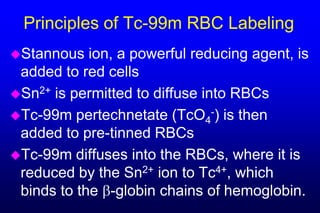
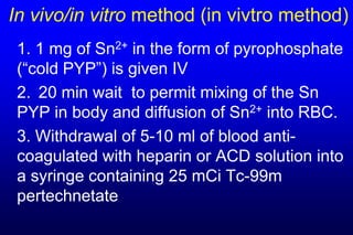
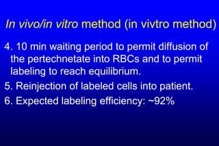
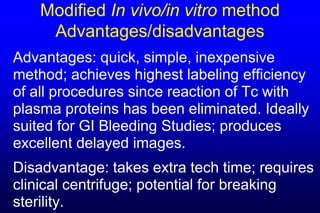
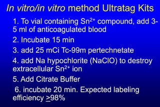
Cell Labeling


Stephen M. Karesh, Ph.D.
Cell Labeling topics to be covered


Red  Cells:    Tc-99m and Cr-51
White Cells:   Tc-99m and In-111
Platelets:     In-111
Uses of Tc-99m Red Cells

FirstPass Cardiac study
Gated Blood Pool study
G.I. Bleed Study
Localization of hepatic hemangioma
Principles of Tc-99m RBC Labeling
Stannous   ion, a powerful reducing agent, is
 added to red cells
Sn2+ is permitted to diffuse into RBCs
Tc-99m pertechnetate (TcO4-) is then
 added to pre-tinned RBCs
Tc-99m diffuses into the RBCs, where it is
 reduced by the Sn2+ ion to Tc4+, which
 binds to the -globin chains of hemoglobin.
Principles of Tc-99m RBC Labeling
Reduced   Tc-99m (Tc4+)cannot bind to the
 heme portion of the hemoglobin molecule
 since the Fe incorporated into the heme
 can not be displaced under physiological
 conditions. Tc4+ therefore binds only to the
 globin portion of the molecule, specifically
 the -globin chains.
NOW HEAR THIS!!!
 Every  container which
  holds a blood sample


                                                                 Full Name
  must be labeled with the


                                                     Full Name
                                         Full Name
  patient’s FULL NAME
 Syringes
                             Full Name
 Test tubes
 Vials
Methods of Preparation of Tc-99m RBCs


          • In vivo/in vivo
          • In vivo/in vitro
          • In vitro/in vitro
Methods of Preparation of Tc-99m RBCs

                    In vivo / in vivo
                    In vivo / in vitro
                   In vitro / in vitro
Refers to                                Refers to
tinning of the                           radiolabeling
cells, i.e.,                             of the cells,
injection of the                         i.e., injection of
stannous                                 the Tc-99m
pyrophosphate                            pertechnetate
In vivo/in vivo method
1. 1 mg of Sn2+ in the form of pyrophosphate
(“cold PYP”) is given IV
2. 20 min wait to permit mixing of the Sn
PYP in body and diffusion of Sn2+ into RBC.
3. IV injection of 25 mCi 99mTc pertechnetate
4. 10 min waiting period to permit diffusion of
pertechnetate into RBC's where radio-
labeling takes place.
In Vivo/In Vivo:
    Advantages/disadvantages

Advantages: quick, simple, inexpensive

Disadvantage: lowest labeling efficiency of
all commonly used procedures, but
perfectly acceptable for routine work, e.g.,
MUGAs.
In vivo/in vitro method (in vivtro method)
 1. 1 mg of Sn2+ in the form of pyrophosphate
 (“cold PYP”) is given IV
 2. 20 min wait to permit mixing of the Sn
 PYP in body and diffusion of Sn2+ into RBC.
 3. Withdrawal of 5-10 ml of blood anti-
 coagulated with heparin or ACD solution into
 a syringe containing 25 mCi Tc-99m
 pertechnetate
In vivo/in vitro method (in vivtro method)
4. 10 min waiting period to permit diffusion of
 the pertechnetate into RBCs and to permit
 labeling to reach equilibrium.
5. Reinjection of labeled cells into patient.
6. Expected labeling efficiency: ~92%
In vivo/in vitro:
    Advantages/disadvantages
Advantages: quick, simple, inexpensive
method; achieves higher labeling efficiency
than in vivo/in vivo technique since
incubation with RBC is extracorporeal. Not
optimal for GI Bleeding Studies.
Disadvantages: takes extra tech time;
potential for breaking sterility
Modified In vivo/in vitro method
1. 1 mg of Sn2+ in the form of pyrophosphate
(“cold PYP”) is given IV.
2. 20 min wait to permit mixing of the Sn
PYP in body and diffusion of Sn2+ into RBC.
3. Withdrawal of 5-10 ml of anti-coagulated
blood (heparin, ACD) into vacutainer.
4. Centrifuge the vacutainer in inverted
position for 5 min at 3000 rpm.
Modified In vivo/in vitro method

5. Removal of 1-2 ml of packed cells
through a 20 ga or larger needle.
6. Aseptic addition of these tinned, packed
cells to a sterile vial containing 35 mCi of
Tc-99m pertechnetate.
7. 10 min incubation to permit labeling
reaction to go to completion. Expected
labeling efficiency: 98-100%
8. Reinjection of Tc RBC
Modified In vivo/in vitro method
     Advantages/disadvantages
Advantages: quick, simple, inexpensive
method; achieves highest labeling efficiency
of all procedures since reaction of Tc with
plasma proteins has been eliminated. Ideally
suited for GI Bleeding Studies; produces
excellent delayed images.
Disadvantage: takes extra tech time; requires
clinical centrifuge; potential for breaking
sterility.
In vitro/in vitro method Ultratag Kits
 1. To vial containing Sn2+ compound, add 3-
 5 ml of anticoagulated blood
 2. Incubate 15 min
 3. add 25 mCi Tc-99m pertechnetate
 4. add Na hypochlorite (NaClO) to destroy
 extracellular Sn2+ ion
 5. Add Citrate Buffer
 6. incubate 20 min. Expected labeling
 efficiency >98%
In vitro/in vitro (Ultratag):
    Advantages/disadvantages
Advantages: quick, simple, inexpensive
method; achieves higher labeling efficiency
than in vivo/in vivo technique since incubation
with RBC is extracorporeal. Ideally suited for
GI Bleeding Studies; produces excellent
delayed images.
Disadvantage: takes extra tech time; slight
potential for breaking sterility
Quality Control for Tc-99m RBCs
Take 0.2 mL of labeled RBC suspension
Add to 2 mL 0.9% NaCl; mix gently
Centrifuge 5 min. Pipette off diluted plasma
Count Radioactivity in plasma and in
 packed RBC
% RBC Labeling = CountsRBC x 100%
                   CountsRBC + CountsPlasma
Heat Damaged Tc-99m RBCs
Used   for spleen imaging
  –Accessory spleen
  –Splenic remnants after splenectomy
Prepare Tc-99m RBCs by one of the
 in vitro methods
Incubate 30 minutes at 50ºC
Heat damaged RBCs are removed
 from circulation by the spleen
Tc-99m Labeled WBCs
Used  for localizing infection and abscesses
Principle of labeling
 –Tc-99m Ceretec is lipophilic and crosses
  lipid bilayer of cell membranes
 –Inside the cell the Ceretec complex is
  broken down and the resulting charged
  Tc-99m species is trapped in the cell
 –All cell types are labeled by Ceretec so
  prior leukocyte separation is necessary
Tc-99m WBC Labeling Procedure
Check  patient’s white count
 (must be > 2K)
Obtain ~50 mL anticoagulated
 whole blood from pt (larger
 volume if WBC count is low)
Optional: add Hetastarch as a
 sedimentation aid
Centrifuge at 15 x g for 10 min
Tc-99m WBC Labeling Procedure
Aseptically remove the platelet-rich &
 leukocyte-rich supernatant
Centrifuge at 200 x g for 10 min.
Decant platelet-rich plasma and then
 wash cells with 0.9% NaCl
Add ~40 mCi freshly prepared Tc-99m
 Ceretec dropwise to leukocyte pellet
 and incubate for 20 min. Do NOT use
 Methylene Blue.
Tc-99m WBC Labeling Procedure
Centrifuge, remove unbound Tc-
 99m, and wash pellet.
Resuspend labeled WBC in either
 platelet poor plasma or 0.9% NaCl
Labeling 50 - 80 %.
Draw up dose and inject patient. If
 patient has been on dialysis, wait
 until procedure is complete
Quality Control of Tc-99m WBC
Place  a drop of labeled cells on
 a hemocytometer
Add a drop of Trypan Blue dye
Add a cover slip and examine
 under microscope
Viable cells exclude dye; dead
 cells are stained blue
If >10% are dead or large
 clumps are present, don’t use
Labeling In-111 WBC
Used  for localizing infection and abscesses
Principle of labeling
 –In-111 Oxine is lipophilic and crosses lipid
  bilayer of cell membranes
 –Inside the cell the Oxine complex is
  broken down and the resulting charged In-
  111 species is trapped in the cell
 –All cell types are labeled so prior
  leukocyte separation is necessary
In-111 WBC Labeling Procedure
Check  patient’s WBC count; adjust if
 necessary
Separate WBC fraction as previously
 described
Add 500-1000 µCi In-111 Oxine.
Radiolabel as previously described.
 Labeling typically 50-95%.
QC is the same as for Tc-99m WBC
WBC Separation by Osmotic Lysis
RBCs  are more susceptible to lysis from
 osmotic pressure than WBCs
Add sterile H2O to WBC pellet
 contaminated w/ RBCs
  – This lowers the concentration of ions, etc.
  – The result is increased pressure in cells
  – RBCs lyse (burst open and die)
Quickly   add 5% NaCl to restore tonicity
WBC Separation by Osmotic Lysis
 Centrifuge
 WBC   sediment, Platelets and RBC
  debris remain in supernatant
 Remove supernatant
 Resuspend WBC
 Label WBC with Tc-99m or In-111
Labeled Platelets
Separate  platelets from whole
 blood by centrifugation
Label by incubation with lipophilic
 complexes of Tc-99m or In-111
Uses
 –Localization of thrombi
 –Platelet survival studies
Cr-51 Labeled RBCs
Uses
 –RBC Mass
 –RBC Survival and Sequestration
Properties of Cr-51
 –Half-life = 28 days
 –Gamma energy = 320 keV
Principle of Labeling Cr-51RBCs

 Cr-51  in the form of Na2CrO4
  (+6 oxidation state) is
  incubated with RBCs
 Cr-51 binds to RBCs
 Reaction terminated by
  adding a reducing agent such
  as ascorbic acid
Procedure for Labeling Cr-51 RBCs
Obtain 25-30 mL of anticoagulated blood
Combine blood with 75 - 150 µCi Cr-51
Incubate 20-30 minutes at 37ºC or at
 room temp
Add 100 mg ascorbic acid and incubate
 5 more minutes
Remove and use the labeled cells
Summary and Conclusions
1. Multiple methods are available for labeling
   RBCs with Tc-99m. All are simple, rapid,
   inexpensive, and reliable
2. GI Bleeding studies are optimally
   performed with Tc-99m RBCs prepared
   using the packed cell technique or the
   UltraTag kit.
Summary and Conclusions
3. It is easy to label WBCs with either Tc-99m
   or In-111. Both preparations involve
   isolation of the WBCs and incubation with
   a lipophilic intermediate to effect labeling.
4. It is very easy to label red cells with Cr-51.
   Isolation of RBCs is not required.
5. Most labeling procedures can be
   performed in any laboratory by a person
   knowledgeable about aseptic technique.

Red blood celllabeling (1)

  • 1.
  • 2.
    Cell Labeling topicsto be covered Red Cells: Tc-99m and Cr-51 White Cells: Tc-99m and In-111 Platelets: In-111
  • 3.
    Uses of Tc-99mRed Cells FirstPass Cardiac study Gated Blood Pool study G.I. Bleed Study Localization of hepatic hemangioma
  • 4.
    Principles of Tc-99mRBC Labeling Stannous ion, a powerful reducing agent, is added to red cells Sn2+ is permitted to diffuse into RBCs Tc-99m pertechnetate (TcO4-) is then added to pre-tinned RBCs Tc-99m diffuses into the RBCs, where it is reduced by the Sn2+ ion to Tc4+, which binds to the -globin chains of hemoglobin.
  • 5.
    Principles of Tc-99mRBC Labeling Reduced Tc-99m (Tc4+)cannot bind to the heme portion of the hemoglobin molecule since the Fe incorporated into the heme can not be displaced under physiological conditions. Tc4+ therefore binds only to the globin portion of the molecule, specifically the -globin chains.
  • 6.
    NOW HEAR THIS!!! Every container which holds a blood sample Full Name must be labeled with the Full Name Full Name patient’s FULL NAME  Syringes Full Name  Test tubes  Vials
  • 7.
    Methods of Preparationof Tc-99m RBCs • In vivo/in vivo • In vivo/in vitro • In vitro/in vitro
  • 8.
    Methods of Preparationof Tc-99m RBCs In vivo / in vivo In vivo / in vitro In vitro / in vitro Refers to Refers to tinning of the radiolabeling cells, i.e., of the cells, injection of the i.e., injection of stannous the Tc-99m pyrophosphate pertechnetate
  • 9.
    In vivo/in vivomethod 1. 1 mg of Sn2+ in the form of pyrophosphate (“cold PYP”) is given IV 2. 20 min wait to permit mixing of the Sn PYP in body and diffusion of Sn2+ into RBC. 3. IV injection of 25 mCi 99mTc pertechnetate 4. 10 min waiting period to permit diffusion of pertechnetate into RBC's where radio- labeling takes place.
  • 10.
    In Vivo/In Vivo: Advantages/disadvantages Advantages: quick, simple, inexpensive Disadvantage: lowest labeling efficiency of all commonly used procedures, but perfectly acceptable for routine work, e.g., MUGAs.
  • 11.
    In vivo/in vitromethod (in vivtro method) 1. 1 mg of Sn2+ in the form of pyrophosphate (“cold PYP”) is given IV 2. 20 min wait to permit mixing of the Sn PYP in body and diffusion of Sn2+ into RBC. 3. Withdrawal of 5-10 ml of blood anti- coagulated with heparin or ACD solution into a syringe containing 25 mCi Tc-99m pertechnetate
  • 12.
    In vivo/in vitromethod (in vivtro method) 4. 10 min waiting period to permit diffusion of the pertechnetate into RBCs and to permit labeling to reach equilibrium. 5. Reinjection of labeled cells into patient. 6. Expected labeling efficiency: ~92%
  • 13.
    In vivo/in vitro: Advantages/disadvantages Advantages: quick, simple, inexpensive method; achieves higher labeling efficiency than in vivo/in vivo technique since incubation with RBC is extracorporeal. Not optimal for GI Bleeding Studies. Disadvantages: takes extra tech time; potential for breaking sterility
  • 14.
    Modified In vivo/invitro method 1. 1 mg of Sn2+ in the form of pyrophosphate (“cold PYP”) is given IV. 2. 20 min wait to permit mixing of the Sn PYP in body and diffusion of Sn2+ into RBC. 3. Withdrawal of 5-10 ml of anti-coagulated blood (heparin, ACD) into vacutainer. 4. Centrifuge the vacutainer in inverted position for 5 min at 3000 rpm.
  • 15.
    Modified In vivo/invitro method 5. Removal of 1-2 ml of packed cells through a 20 ga or larger needle. 6. Aseptic addition of these tinned, packed cells to a sterile vial containing 35 mCi of Tc-99m pertechnetate. 7. 10 min incubation to permit labeling reaction to go to completion. Expected labeling efficiency: 98-100% 8. Reinjection of Tc RBC
  • 16.
    Modified In vivo/invitro method Advantages/disadvantages Advantages: quick, simple, inexpensive method; achieves highest labeling efficiency of all procedures since reaction of Tc with plasma proteins has been eliminated. Ideally suited for GI Bleeding Studies; produces excellent delayed images. Disadvantage: takes extra tech time; requires clinical centrifuge; potential for breaking sterility.
  • 17.
    In vitro/in vitromethod Ultratag Kits 1. To vial containing Sn2+ compound, add 3- 5 ml of anticoagulated blood 2. Incubate 15 min 3. add 25 mCi Tc-99m pertechnetate 4. add Na hypochlorite (NaClO) to destroy extracellular Sn2+ ion 5. Add Citrate Buffer 6. incubate 20 min. Expected labeling efficiency >98%
  • 18.
    In vitro/in vitro(Ultratag): Advantages/disadvantages Advantages: quick, simple, inexpensive method; achieves higher labeling efficiency than in vivo/in vivo technique since incubation with RBC is extracorporeal. Ideally suited for GI Bleeding Studies; produces excellent delayed images. Disadvantage: takes extra tech time; slight potential for breaking sterility
  • 19.
    Quality Control forTc-99m RBCs Take 0.2 mL of labeled RBC suspension Add to 2 mL 0.9% NaCl; mix gently Centrifuge 5 min. Pipette off diluted plasma Count Radioactivity in plasma and in packed RBC % RBC Labeling = CountsRBC x 100% CountsRBC + CountsPlasma
  • 20.
    Heat Damaged Tc-99mRBCs Used for spleen imaging –Accessory spleen –Splenic remnants after splenectomy Prepare Tc-99m RBCs by one of the in vitro methods Incubate 30 minutes at 50ºC Heat damaged RBCs are removed from circulation by the spleen
  • 21.
    Tc-99m Labeled WBCs Used for localizing infection and abscesses Principle of labeling –Tc-99m Ceretec is lipophilic and crosses lipid bilayer of cell membranes –Inside the cell the Ceretec complex is broken down and the resulting charged Tc-99m species is trapped in the cell –All cell types are labeled by Ceretec so prior leukocyte separation is necessary
  • 22.
    Tc-99m WBC LabelingProcedure Check patient’s white count (must be > 2K) Obtain ~50 mL anticoagulated whole blood from pt (larger volume if WBC count is low) Optional: add Hetastarch as a sedimentation aid Centrifuge at 15 x g for 10 min
  • 23.
    Tc-99m WBC LabelingProcedure Aseptically remove the platelet-rich & leukocyte-rich supernatant Centrifuge at 200 x g for 10 min. Decant platelet-rich plasma and then wash cells with 0.9% NaCl Add ~40 mCi freshly prepared Tc-99m Ceretec dropwise to leukocyte pellet and incubate for 20 min. Do NOT use Methylene Blue.
  • 24.
    Tc-99m WBC LabelingProcedure Centrifuge, remove unbound Tc- 99m, and wash pellet. Resuspend labeled WBC in either platelet poor plasma or 0.9% NaCl Labeling 50 - 80 %. Draw up dose and inject patient. If patient has been on dialysis, wait until procedure is complete
  • 25.
    Quality Control ofTc-99m WBC Place a drop of labeled cells on a hemocytometer Add a drop of Trypan Blue dye Add a cover slip and examine under microscope Viable cells exclude dye; dead cells are stained blue If >10% are dead or large clumps are present, don’t use
  • 26.
    Labeling In-111 WBC Used for localizing infection and abscesses Principle of labeling –In-111 Oxine is lipophilic and crosses lipid bilayer of cell membranes –Inside the cell the Oxine complex is broken down and the resulting charged In- 111 species is trapped in the cell –All cell types are labeled so prior leukocyte separation is necessary
  • 27.
    In-111 WBC LabelingProcedure Check patient’s WBC count; adjust if necessary Separate WBC fraction as previously described Add 500-1000 µCi In-111 Oxine. Radiolabel as previously described. Labeling typically 50-95%. QC is the same as for Tc-99m WBC
  • 28.
    WBC Separation byOsmotic Lysis RBCs are more susceptible to lysis from osmotic pressure than WBCs Add sterile H2O to WBC pellet contaminated w/ RBCs – This lowers the concentration of ions, etc. – The result is increased pressure in cells – RBCs lyse (burst open and die) Quickly add 5% NaCl to restore tonicity
  • 29.
    WBC Separation byOsmotic Lysis Centrifuge WBC sediment, Platelets and RBC debris remain in supernatant Remove supernatant Resuspend WBC Label WBC with Tc-99m or In-111
  • 30.
    Labeled Platelets Separate platelets from whole blood by centrifugation Label by incubation with lipophilic complexes of Tc-99m or In-111 Uses –Localization of thrombi –Platelet survival studies
  • 31.
    Cr-51 Labeled RBCs Uses –RBC Mass –RBC Survival and Sequestration Properties of Cr-51 –Half-life = 28 days –Gamma energy = 320 keV
  • 32.
    Principle of LabelingCr-51RBCs Cr-51 in the form of Na2CrO4 (+6 oxidation state) is incubated with RBCs Cr-51 binds to RBCs Reaction terminated by adding a reducing agent such as ascorbic acid
  • 33.
    Procedure for LabelingCr-51 RBCs Obtain 25-30 mL of anticoagulated blood Combine blood with 75 - 150 µCi Cr-51 Incubate 20-30 minutes at 37ºC or at room temp Add 100 mg ascorbic acid and incubate 5 more minutes Remove and use the labeled cells
  • 34.
    Summary and Conclusions 1.Multiple methods are available for labeling RBCs with Tc-99m. All are simple, rapid, inexpensive, and reliable 2. GI Bleeding studies are optimally performed with Tc-99m RBCs prepared using the packed cell technique or the UltraTag kit.
  • 35.
    Summary and Conclusions 3.It is easy to label WBCs with either Tc-99m or In-111. Both preparations involve isolation of the WBCs and incubation with a lipophilic intermediate to effect labeling. 4. It is very easy to label red cells with Cr-51. Isolation of RBCs is not required. 5. Most labeling procedures can be performed in any laboratory by a person knowledgeable about aseptic technique.